Follicular Bronchiolitis Associated with Rheumatoid Arthritis
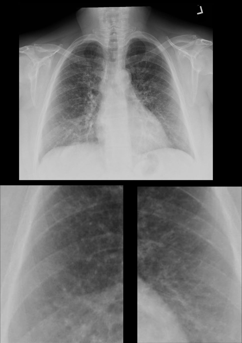
70-year-old female former smoker with long standing history of RA presents with chronic dyspnea. Frontal view of the chest reveals a coarsened nodular interstitial pattern with magnified views showing the micronodularity in the lower panels.
Ashley Davidoff MD TheCommonVein.net 132Lu 136650c01
Follicular Bronchiolitis,, Centrilobular Nodules, Air Trapping, Ground Glass Opacities (GGO) in Upper Lobes
70-year-old female former smoker with long standing history of RA presents with chronic dyspnea.
Axial CT of the chest at the level of the aortic arch reveals centrilobular nodules, ground-glass opacities, and mosaic attenuation (likely due to air trapping in this context) and bronchial wall thickening. In the context of a patient with rheumatoid arthritis a diagnosis of follicular bronchiolitis is likely. However radiologically fibrotic hypersensitivity pneumonitis (HP) is included in the differential diagnosis
Ashley Davidoff MD TheCommonVein.net 132Lu 136652
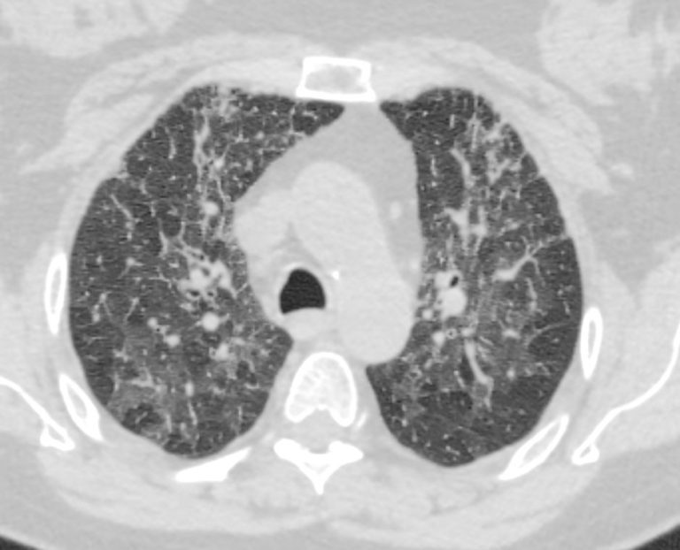
70-year-old female former smoker with long standing history of RA presents with chronic dyspnea.
Axial CT of the chest at the level of the aortic arch reveals centrilobular nodules (b, white arrowheads) , ground-glass opacities, and mosaic attenuation (b, white rings) likely due to air trapping in this context, and bronchial wall thickening (b, c teal rings). There is some irregular thickening of the interlobular septa. In the context of a patient with rheumatoid arthritis a diagnosis of follicular bronchiolitis is likely. However radiologically fibrotic hypersensitivity pneumonitis (HP) is included in the differential diagnosis
Ashley Davidoff MD TheCommonVein.net 132Lu 136652cL
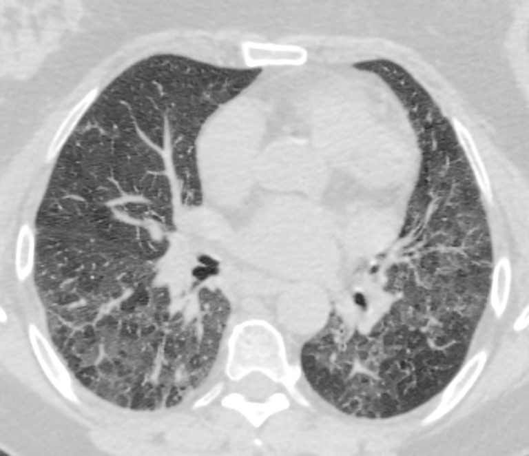
70-year-old female former smoker with long standing history of RA presents with chronic dyspnea.
Axial CT of the chest at the level of the lower lung fields reveals centrilobular nodules, ground-glass opacities, and mosaic attenuation (likely due to air trapping in this context). In the context of a patient with rheumatoid arthritis a diagnosis of follicular bronchiolitis is likely. However radiologically fibrotic hypersensitivity pneumonitis (HP) is included in the differential diagnosis
Ashley Davidoff MD TheCommonVein.net 132Lu 136657
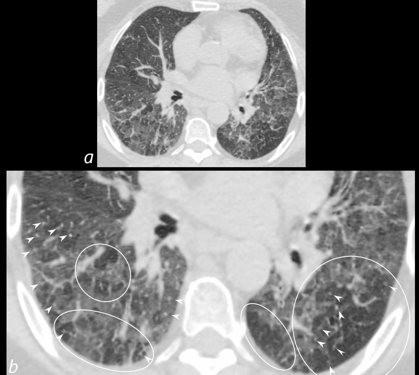
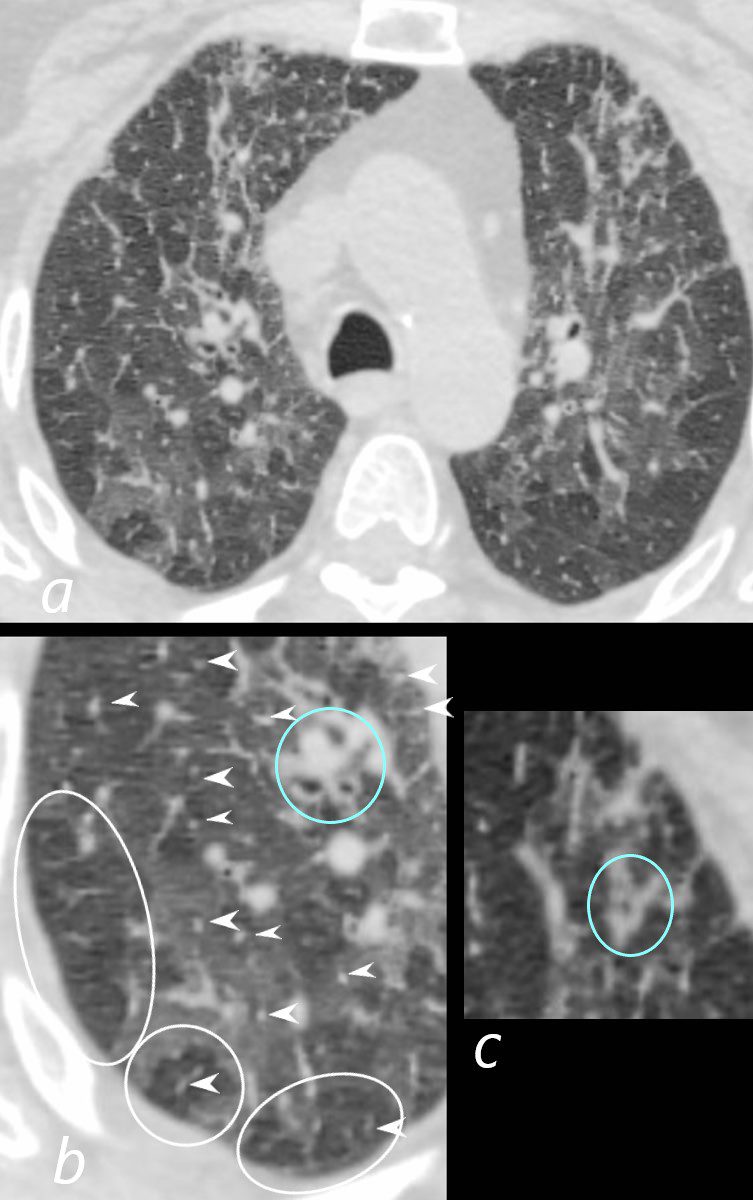
70-year-old female former smoker with long standing history of RA presents with chronic dyspnea.
Axial CT of the chest at the level of the lower lung fields reveals centrilobular nodules (b white arrowheads), ground-glass opacities, and mosaic attenuation (b, white rings) likely due to air trapping in this context.
In the context of a patient with rheumatoid arthritis a diagnosis of follicular bronchiolitis is likely. However radiologically fibrotic hypersensitivity pneumonitis (HP) is included in the differential diagnosis
Ashley Davidoff MD TheCommonVein.net 132Lu 136657cL
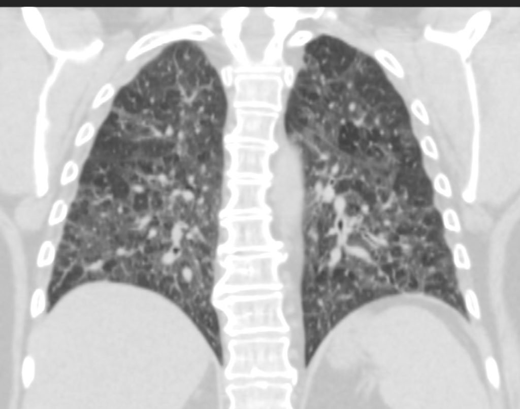
70-year-old female former smoker with long standing history of RA presents with chronic dyspnea.
CT in the coronal plane of the chest at the level of the spine reveals bilateral diffuse changes in the lungs characterized by centrilobular nodules, ground-glass opacities, mosaic attenuation (likely due to air trapping in this context) and irregular thickening of the interlobular septa.
In the context of a patient with rheumatoid arthritis a diagnosis of follicular bronchiolitis is likely. However radiologically fibrotic hypersensitivity pneumonitis (HP) is included in the differential diagnosis
Ashley Davidoff MD TheCommonVein.net 132Lu 136663
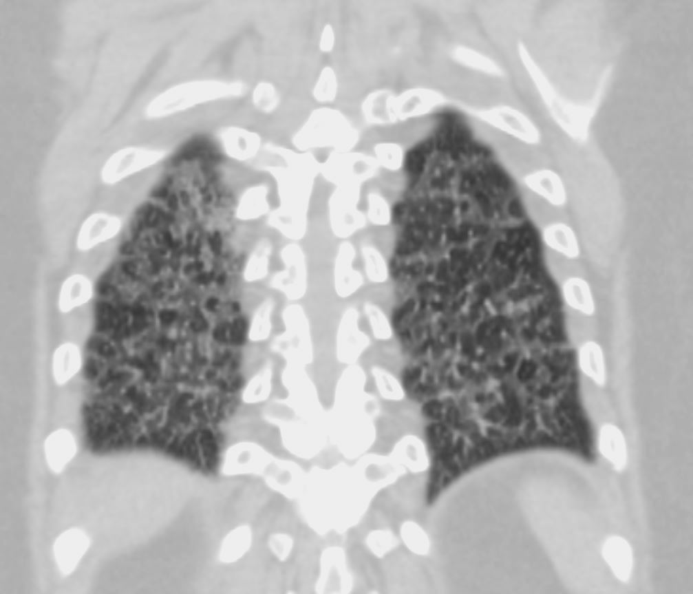
70-year-old female former smoker with long standing history of RA presents with chronic dyspnea.
CT in the coronal plane of the chest at the level of the spine reveals bilateral diffuse changes in the lungs characterized by centrilobular nodules, ground-glass opacities, mosaic attenuation (likely due to air trapping in this context) and irregular thickening of the interlobular septa.
In the context of a patient with rheumatoid arthritis a diagnosis of follicular bronchiolitis is likely. However radiologically fibrotic hypersensitivity pneumonitis (HP) is included in the differential diagnosis
Ashley Davidoff MD TheCommonVein.net 132Lu 136664
-

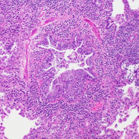
Hematoxylin and eosin (H&E) stain from surgical lung biopsy showing airway narrowing with enlarged peribronchiolar lymphoid follicles, consistent with follicular bronchiolitis.
Assaad M, Aqeel A, Walsh J (February 13, 2022) Follicular Bronchiolitis Associated With Primary IgG2/IgG4 Deficiency in a Previously Healthy 40-Year-Old Woman. Cureus 14(2): e22183. doi:10.7759/cureus.22183- aka bronchiolar nodular lymphoid hyperplasia,
- aka hyperplasia of the bronchial associated lymphoid tissue (BALT),
- is a
- reactive pulmonary lymphoid disorders
- part of as group of
- lymphoproliferative pulmonary diseases (LPDs).
- characterized by the
- accumulation of lymphoid cells in the
- walls of small airways.
- caused by
- antigenic stimulation of BALT, followed by a
- polyclonal lymphoid hyperplasia. It is currently classified as one of the reactive pulmonary lymphoid disorders in a group known as the lymphoproliferative pulmonary diseases (LPDs).
- primary
- secondary
- Connective Tissue Disorders
- Sjogren’s syndrome,
- rheumatoid arthritis, and
- systemic lupus erythematosus
- Infection
- Pneumocystis jirovecii,
- Legionella pneumonia, and
- acute viral hepatitis
- ILD
- lymphoid interstitial pneumonia,
- respiratory bronchiolitis-associated interstitial lung disease,
- desquamative interstitial pneumonia,
- cryptogenic organizing pneumonia, and
- Immunodeficiencies
- CVID and AIDS
- Connective Tissue Disorders