
- A pleural lipoma
- benign tumor of the the pleura, t\
- asymptomatic
- found incidentally on imaging studies,
- Clinical
- if they compress adjacent structures
- some may cause chest pain,
- coughing, or
- shortness of breath .
- if they compress adjacent structures
- Arise from
- mesenchymal cells that
- differentiate into adipose tissue
- during embryonic development.
- Diagnosis of pleural lipomas typically involves imaging studies such as chest X-ray, CT scan, or MRI, which can show a well-circumscribed mass composed of fat.
- In most cases, pleural lipomas do not require treatment unless they cause symptoms or enlarge over time. Surgical excision may be considered for symptomatic or growing tumors, but the risk of complications such as pneumothorax or bleeding should be weighed against the potential benefits.
- The prognosis of pleural lipomas is excellent, as they are benign and do not metastasize. However, long-term follow-up may be recommended to monitor for recurrence or growth.
Ashley Davidoff MD TheCommonVein.net 22663c
Ashley Davidoff MD TheCommonVein.net
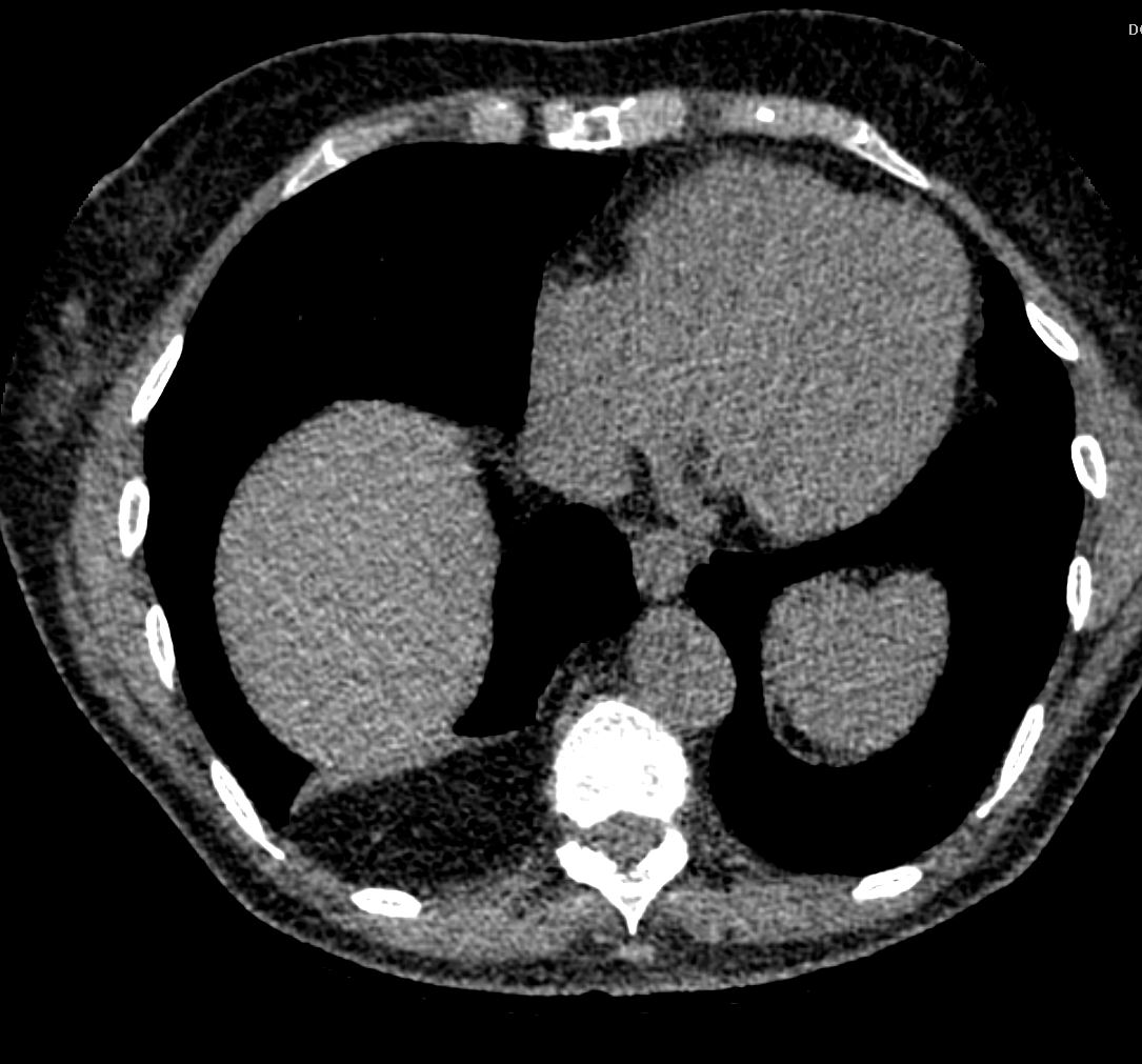
Diaphragm Intact ie not a Bochdalek hernia
Ashley Davidoff MD TheCommonVein.net
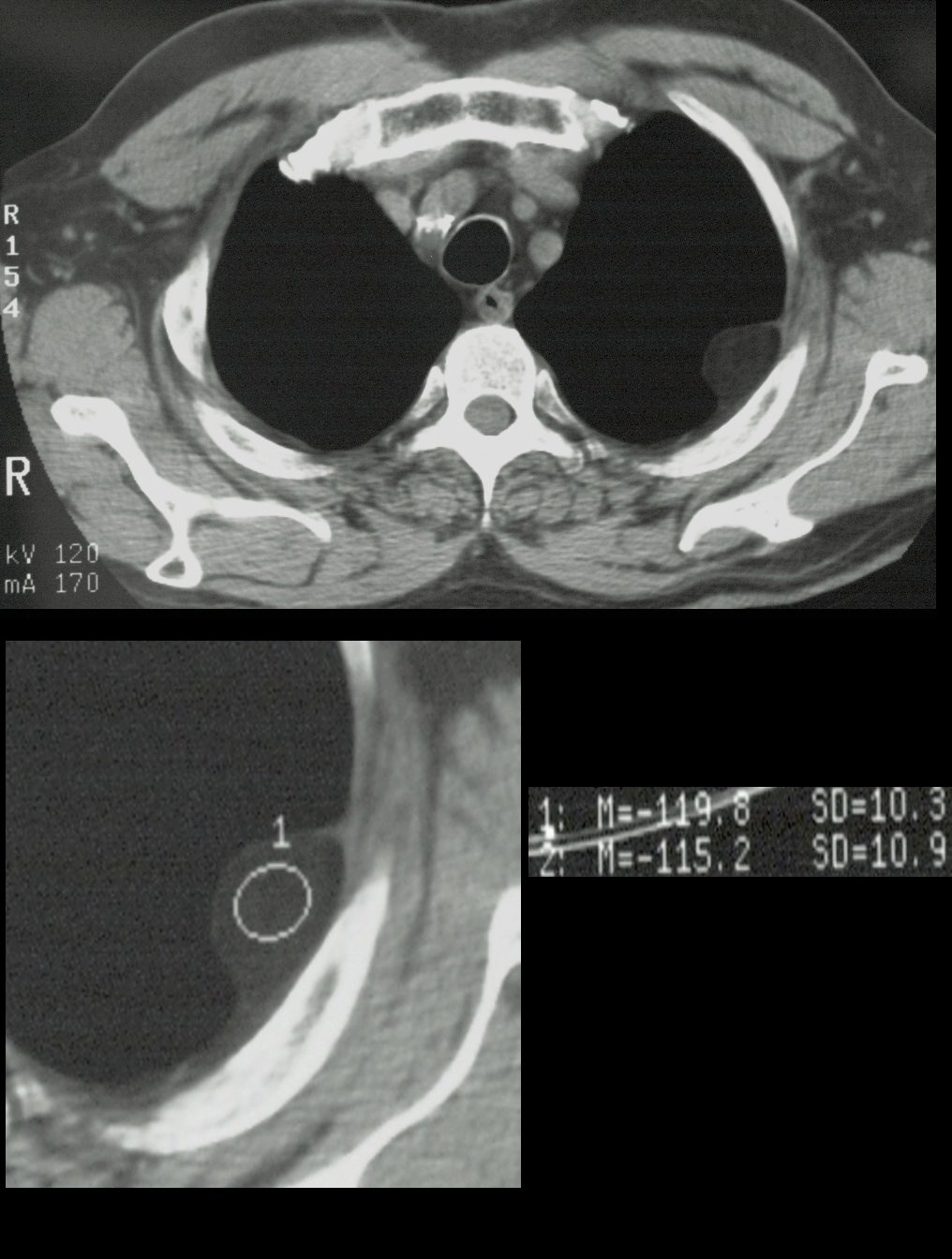
53-year-old male presents with chest pain
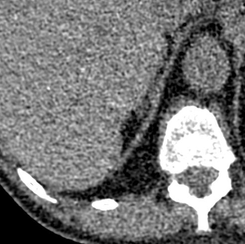
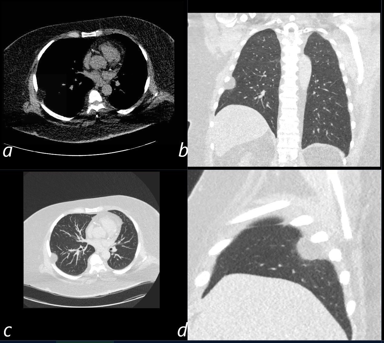
CT without contrast shows a low density pleural based mass with obtuse angles with the pleural interface. Asial CT on ST windows shows a 1.8cms mass that has the same density as the subcutaneous fat. The lung windows reveal the fatty tumor in coronal (b) axial (c) and sagittal view (d).
These findings are consistent with a pleural lipoma
Ashley Davidoff MD TheCommonVein.net 136618c
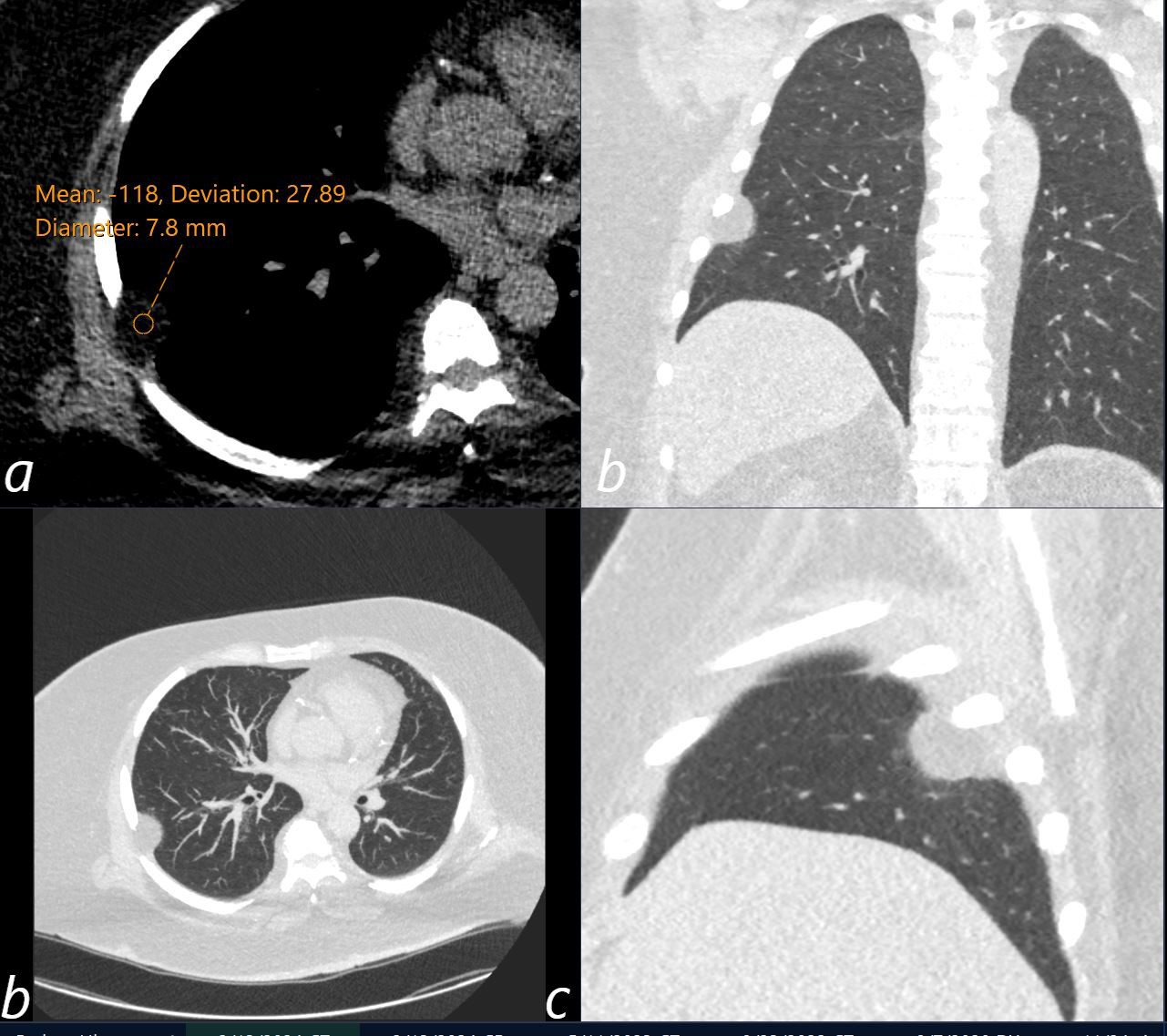
53-year-old male presents with chest pain.
CT without contrast shows a low density pleural based 1.8cms mass with obtuse angles with the pleural interface. The density of the mass is -118HU consistent with a pleural lipoma (a)
The lung windows reveal the fatty tumor in coronal (b) axial (c) and sagittal view (d).
These findings are consistent with a pleural lipoma
Ashley Davidoff MD TheCommonVein.net 136619cL