
Ashley Davidoff MD TheCommonvein.net lungs-0770
Infection
TB
Ashley Davidoff MD TheCommonVein.net
PCP
Inflammation
Sarcoidosis
Ashley Davidoff TheCommonVein.net

Courtesy Maegan Lu, Jonathan Scalera, MD
Hypersensitivity Pneumonitis

Crack Lung
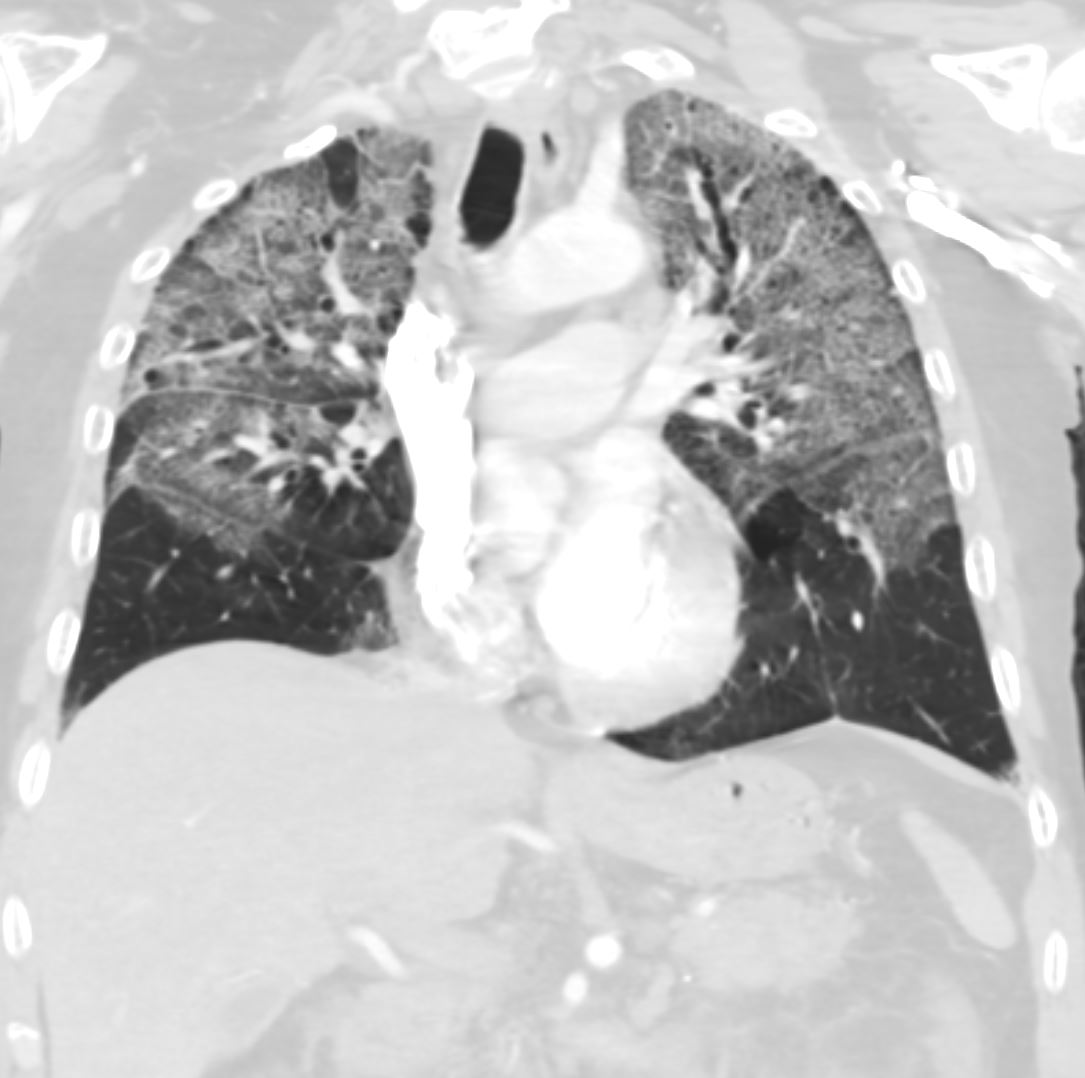
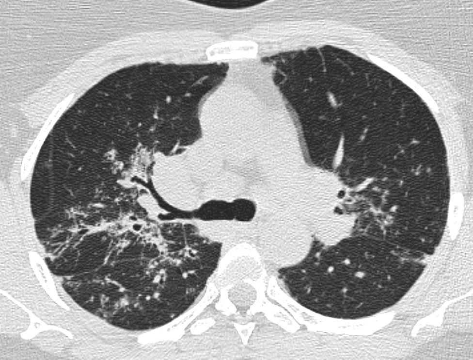
55-year-old male with substance use disorder presents with progressive and now more severe dyspnea. Coronal CT through the mid lung fields shows upper lobe predominant ground glass changes with thickening of the interlobular septa and a “crazy paving” appearance is suggested. The superior segments of the lower lobes are also involved. Thickening and irregularity of the right and left major fissures and the transverse fissure are noted. LVH is suggested.
Progressive inhalational pneumonitis from smoking or cocaine inhalation was suspected. DIP and hypersensitivity pneumonitis remained in the differential diagnosis
Ashley Davidoff MD TheCommonVein.net 251Lu 135934
Malignancy Mechanical/Atelectasis Trauma
Metabolic
Alveolar Proteinosis
Follow up Ashley Davidoff TheCommonVein.net 117528.8

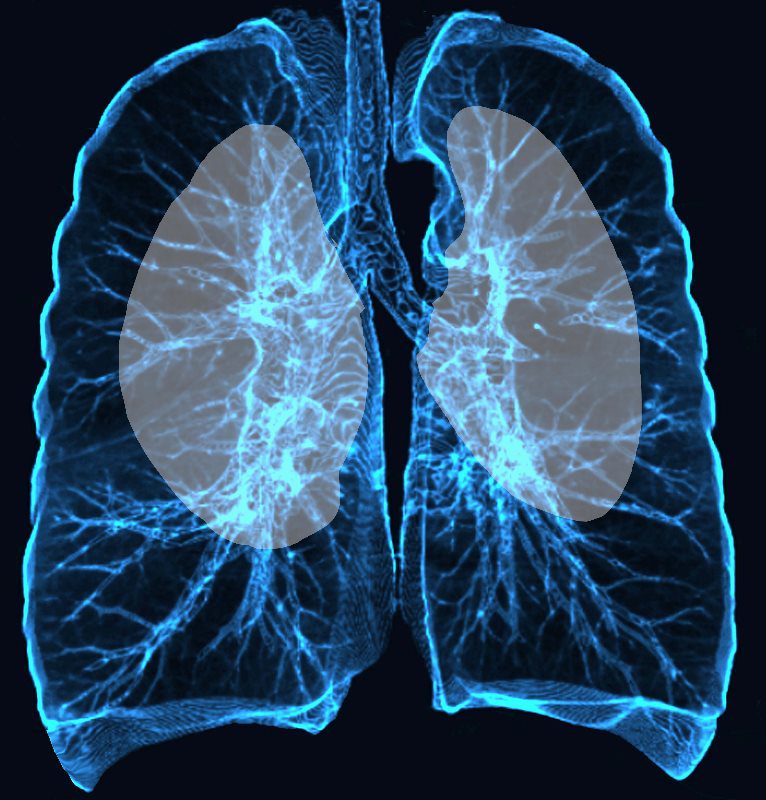
central distribution
Ashley Davidoff TheCommonVein.net
Circulatory- CHF
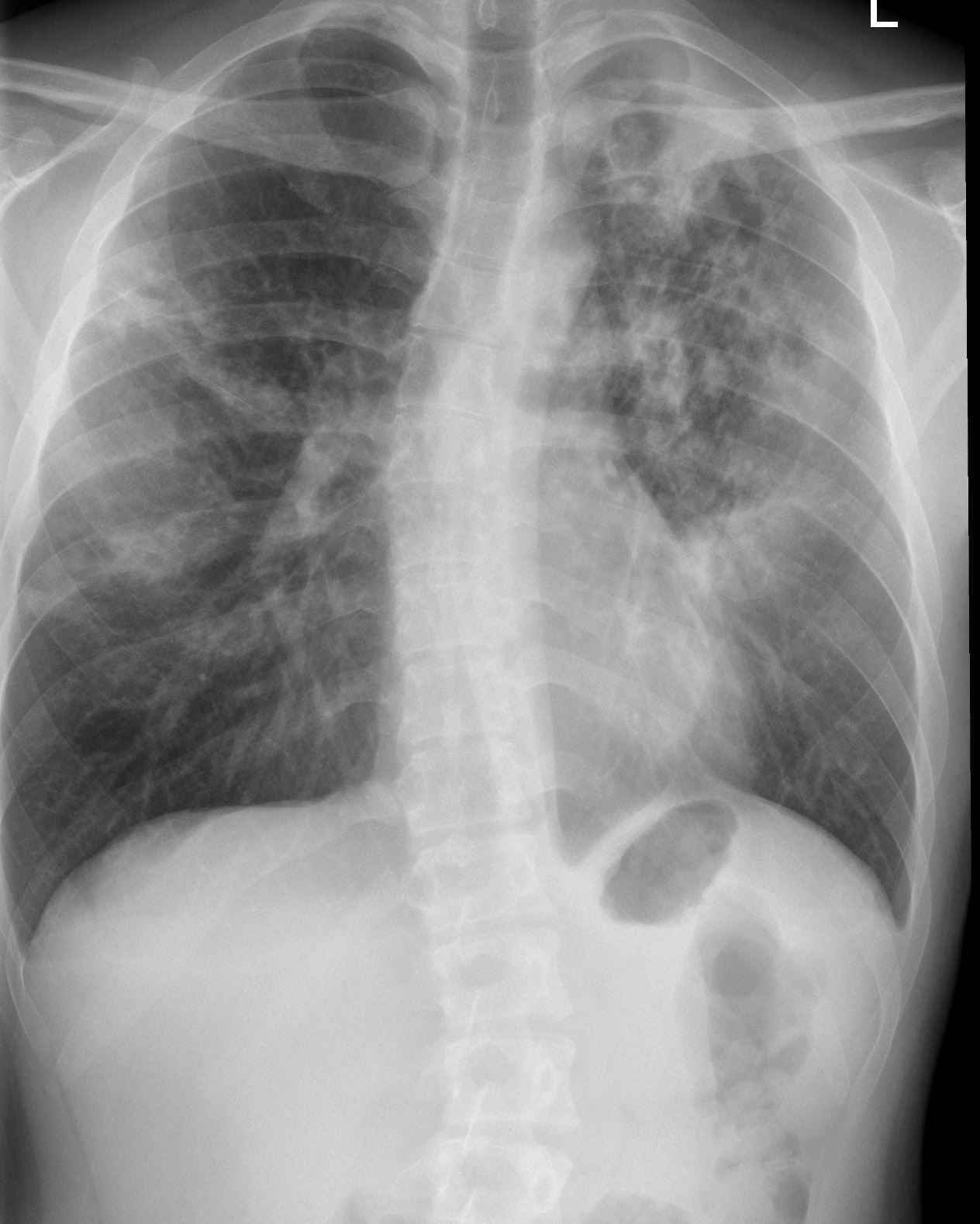
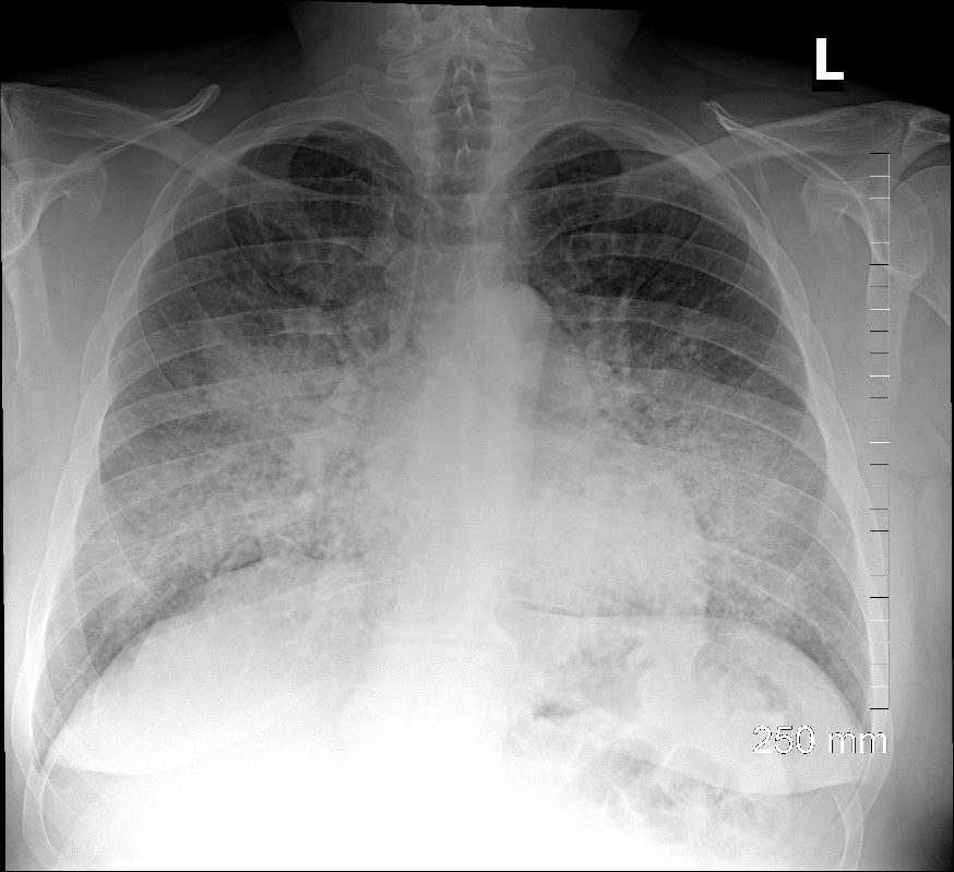
70 year old female s/p cardiac arrest and ROSC. CXR shows centralized alveolar edema as a result of intra- alveolar accumulation of transudate. The centralized distribution of the infiltrates is characteristic of severe heart failure – batwing distribution
Ashley Davidoff MD TheCommonVein.net
Mild early Severe CHF
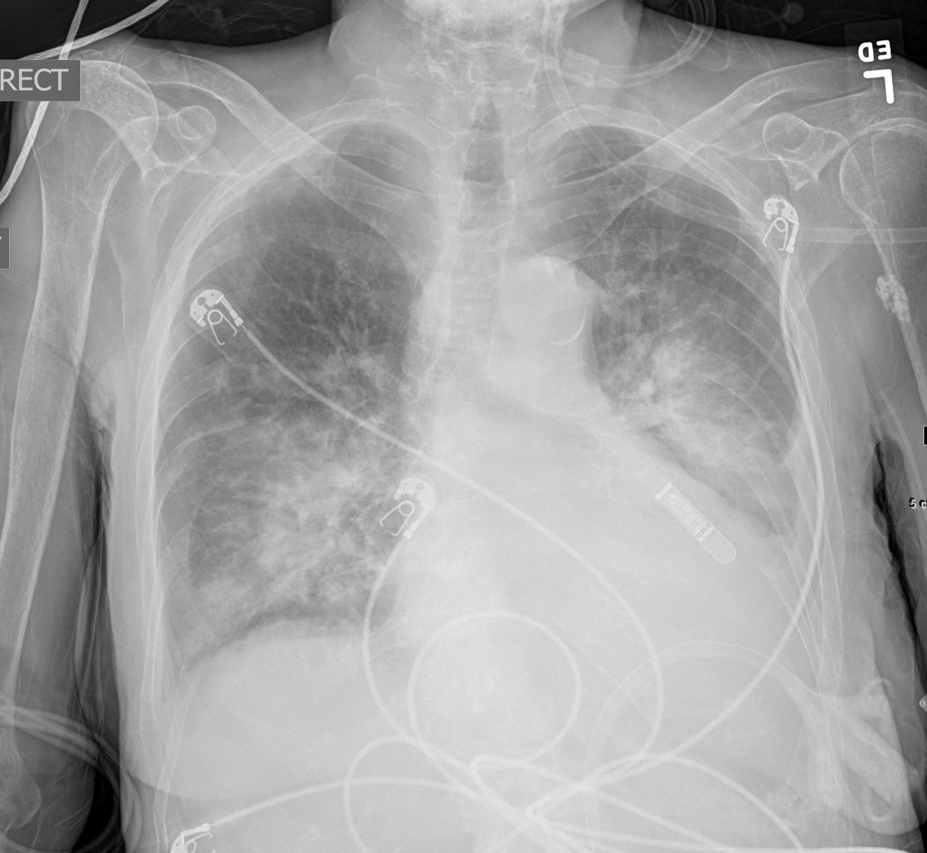
50 year-old male presents with severe dyspnea and orthopnea. CXR in the frontal projection shows perihilar congestion with batwing distribution, left atrial enlargement and left ventricular configuration of the heart. These findings are consistent with severe heart failure with a projected LVEDP of greater than 30mmHg
Ashley Davidoff MD TheCommonVein.net 285Lu 135759
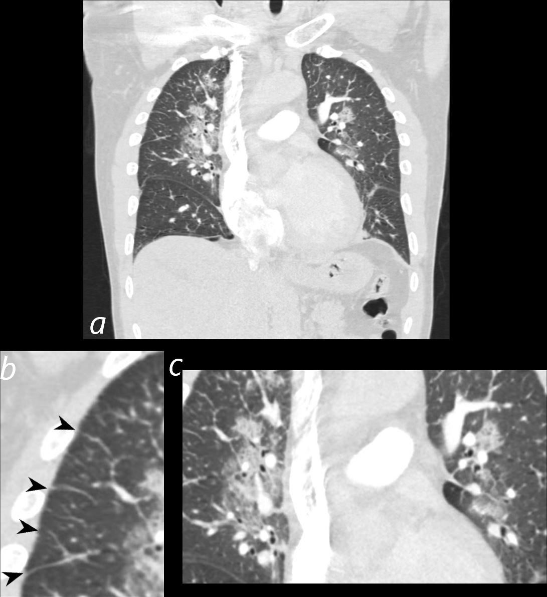
50 year-old male presents with severe dyspnea and orthopnea. CT in the coronal plain shows early severe CHF, with perihilar ground glass changes (magnified in c) and Kerley B lines (b, black arrowheads) These findings are consistent with early severe heart failure with a projected LVEDP of greater than 30mmHg
Ashley Davidoff MD TheCommonVein.net 285Lu 135761cL
Hemorrhage Immune Infiltrative Idiopathic Iatrogenic Idiopathic