
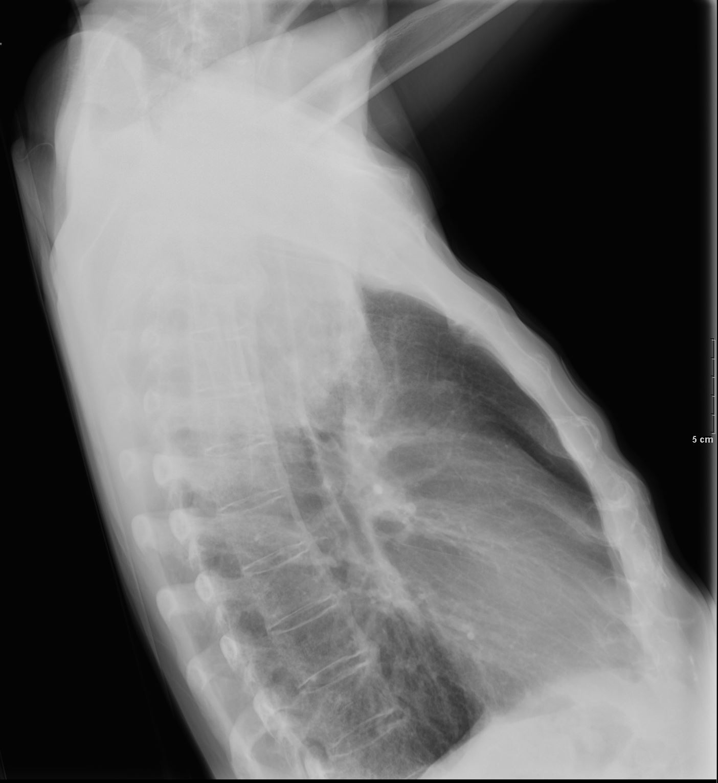
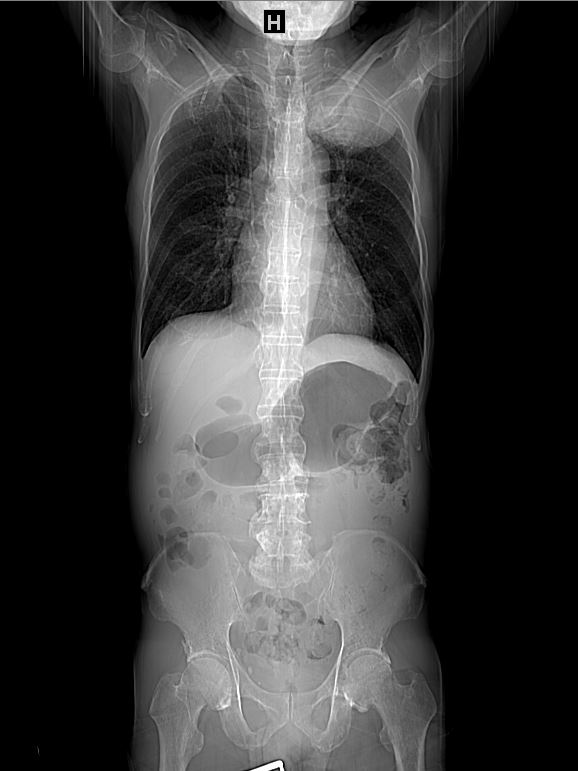
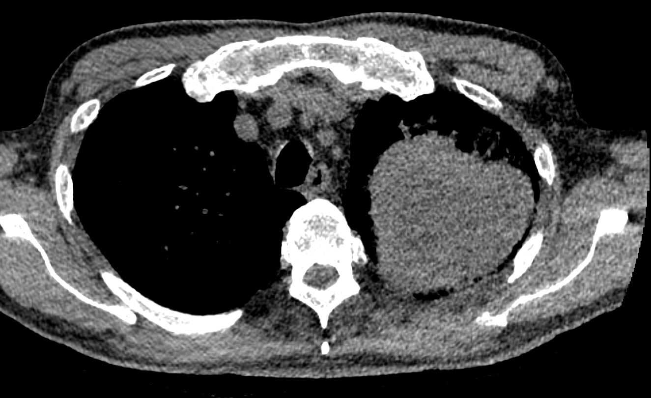
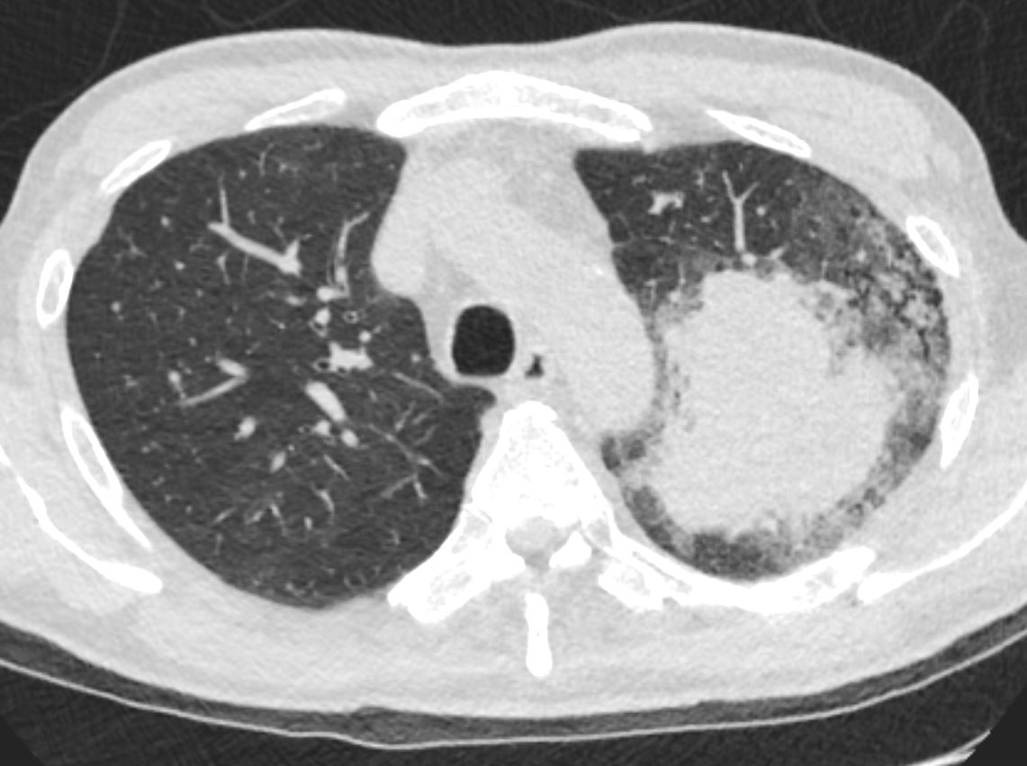
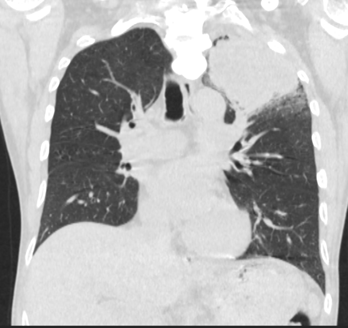
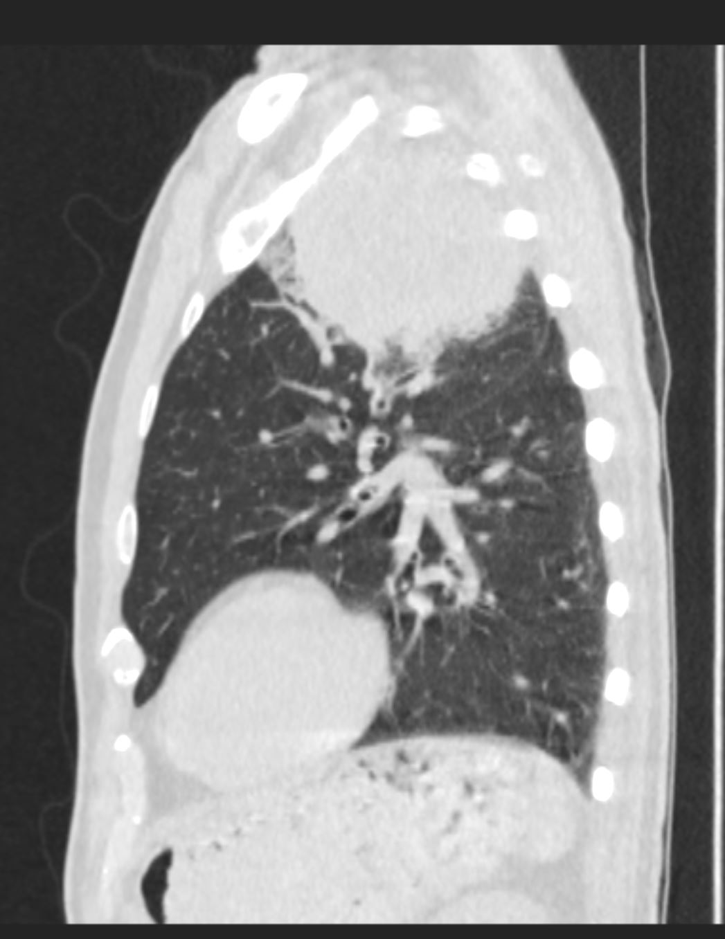
53 year old male history of stage IIIB adeno carcinoma of the left upper lobe with prevascular and AP window lymph node involvement based on PET/CT with presumed lymphangitic spread surrounding the large mass. Treatment has included both radiation and chemotherapy
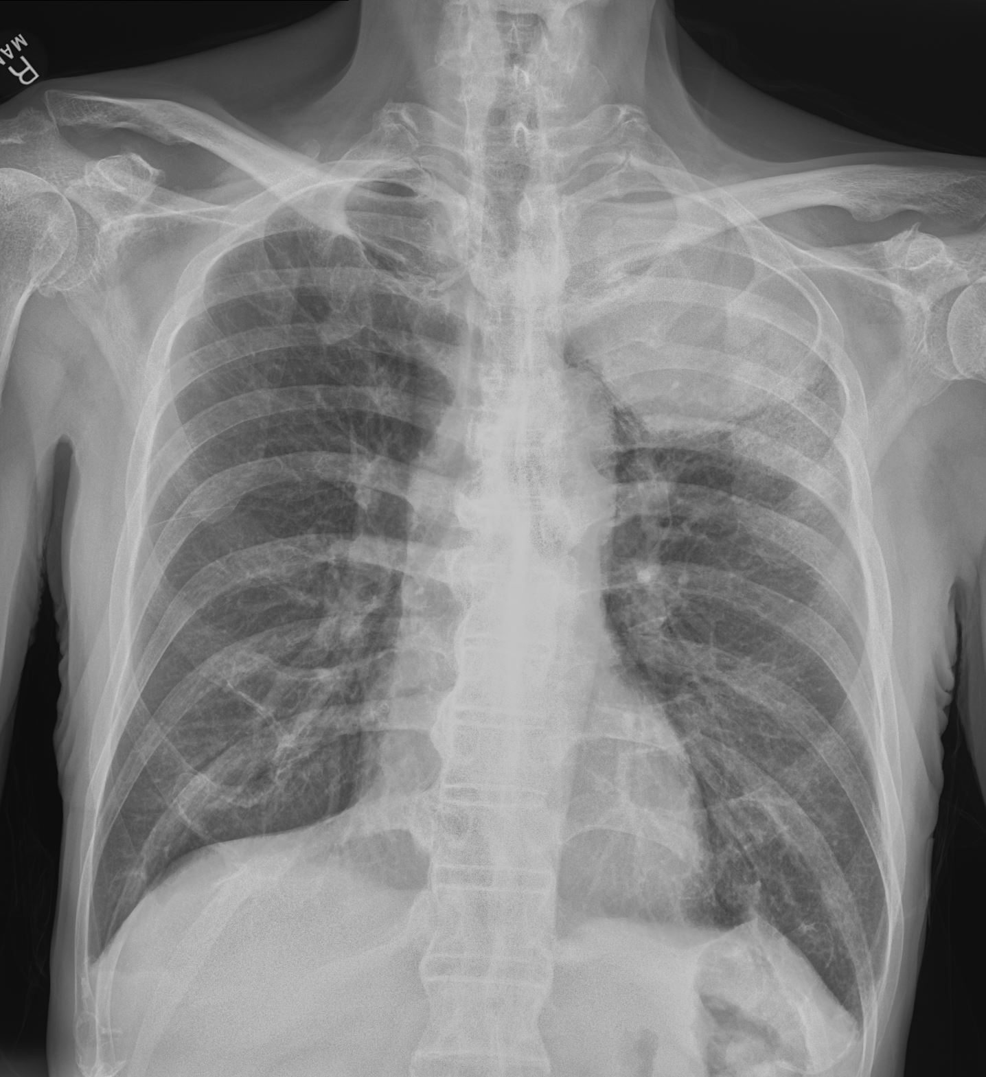
Diagnosis – adenocarcinoma of the lung with extensive necrosis of the tumor
Ashley Davidoff MD The CommonVein.net
Diagnosis – adenocarcinoma of the lung with extensive necrosis of the tumor
Ashley Davidoff MD The CommonVein.net
Diagnosis – adenocarcinoma of the lung with extensive necrosis of the tumor
Ashley Davidoff MD The CommonVein.net
Diagnosis – adenocarcinoma of the lung with extensive necrosis of the tumor
Ashley Davidoff MD The CommonVein.net
Diagnosis – adenocarcinoma of the lung with extensive necrosis of the tumor
Ashley Davidoff MD The CommonVein.net
Diagnosis – adenocarcinoma of the lung with extensive necrosis of the tumor
Ashley Davidoff MD The CommonVein.net
Diagnosis – adenocarcinoma of the lung with extensive necrosis of the tumor
Ashley Davidoff MD The CommonVein.net
Diagnosis – adenocarcinoma of the lung with extensive necrosis of the tumor
Ashley Davidoff MD The CommonVein.net
Diagnosis – adenocarcinoma of the lung with extensive necrosis of the tumor
Ashley Davidoff MD The CommonVein.net
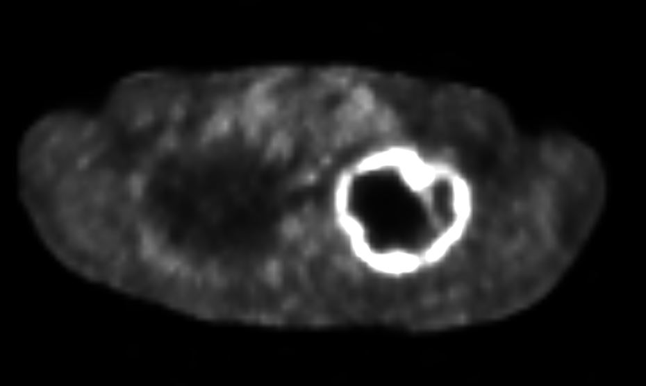
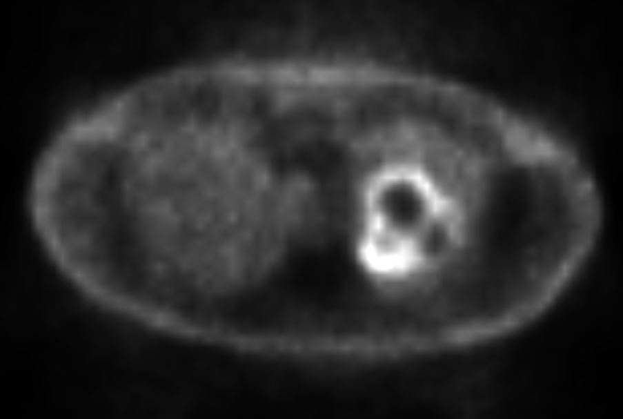
53 year old male with history of smoking presents with a cough PET CT shows a large 8.9 cm left upper lobe apical lung mass which is peripherally intensely hypermetabolic, centrally photopenic there is suspicious for lung malignancy with central necrosis The tumor circumferentially shows mild tracer uptake and becomes suspicious for lymphangitis carcinomatosa
Diagnosis – adenocarcinoma of the lung with extensive necrosis of the tumor
Ashley Davidoff MD The CommonVein.net