
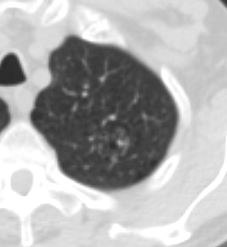
6 Years Prior left Upper Lobe Complex Cystic Lesion
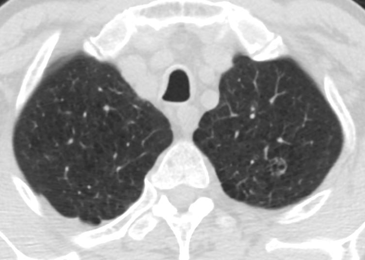
Final diagnosis Squamous Cell Carcinoma
Ashley Davidoff MD TheCommonVein.net
Final diagnosis Squamous Cell Carcinoma
Ashley Davidoff MD TheCommonVein.net
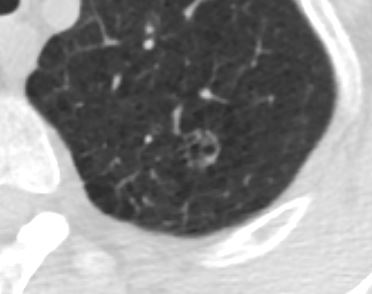
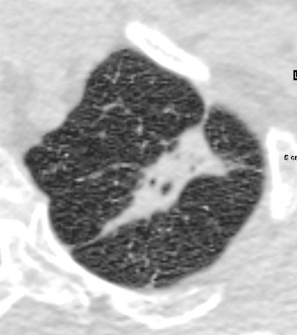
Growth of Cystic Lesion Over 6months
Final diagnosis Squamous Cell Carcinoma
Ashley Davidoff MD TheCommonVein.net
Final diagnosis Squamous Cell Carcinoma
Ashley Davidoff MD TheCommonVein.net
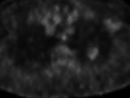
PET CT One Month Later
Final diagnosis Squamous Cell Carcinoma
Ashley Davidoff MD TheCommonVein.net
Final diagnosis Squamous Cell Carcinoma
Ashley Davidoff MD TheCommonVein.net
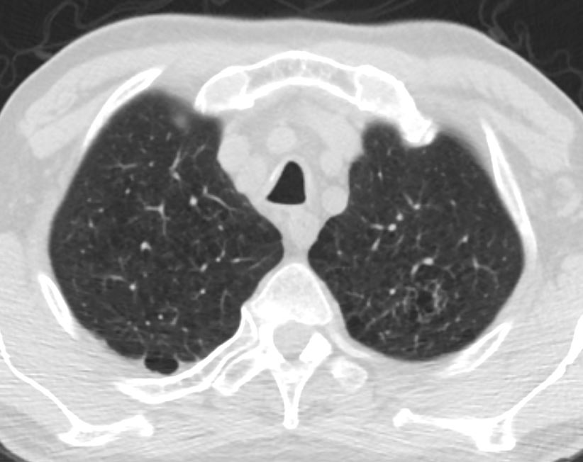
Significant Progression of Soft Tissue Growth
2 Months Later
Prior to Biopsy
Final diagnosis Squamous Cell Carcinoma
Ashley Davidoff MD TheCommonVein.net
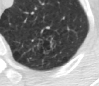
Lung Biopsy
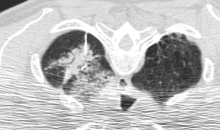
CT scan performed during the the lung biopsy with the patient in prone position performed 2 months later shows appropriate positioning of the needle, with evidence of small amount of hemorrhage around the lesion caused by the procedure
Final diagnosis Squamous Cell Carcinoma
Ashley Davidoff MD TheCommonVein.net