
Ashley Davidoff MD TheCommonVein.net lungs-0696
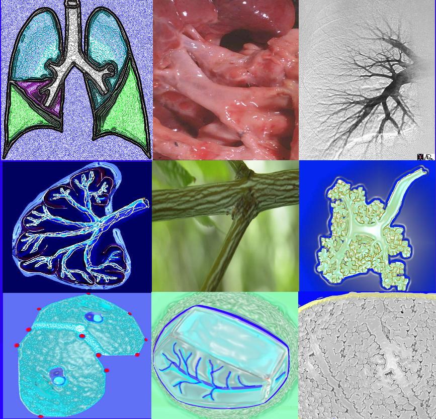
The secondary lobule is subtended by the lobular arteriole (a) and the lobular bronchiole (b) which which in turn branches into the respiratory bronchioles, alveolar ducts, and nd alveolar sacs (c) The acinus (d) consists of a respiratory bronchiole and its associated alveolar ducts, sacs, and alveoli and represents the functional unit of the lung.
The secondary lobule is drained by the pulmonary venule (e) which runs in the interlobular septum also containing the lymphatics (f). The whole unit is housed and surrounded by a connective tissue framework (g) . The latter 3 structures form the interlobular septum.
Ashley Davidoff MD TheCommonVein.net lungs-0751
Parts and Bonds
Ashley Davidoff MD TheCommonVein.net lungs-0060
by Ashley Davidoff MD TheCommonVein.net lungs-0023
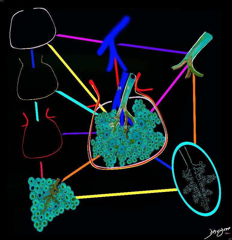
This is a collage illustrating the axial fiber system starting at the hilum, (1,2) coursing along the pulmonary artery (3) and bronchovascular system, (3,4,6) surrounded by a basket of connective tissue (4,5) extending into the polygonal secondary lobule (7,8) and ending in the alveolar ducts and sacs. (9) .
Ashley Davidoff MD TheCommonVein.net
01collageaxialfiber
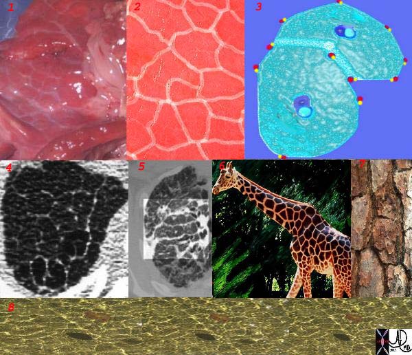
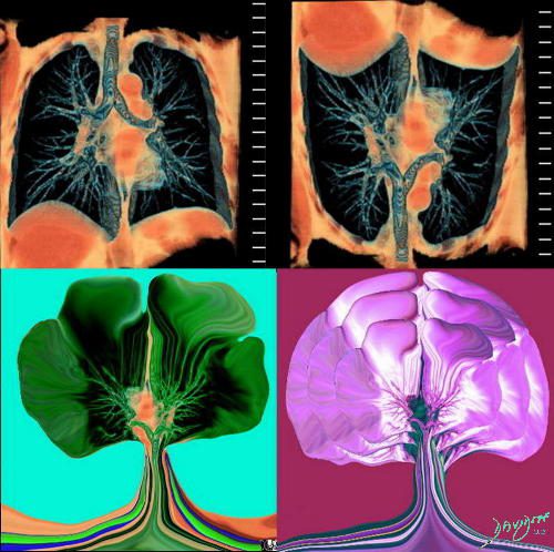
This is a series of images demonstrating the shape of the secondary lobule. The first image (1) is a post mortem specimen with congested lungs showing the interlobular septa, while the next (2), is an overlay of the septa in white showing their polygonal shape. The next drawing reveals side-by-side secondary lobules with central bronchovascular bundles and peripheral lympho-vascular bundles. Image 4 is a CT image through the apex of the lung showing thickened secondary lobules in a patient with mild emphysema, and 5 shows marked thickening of the interlobular septa in a patient with end stage sarcoidosis. 6,7,8 show the shape of the secondary lobules in the skin of a giraffe, the bark of a pine, and the ripples of the water respectively.
Ashley Davidoff MD TheCommonVein.net 31866collage
Ashley Davidoff MD TheCommonVein.net lungs-0013 aka 42444b18.8
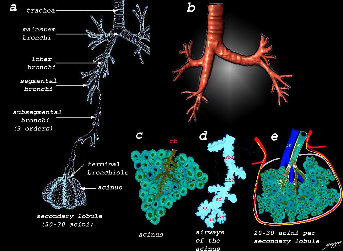
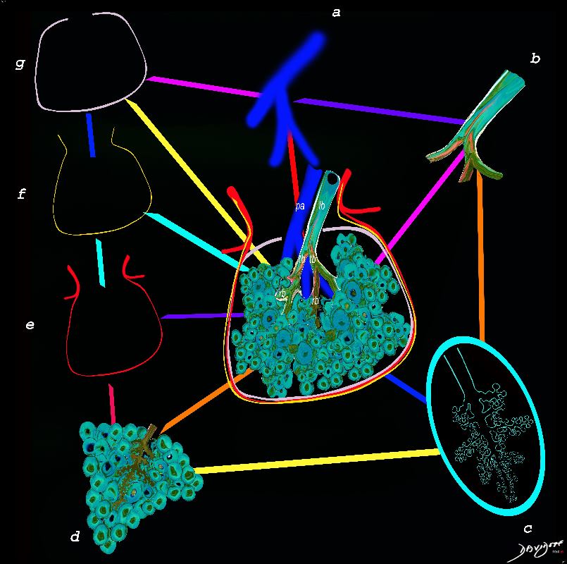
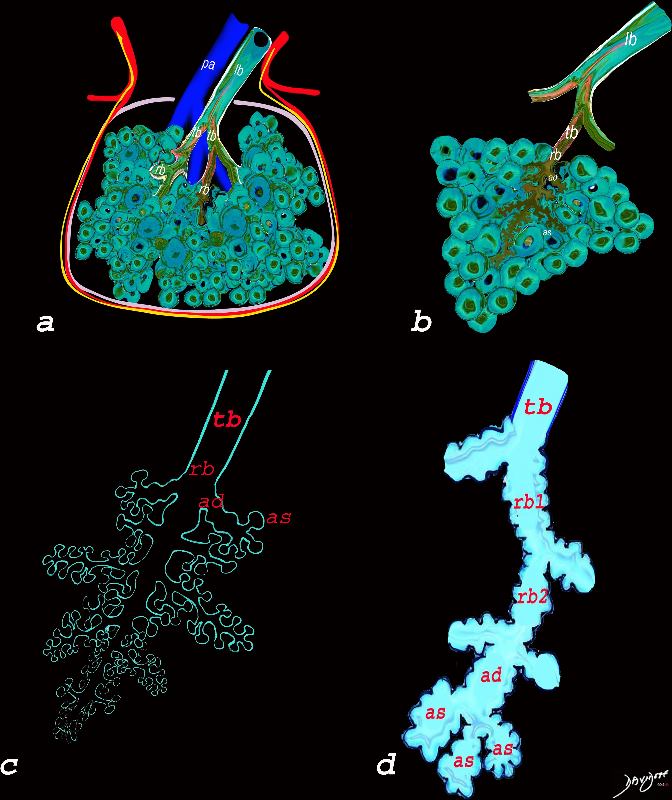
Image a shows the airways starting in the trachea and continuing to the mainstem bronchi, lobar bronchi, segmental bronchi, and subsegmental bronchi,. The subsegmental bronchi have 3 subsequent generations until the bronchiole is reached. The terminal bronchiole is the last of the transporting airways and is considered the most proximal small airway with a diameter of 2mm or less, and it gives rise to the respiratory bronchiole which is the feeding airway for the acinus . The acinus is the functional unit of the lung.
Image b is a 3D reconstruction of a CT scan showing the proximal airways from the trachea to the segmental airways.
Image c shows the structures that make up the acinus and the other parts of the small airways, starting with the respiratory bronchiole (rb) . The diagram in d, shows the detail of the small airways that participate in gas exchange, including the respiratory bronchiole, (rb) alveolar duct, (ad) and alveolar sac (as)
Image e shows the secondary lobule made from about 20-30 acini, arising from a single lobular bronchiole accompanied by a single pulmonary arteriole (pa).. Structure that surround and enclose the secondary lobule include the pulmonary venule, (red) lymphatics,(yellow) and a fibrous septum (pink).
Ashley Davidoff MD TheCommonVein.net
lungs-0739
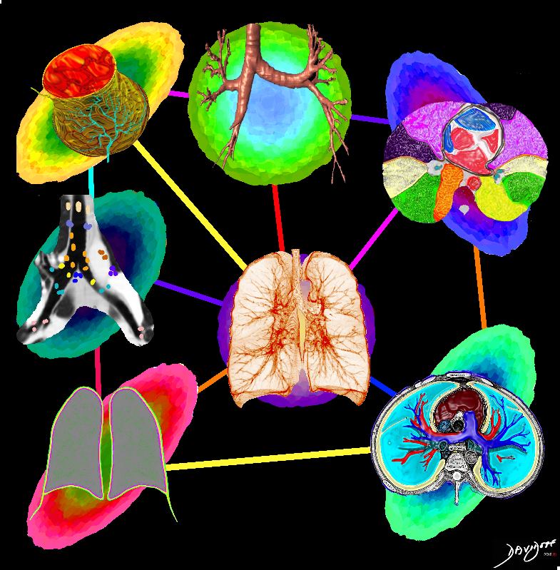
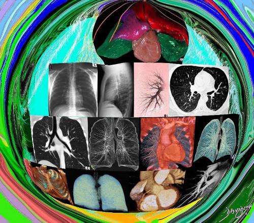
The image shows some of the major components of the lung that when bonded create a new and powerful unit – a vital organ. In the center is an example of the airways and parenchyma making up the 2 lungs. At 12 oclock the tracheo-bronchial tree with segmental and subsegmental airways. At 1 o’cloclock, is a cross section of the lungs showing some of the segments of the lung. At 5o’clock a cross section shows the arteries and veins of the lungs. At 7o’clock the drawing shows the pleura and pleural space of the lungs. At 9o’clock, a coronal reformat of the tracheobronchial tree shows the lymph node stations of the lungs. At 11 o’clock is the golden alveolus, the epicentral unit where gas exchange takes place
Ashley Davidoff MD TheCommonVein.net lungs-0696-lo res
The secondary lobule is subtended by the lobular arteriole (a) and the lobular bronchiole (b) which which in turn branches into the respiratory bronchioles, alveolar ducts, and nd alveolar sacs (c) The acinus (d) consists of a respiratory bronchiole and its associated alveolar ducts, sacs, and alveoli and represents the functional unit of the lung.
The secondary lobule is drained by the pulmonary venule (e) which runs in the interlobular septum also containing the lymphatics (f). The whole unit is housed and surrounded by a connective tissue framework (g) . The latter 3 structures form the interlobular septum.
Ashley Davidoff MD TheCommonVein.net lungs-0751
The secondary lobule is housed in a connective tissue framework in which run the lymphatic and venular tributaries . Together these 3 structures form the interlobular septum.
The lobular arteriole enters the framework, accompanied by the lobular bronchiole, and they all run together and form the interlobular septa. This structure measures between .5cms and 2cms and is visible on CT scan.
It is important in clinical radiology since many of the structures can be identified in health, and more particularly in disease, enabling the identification and characterization of many pathological processes.
Courtesy Ashley Davidoff MD The CommonVein.net lungs-0751
Ashley Davidoff MD TheCommonVein.net lungs-0744
Ashley Davidoff MD TheCommonvein.net lungs-0009
Diseases
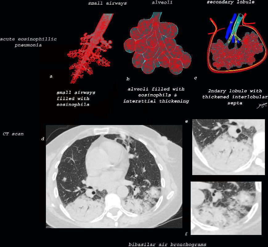
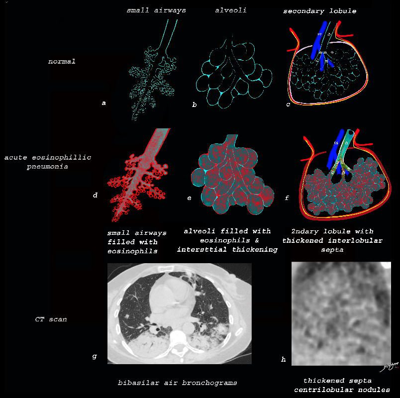
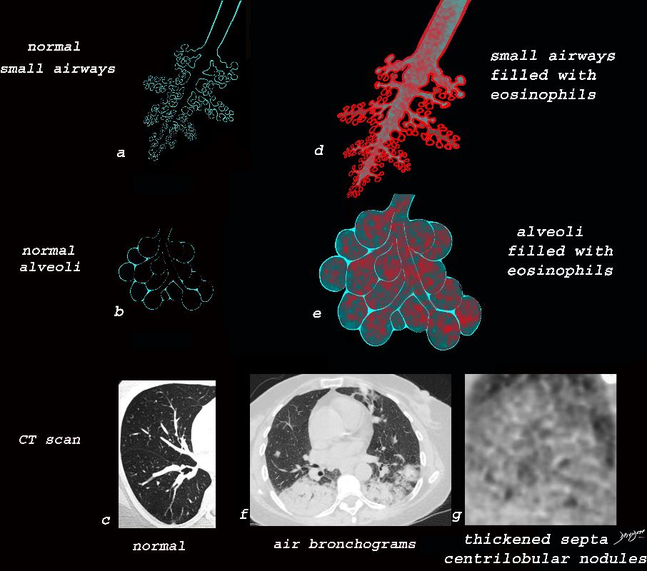
As the disease advances the small airways, and alveoli, get progressively filled with eosinophils, inflammatory cells and fluids resulting in consolidation. This image reveals progressive filling of the small airways, (a) alveoli, (b) and secondary lobules (c) with eosinophils and products of inflammation resulting in multi-segmental consolidations (d), in the lung bases, with air bronchograms at the right base (e), and less dense consolidation at the left base (f)
Ashley Davidoff MD The CommonVein.net lungs-0763
Ashley Davidoff TheCommonVein.net lungs-0757b
Ashley Davidoff TheCommonVein.net lungs-0757
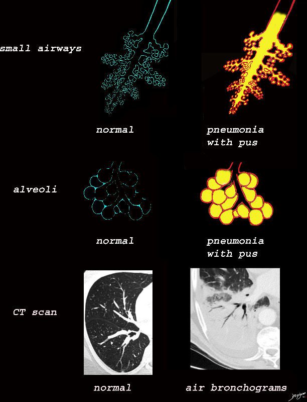
The collage provides a perspective of purulent accumulation in the small airways and the alveoli that results in consolidation. A process that increases the density of the lungs to a net “white” regional density will result in a consolidation and in this case when the fluid is infected it is labelled “pneumonia” The net result on CT is air bronchograms within the non aerated dense lung tissue.
Ashley Davidoff MD TheCommonVein.net lungs-0734
Ashley Davidoff MD TheCommonVein.net lungs-0733
Ashley Davidoff MD TheCommonvein.net lungs-0732b01
Ashley Davidoff MD TheCommonVein.net lungs-0702d- lo res
Black White and Gray Densities
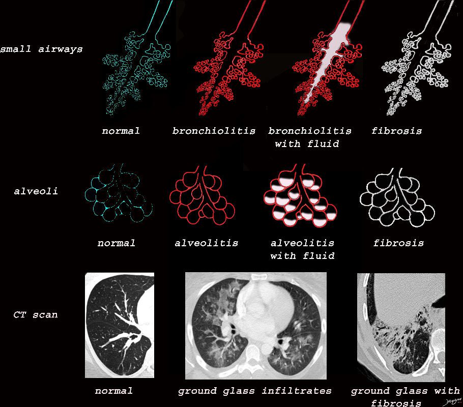
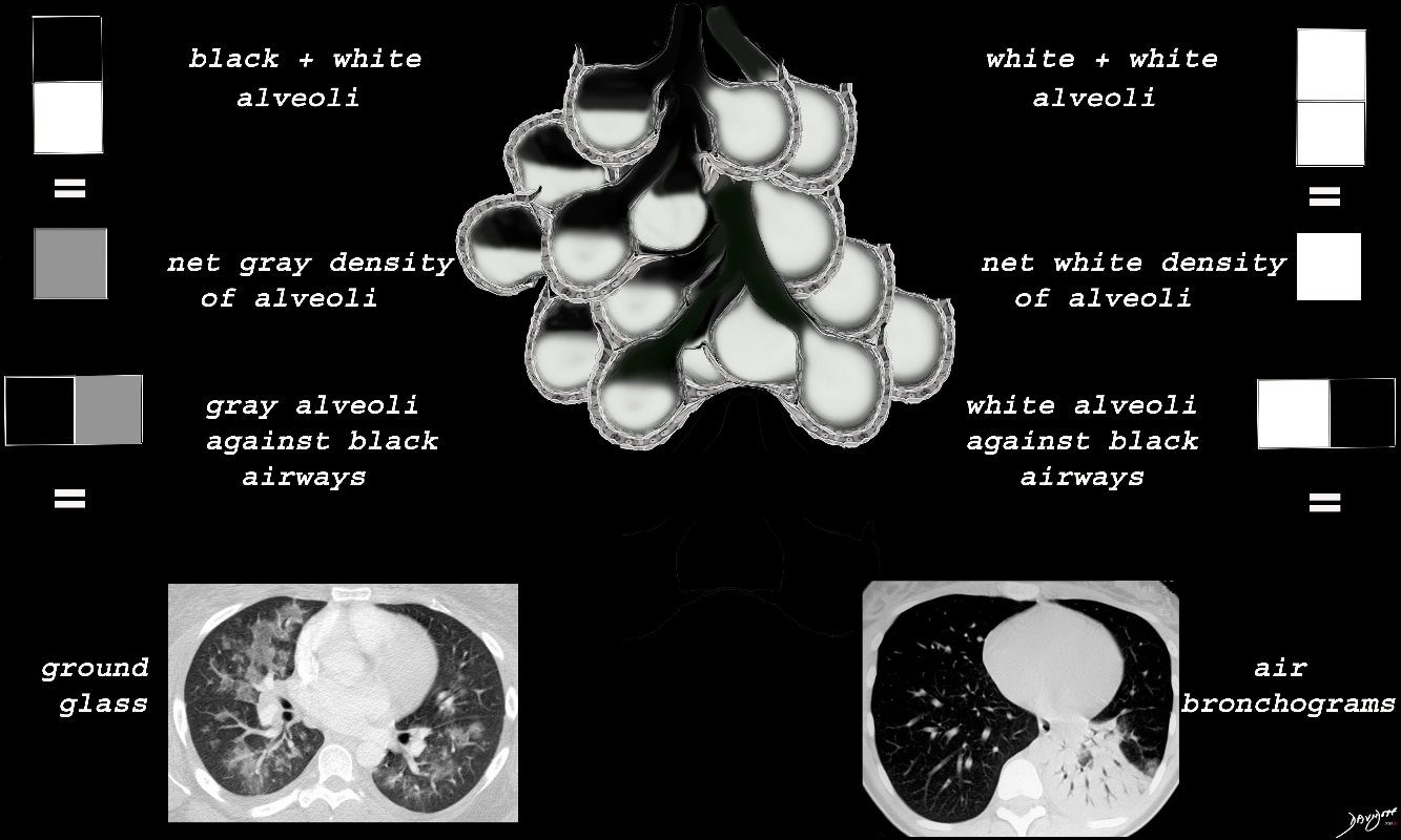
The filling of alveoli with fluids or cells results in a density that is “white” on X-ray and CT scan and is in distinct contrast to the black of the air filled airways. This contrast results in an air bronchogram. The smaller airways in a normal patient are not usually visualized because the “black” of the of the airways and the black of the air filled alveoli does not create a contrast.
Ashley Davidoff MD TheCommonvein.net
lungs-0708d
Ashley Davidoff MD TheCommonVein.net
lungs-0704d
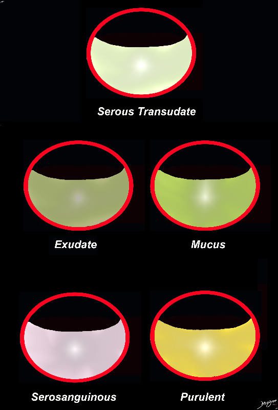
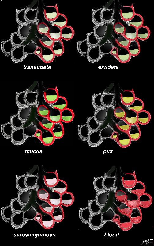
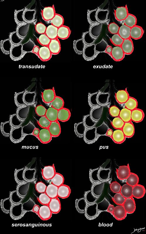
The acute inflammatory process results in fluid exudation into the alveoli which can take the form of a serous transudate, and exudate or in the form of mucus, and when severe (eg ARDS) can result in tissue and vessel destruction and could be be blood tinged. Infected fluid could be mucoid or purulent. The extent of filling the alveoli results either in a ground glass appearance when partially filled or a consolidation when filled.
Ashley DAvidoff MD TheCommonVein.net
lungs-0701d- lo res

Black White and Gray Densities
The filling of alveoli with fluids or cells results in a density that is “white” on X-ray and CT scan and is in distinct contrast to the black of the air filled airways. This contrast results in an air bronchogram. The smaller airways in a normal patient are not usually visualized because the “black” of the of the airways and the black of the air filled alveoli does not create a contrast.
Ashley Davidoff MD TheCommonvein.net
lungs-0708
Black White and Gray Densities
An air filled alveolus appears as black, a fluid filled alveolus appears as white and a a half filled alveolus appears as gray
Ashley Davidoff MD TheCommonvein.net lungs-00688b
Ashley Davidoff MD TheCommonVein.net lungs-0057
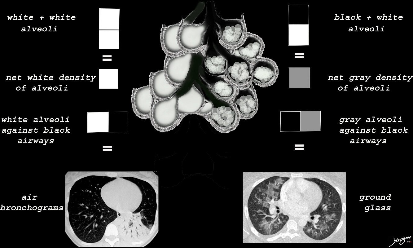
When there are extensive ceelular accumulations in the alveoli, such as adenocarcinoma with lepidic growth, Langerhans cells or other macrophages, the overall net density of the region of involvement will be gray, and when adjacent to the black air filled airways, a ground glass appearance will be apparent
Ashley Davidoff
TheCommonVein.net
ssb = subsegmental bronchiole
tb = terminal bronchiole
rb = respiratory bronchiole
as = alveolar duct
as = alvelar sac
is = anteralveolar septum
lungs-00688

When the alveoli are fully filled with fluid, tumor, or pus for example, the overall net density will be white, and when adjacent to air filled airways, air bronchograms are visible (left side of image)
When the alveoli are only partially filled, the density of the fluid added to the density of the air results in an overall gray density, and when positioned next to air filled bronchi, there is insufficient contrast to create an air bronchogram and sufficient to enable visualization of the blood vessels. This is called ground glass opacification
Ashley Davidoff
TheCommonVein.net lungs-00681