28 y.o. male who recently underwent biopsy of a non-healing ulcer in the perianal region Pathology showed granulomatous inflammation and cultures have now isolated M. Tuberculsosis and referred to TB clinic.
3 months prior
Cough which started for about 8 months which lasted approx a few weeks; non productive.
Cough resolved and then about 2 months later the cough recurred and was productive, which initially appeared to improve but now says over the past 2 months he has had a persistent cough, worse in the AM.
Sputum is brownish and thick in the AM and then later turns yellow. No blood
Ashley Davidoff MD
TheCommonVein.net
Ashley Davidoff MD
TheCommonVein.net
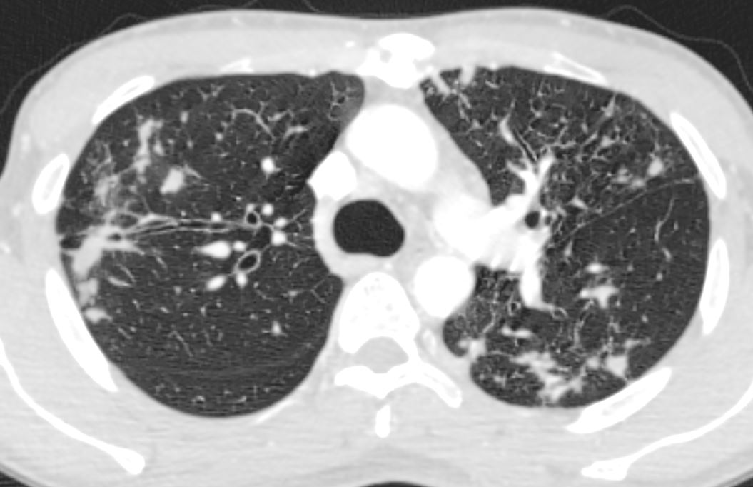
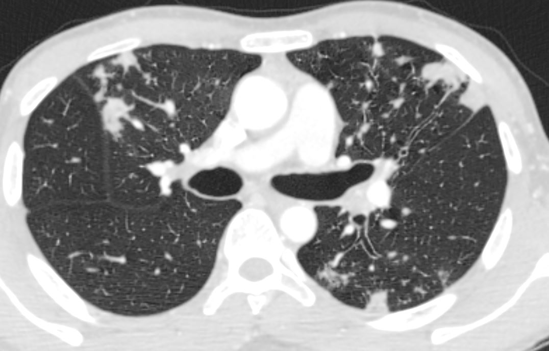
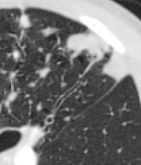
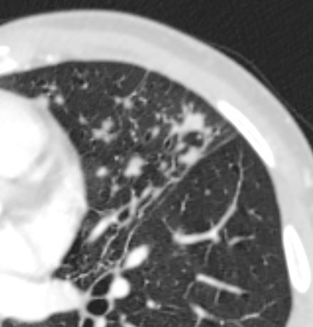
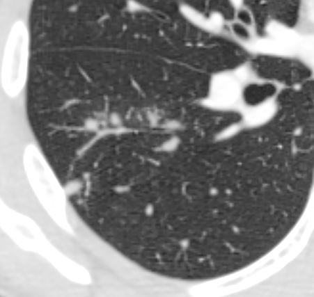
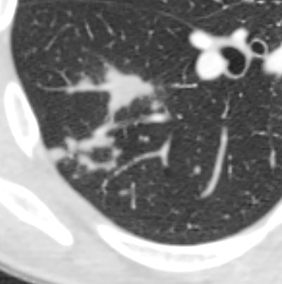
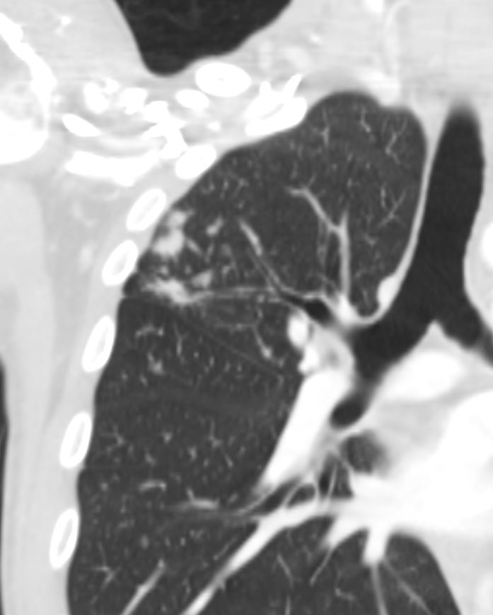
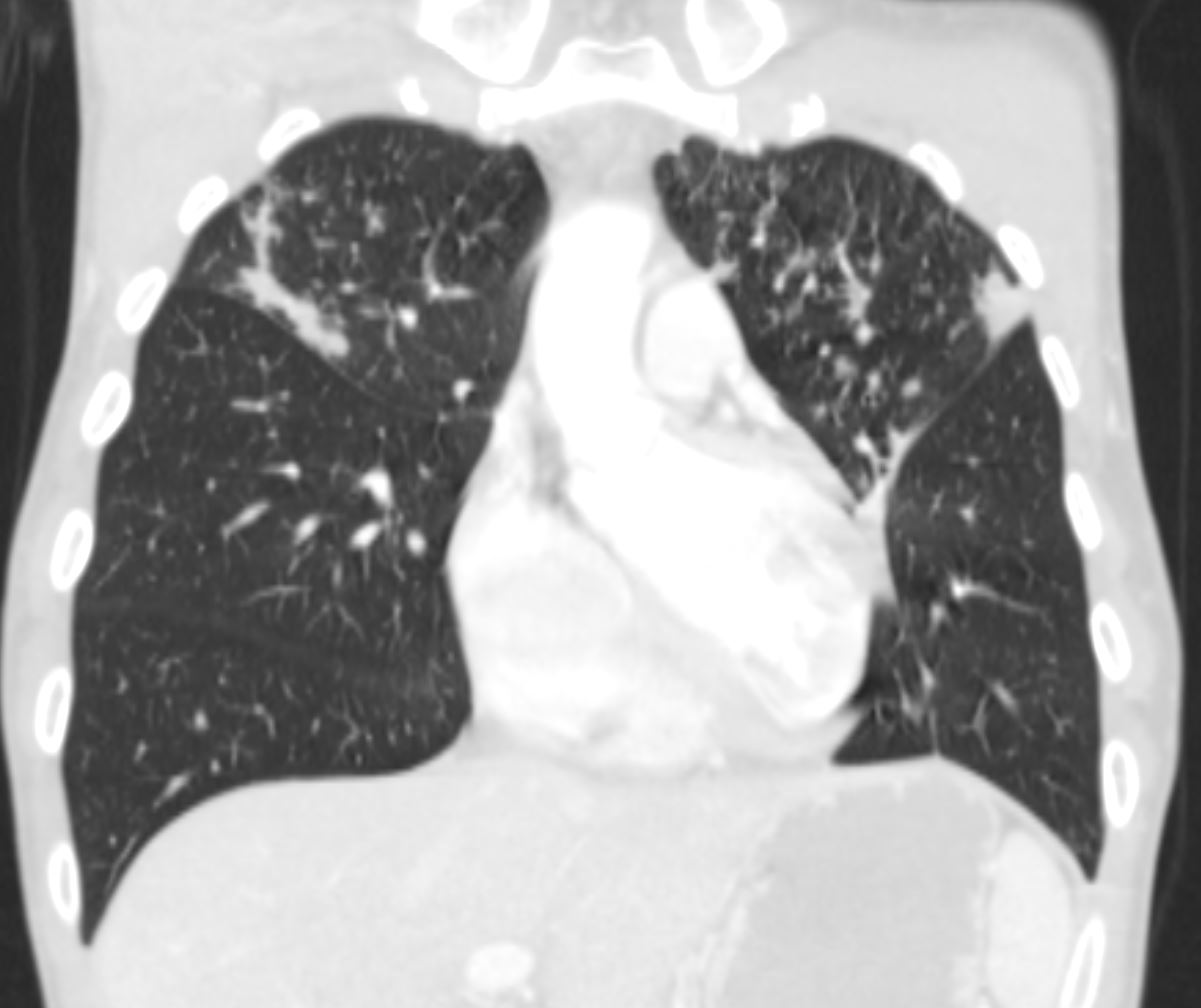
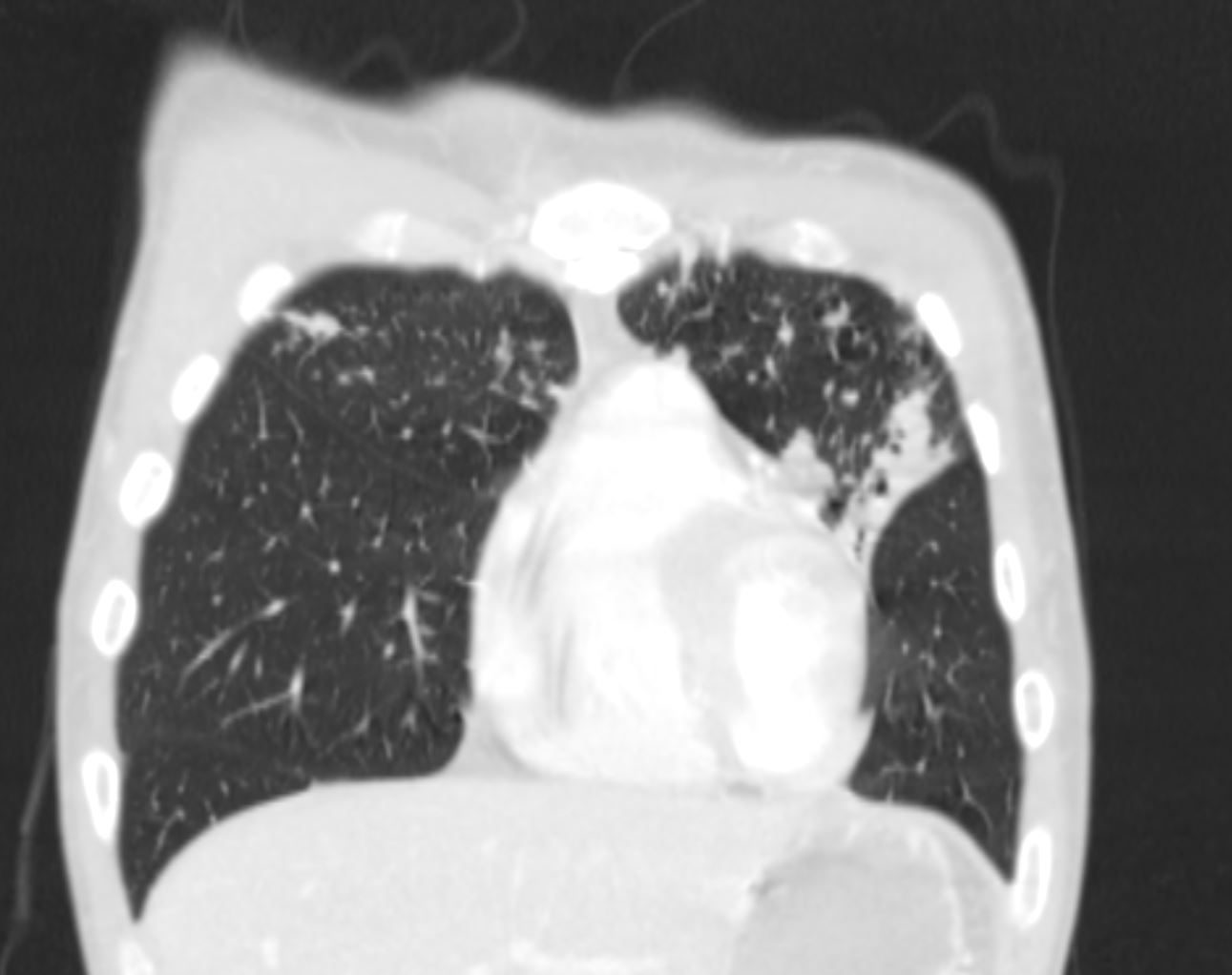
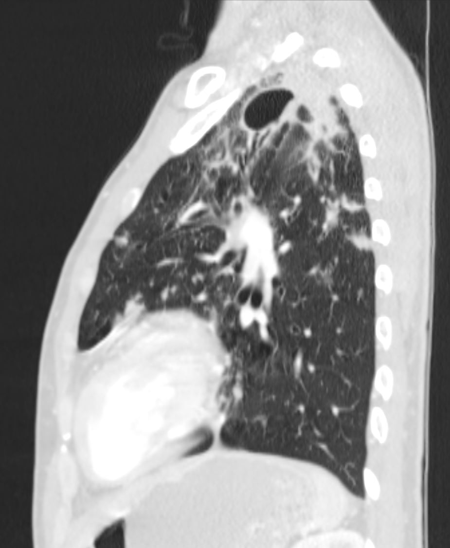
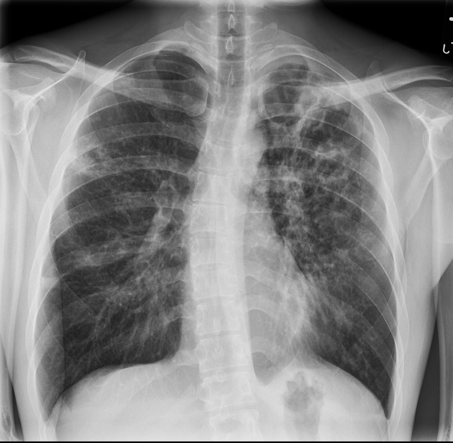
- Chest x-ray shows bilateral patchy opacities with multiple lesions that appear cavitary, consistent with pulmonary TB
- extrapulmonary disease involving the perianal area.
- Sigmoidoscopy did not reveal any evidence of fistula.
- Given perianal ulcer, query extension from the rectum vs genitourinary tract despite lack of symptoms.
- HIV negative.
- 4+ smear positive and isolated M. TB
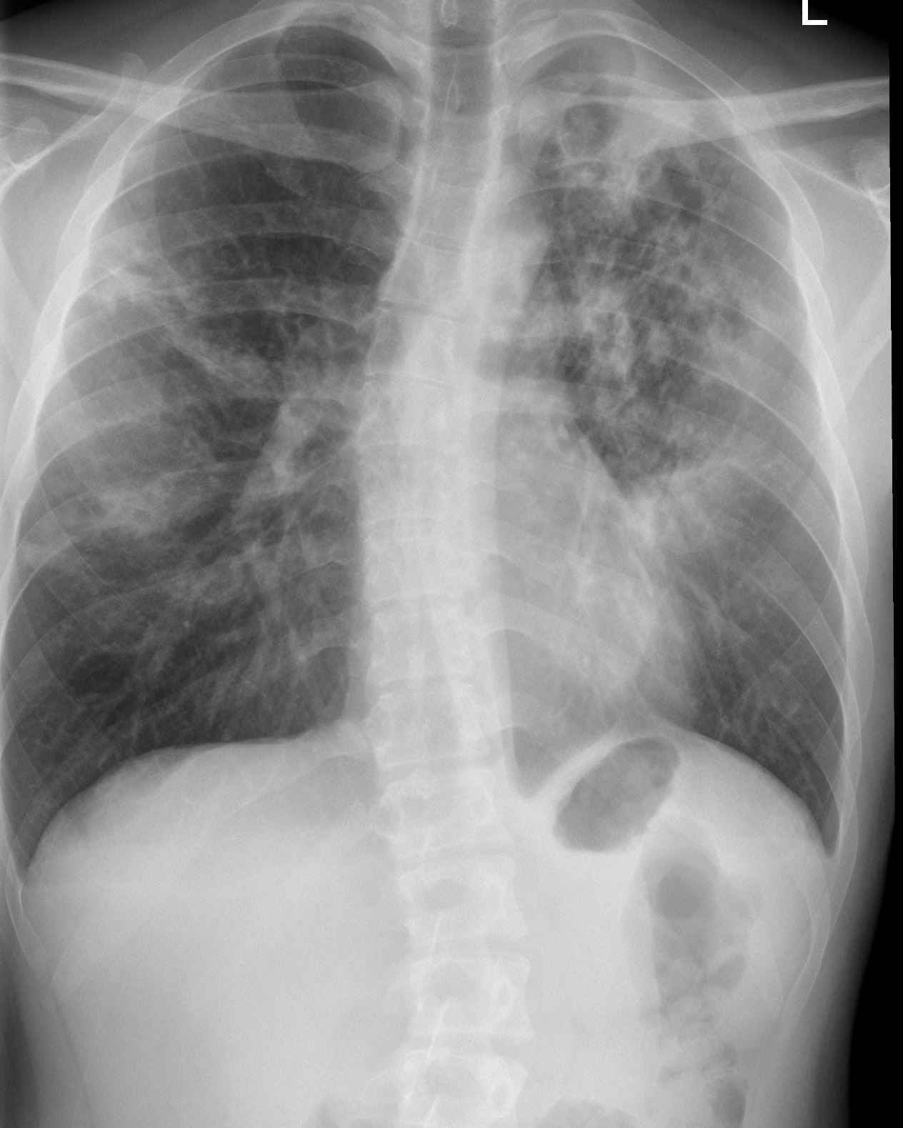
2 Months Later
Ashley Davidoff MD
TheCommonVein.net
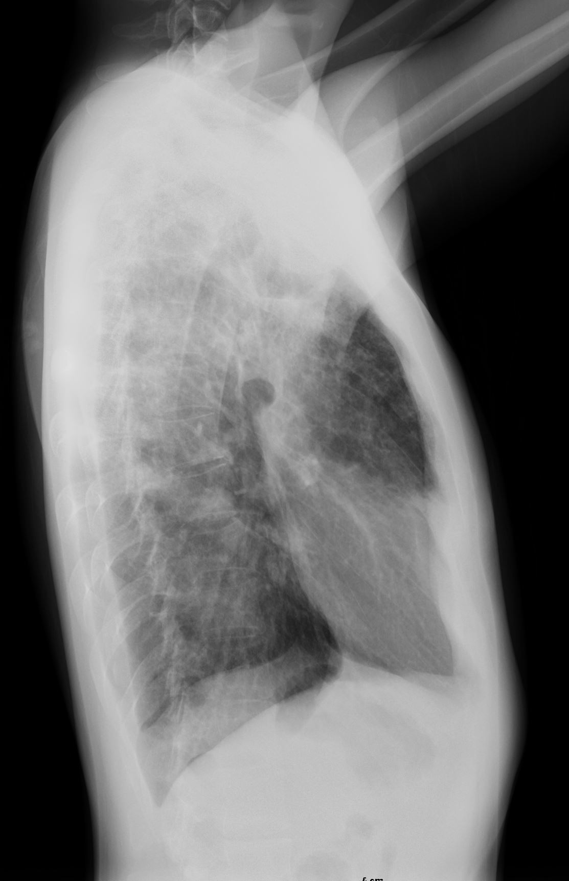
1 Month Later
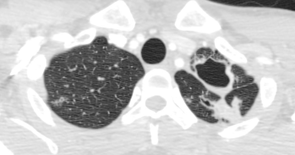
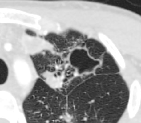
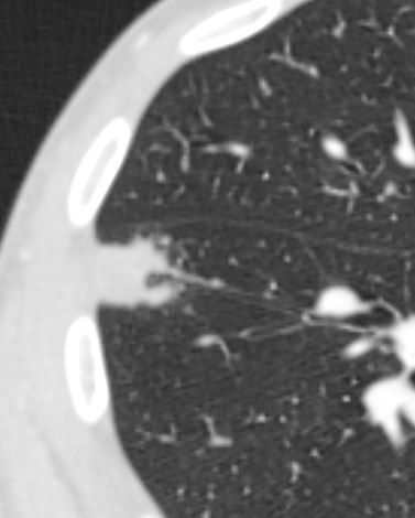
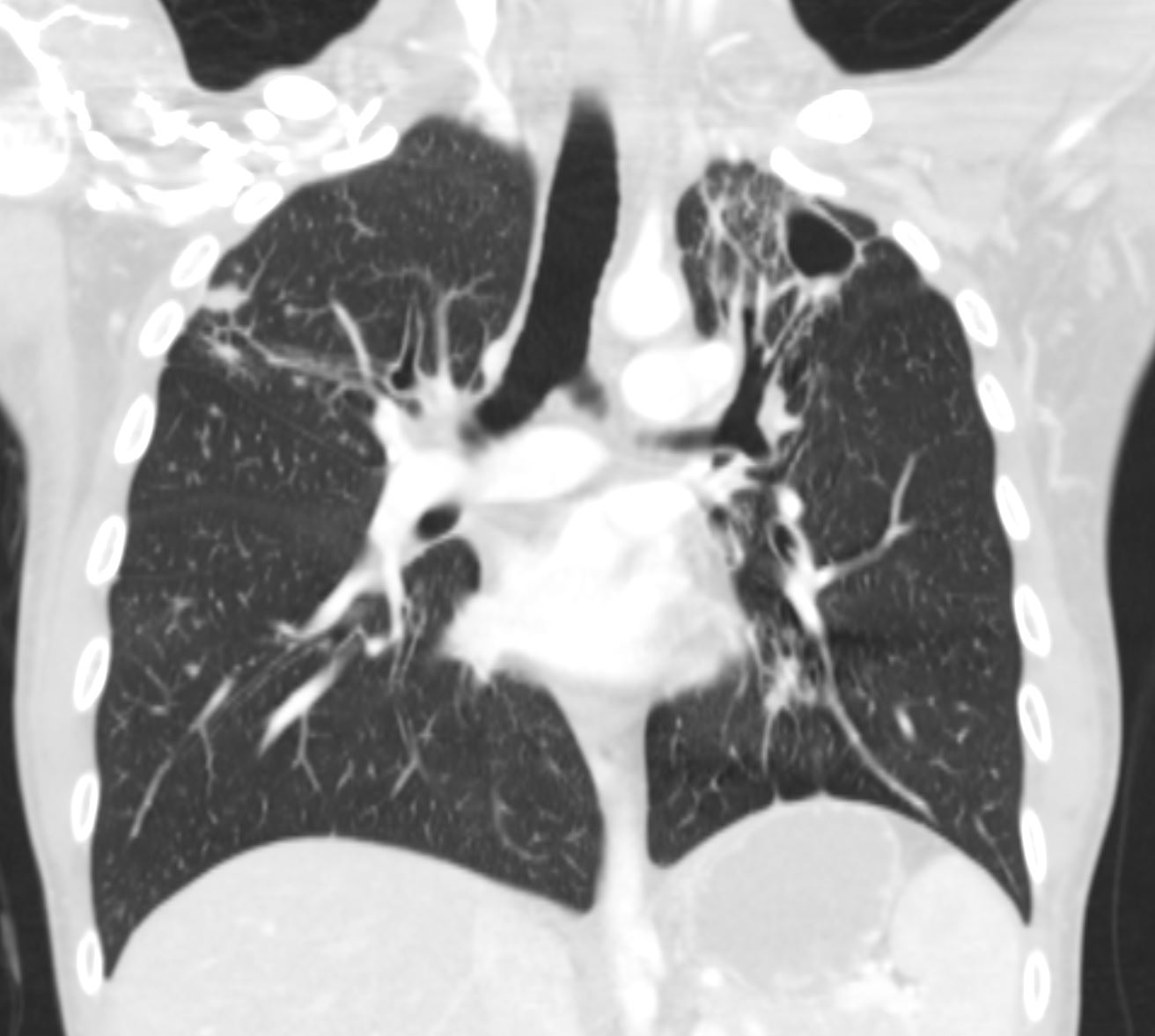
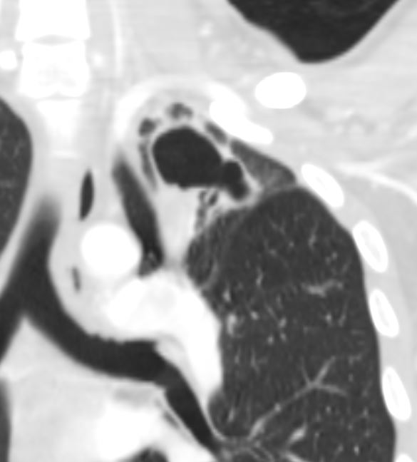
CAvitation Atelectasis Bronchovascular Involvement
Large cavitary lesion in the left upper lobe and a smaller cavitary lesionin the right upper lobe concerning for granulomatous
disease such as tuberculosis.
Additional numerous patchy nodular opacities in bilateral lungs as
described above with tree-in-bud appearance representing endobronchialspread .