
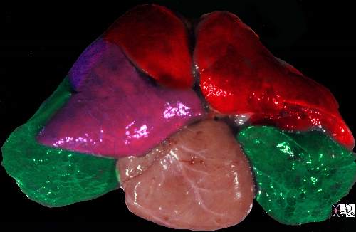
The post mortem specimen is viewed from the anterior aspect showing the upper lobes in red, the right middle lobe in pink and the lower lobes in green.
Ashley Davidoff MD TheCommonVein.net 32558b02
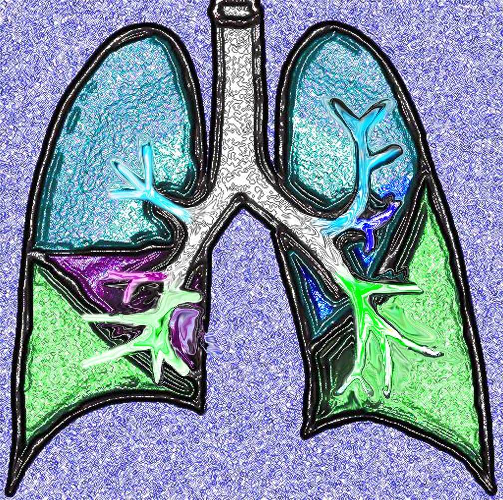
This diagram shows the basic division of the tracheobronchial tree into lobes. The right lung is divided into right upper (RUL) (teal) right middle, (RML pink) and right lower lobe (RLL green). The left lung is divided into left upper (LUL teal), which includes the lingula(dark blue), and left lower lobe (LLL= green). Note that the two mainstem bronchi are of unequal length and size. The right mainstem is short and fat while the left is long and thin. This irregular dichotomous branching pattern is characteristic of the branching pattern of all the conducting systems within the lungs.
Ashley Davidoff
TheCommonVein.net 32686b05

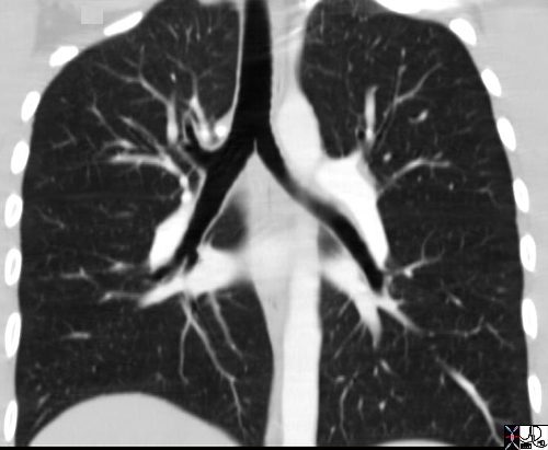
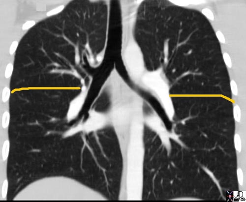
This diagram of the airways reveals the approximate lengths of the airways. Note that the left main stem bronchus is about twice the length of the right whose length is truncated by the take off of the right upper lobe bronchus.
Courtesy Ashley Davidoff MD
TheCommonVein.net
32686b05L04b
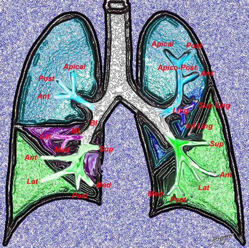
Ashley Davidoff TheCommonvein.net 32686b05L segmental bronchi.8
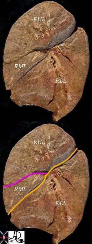
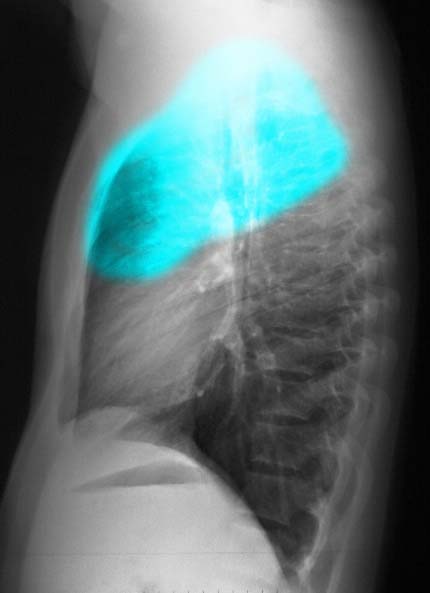
The right lung has a small right upper lobe (RUL) separated from the middle lobe (RML) by the minor fissure (pink,lower image) . Both the RUL and RML are anterior and are separated from the lower lober by the major fissure (orange line)
Ashley Davidoff MD
Ashley Davidoff M.D.
TheCommonVein.net 32682
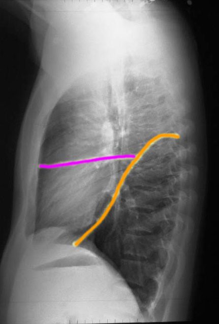
This coronal reformats through the tracheobronchial tree show the major fissures bilaterally. They are usually quite difficult to see and unless you know where to look you may miss them altogether. Usually there is a hint of hypovascularity along the fissure resulting in a relative lucency as can be appreciated in this examination. The fissures have been overlaid in orange. As the coronal cut proceeds posteriorly the lower lobe becomes more prominent and the upper lobes less so. If you review the lateral chest x-ray and project the cuts you would get a better sense of this concept of the dominance of the RLL and its posterior location .
Ashley Davidoff MD. TheCommonVein.net 32682b01.jpg
Ashley Davidoff MD
Ashley Davidoff MD
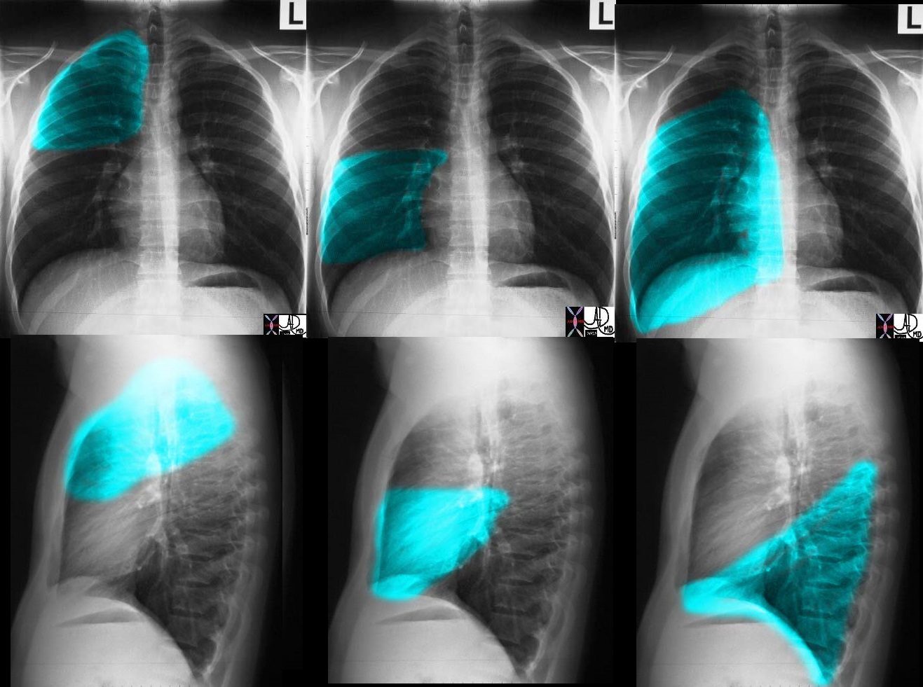
These images (left to right) show the shapes of the RUL, RML and RLL. In Fig 1a and 1b the overlay represents the upper lobe. Image 2a and 2b represent the middle lobe while in 3a and 3b the lower lobe is represented. Note how the middle lobe hugs the right heart border and how much larger the RLL is compared to the RML and the RUL.
Ashley Davidoff TheCommonVein.net 30398c01
Ashley Davidoff MD
The right lung has a relatively small right upper lobe (RUL) separated from the middle lobe (RML) by the minor fissure (pink,lower image). Both the RUL and RML are anterior and are separated from the lower lobe by the major fissure (orange line)
Ashley Davidoff MD. TheCommonVein.net 30398b06b
The secondary lobules, connect, and unite, linked through the airways, blood vessels, lymphatics and nerves, to form segments in the lungs
There are ten bronchopulmonary segments in the right lung: three in the upper lobe, two in the middle lobe, and five in the lower lobe. Some of the segments may fuse in the left lung to form usually eight to nine segments (four to five in the upper lobe and four to five in the lower lobe.
Ashley Davidoff MD
TheCommonVein.net lungs-0015
Infection
TB
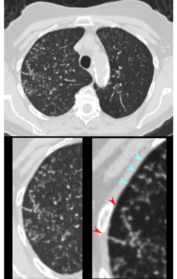
CT in the axial plane shows extensive small airway disease dominant in the right upper lobe characterized by innumerable, ground glass micronodules, centrilobular nodules (teal blue arrowheads) and nodular thickening of the interlobular septa likely reflecting lymphatic involvement (red arrowheads)
Ashley Davidoff MD TheCommonVein.net 135825cL 192Lu
Inflammation
Malignancy
Halo Sign Around a Malignant Mass
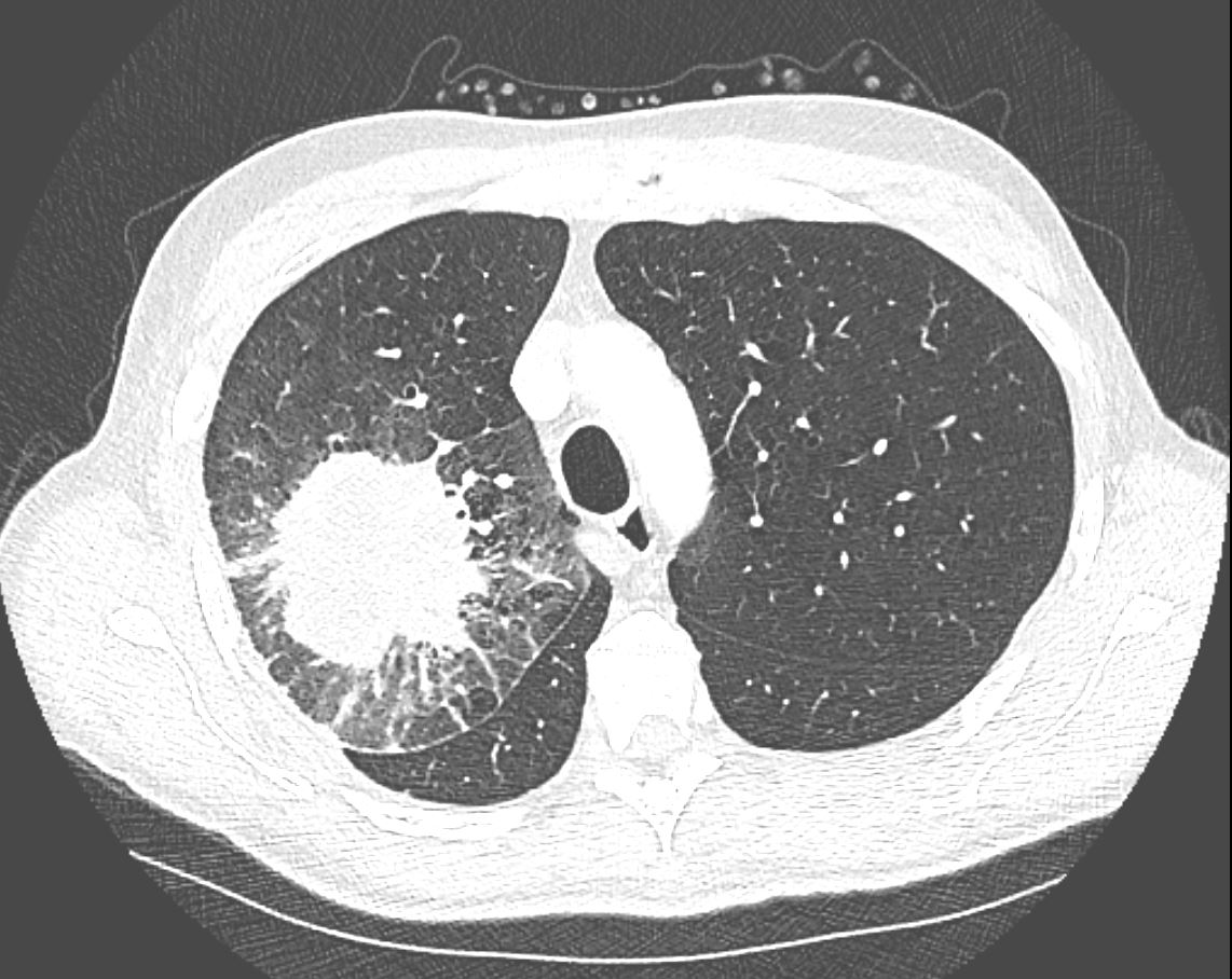
CT in the axial plane demonstrates a large, spiculated mass in the right upper lobe with surrounding halo likely reflecting hemorrhage or lymphatic edema around the mass. In addition, there is evidence of irregular interlobular septal thickening likely reflecting lymphatic invasion and indicating lymphangitis carcinomatosa. There is irregular thickening of the major fissure suggesting involvement.
Ashley Davidoff MD TheCommonVein.net 135865
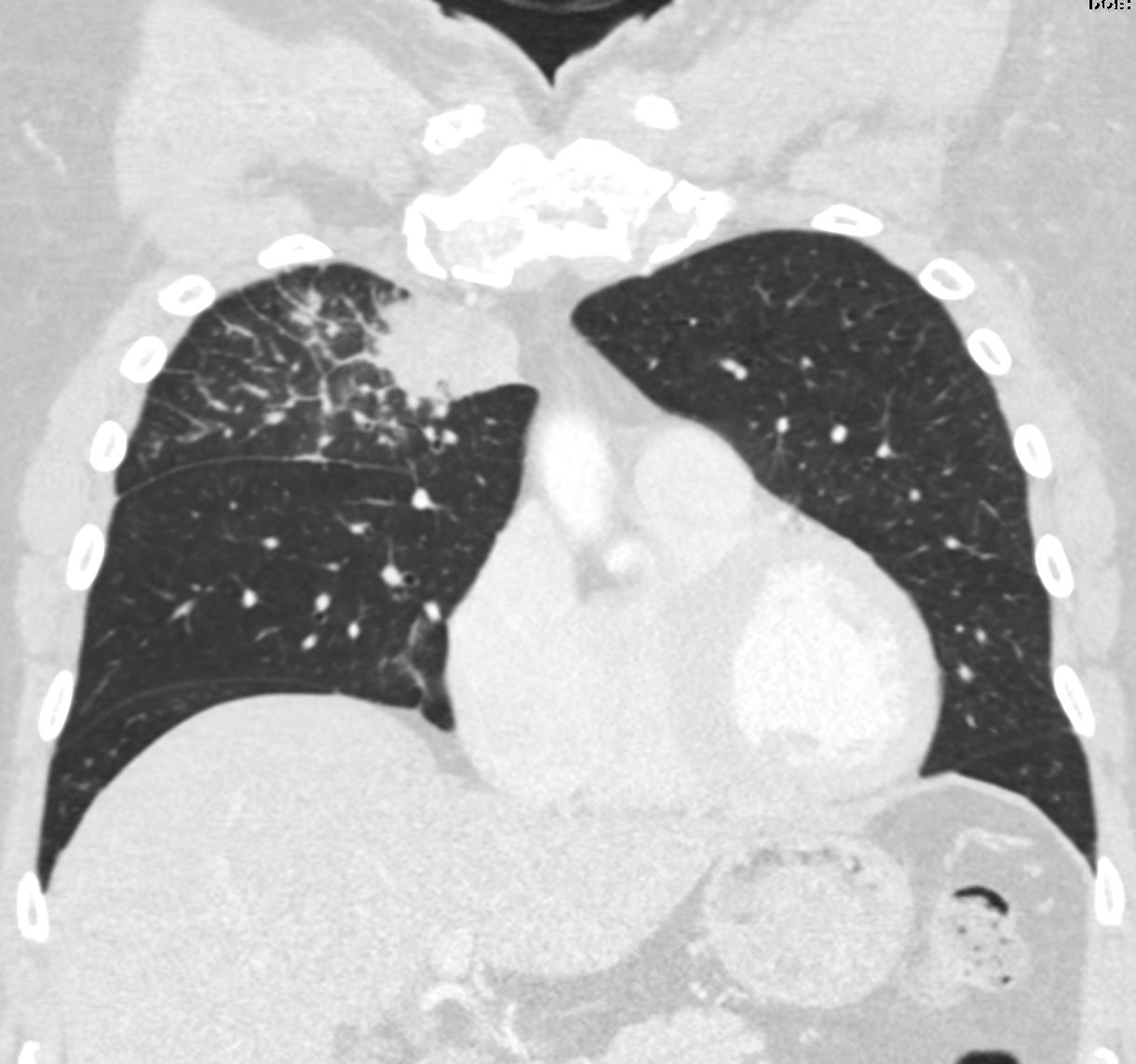
3months later the patient presented with chest pain and a cough. CT of the chest in the coronal plane showed a right upper lobe mass abutting the mediastinum in the previous region of abnormality characterised by abnormal secondary lobules. The findings are suggestive of a rapidly developing metastasis in the right upper lobe. The thickened septa likely reflect lymphangitis carcinomatosa
Ashley Davidoff MD TheCommonVein.net 013Lu 136064
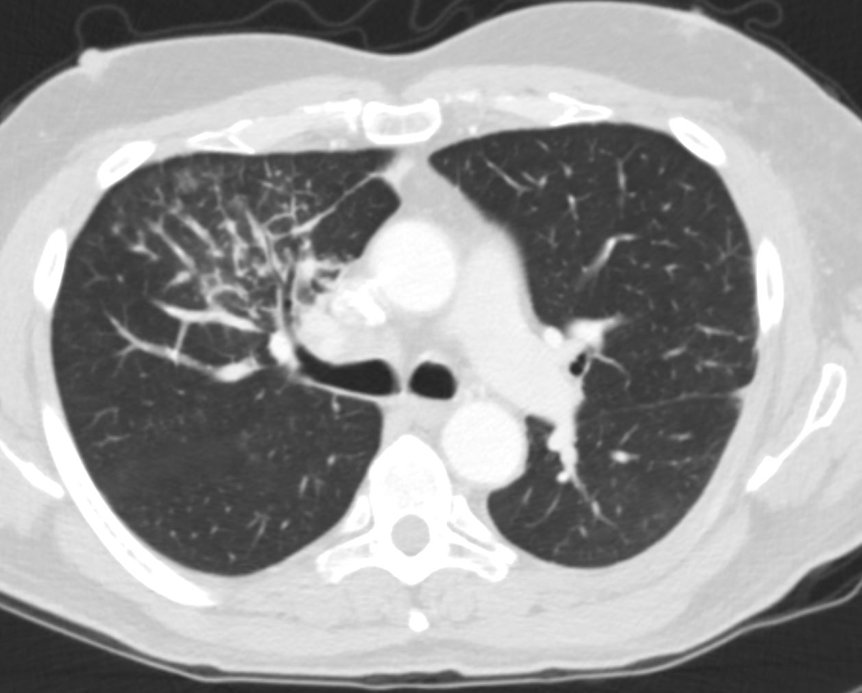
3 months later the patient presented with chest pain and a cough. CT of the chest in the axial plane showed a right upper lobe mass abutting the mass There is interlobular involvement of secondary lobules. The findings are suggestive of a rapidly developing metastasis in the right upper lobe with lymphangitis carcinomatosa
Recurrence
Non Small Cancer of the lung s/p Radiation and 6 Months Later
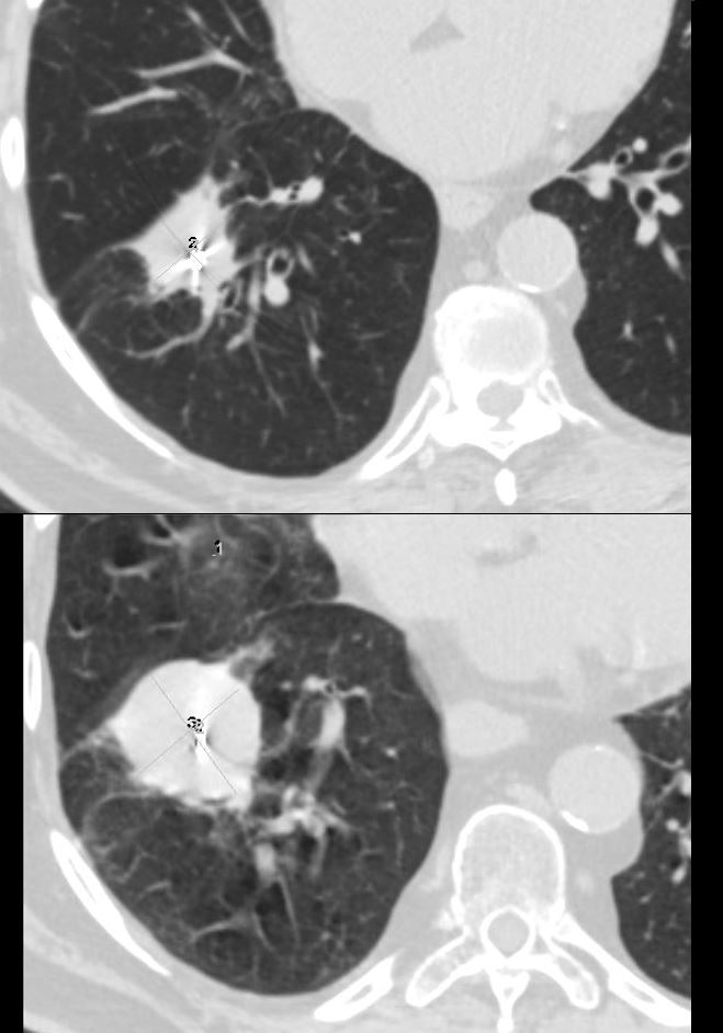
The top image shows a soft tissue nodule with fiducial markers in the right upper lobe. The lesion had been radiated and stable for a few years
The bottom image shows significant growth over a 6month period representing recurrence of the carcinoma
Ashley Davidoff MD TheCommonVein.net
Mechanical
Atelectasis
Trauma
Metabolic
Circulatory-
Hemorrhage
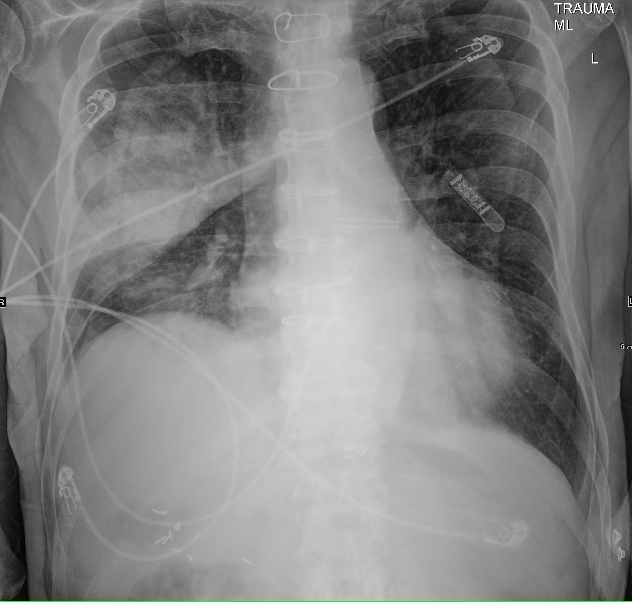
75-year-old man on blood thinners s/p aortic valve replacement s/p trauma, presents with hemoptysis. He was afebrile and without an elevated white count
CXR shows an elevated right hemidiaphragm and inferior displacement of the major fissure with a dense right upper lobe consolidation. The mass effect on the major fissure likely results from a hematoma, and the hemorrhage results in air bronchograms and groundglass changes.
Skin folds manifest as bilateral pseudo-pneumothoraces. A loop recorder is noted overlying the left upper chest.
Ashley Davidoff MD TheCommonVein.net 165Lu 135849
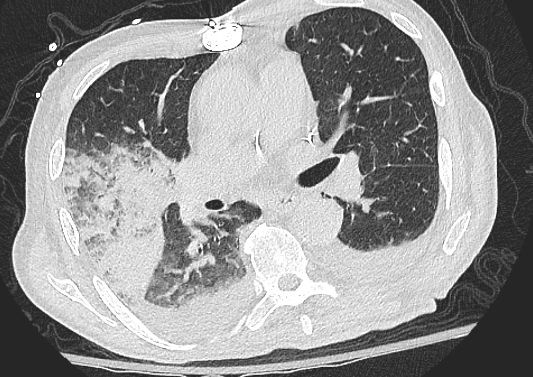
75-year-old man on blood thinners s/p aortic valve replacement s/p trauma, presents with hemoptysis. He was afebrile and without an elevated white count
Axial CT at the level below the carina shows medial displacement of the major fissure by a dense right upper lobe consolidation. The mass effect on the major fissure likely results from a hematoma. Anterior to the consolidation there is a combination of ground glass opacity with thickened interlobular septa, and minor region of subsegmental consolidation with air bronchograms, likely resulting from hemorrhage.
There are bilateral effusions
Ashley Davidoff MD TheCommonVein.net 165Lu 135851
Immune Infiltrative Idiopathic Iatrogenic
Ashley Davidoff MD TheCommonVein.net 013Lu 136061