
Pulmonary hypertension is characterized by enlarging arteries. The margins of the main arteries are usually quite distinct on the plain film. The lower lobe arteries should not measure more than 16mms in the male and more than 14mms in the femaleThey become blurred when there is interstitial edema, most commonly caused by heart failure.
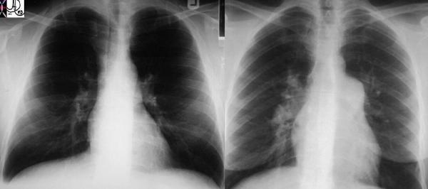
These two P-A chest X-rays show a normal cardio-mediastinal silhouette on the left and an abnormally enlarged MPA and RPA on the right in this patient with pulmonary hypertension.
Ashley Davidoff MD TheCommonVein.net 22089
Normal Pulmonary Artery
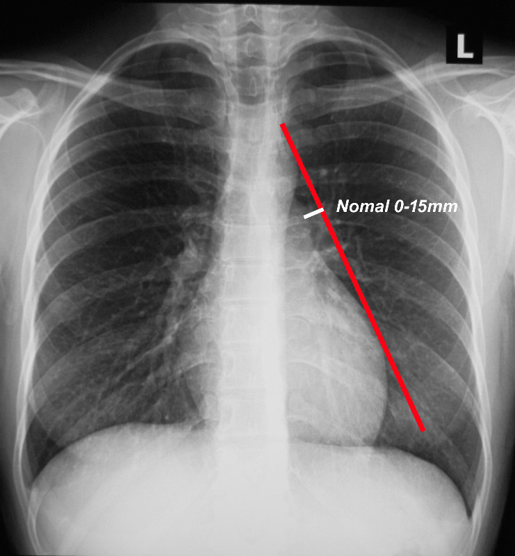
When a line is drawn from the aortic knob to the left edge of the heart, (red line) the pulmonary artery should lie medial to that line (ie along the line drawn to 1.5cms medial to the line)
Ashley Davidoff MD TheCommonVein.net
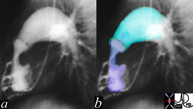
Pulmonary Hypertension
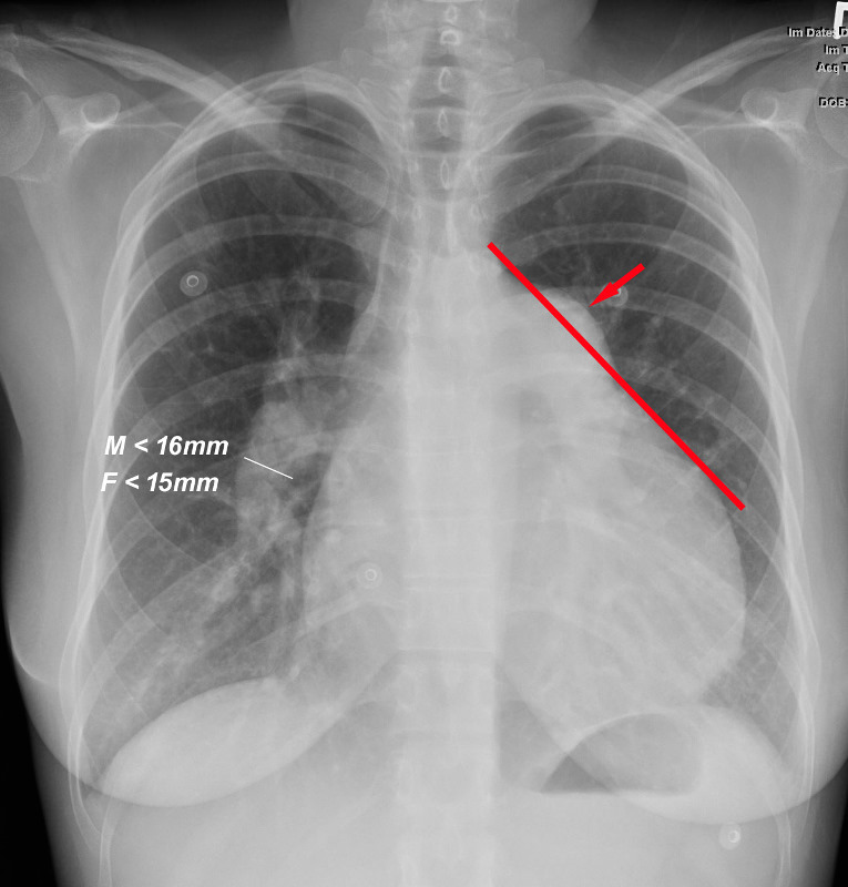
When a line is drawn from the aortic knob to the left edge of the heart, (red line) the pulmonary artery lies lateral to that line indicating an enlarged pulmonary artery most commonly caused by hypertension . In this instance the size of the descending right pulmonary artery is greater than 15 mms confirming the presence of pulmonary hypertension
Ashley Davidoff MD TheCommonVein.net
The other arterial circulation to the lungs – the bronchial circulation, can also be the source of disease. In chronic disease states such as cystic fibrosis, and bronchiectasis for example, chronic increase flow due to the inflammation and infection occurs, and the combination of an infected and friable mucosa with enlarged arteries, is a cause for hemoptysis. The treatment of choice for recurrent hemoptysis is embolization of the bronchial arteries.
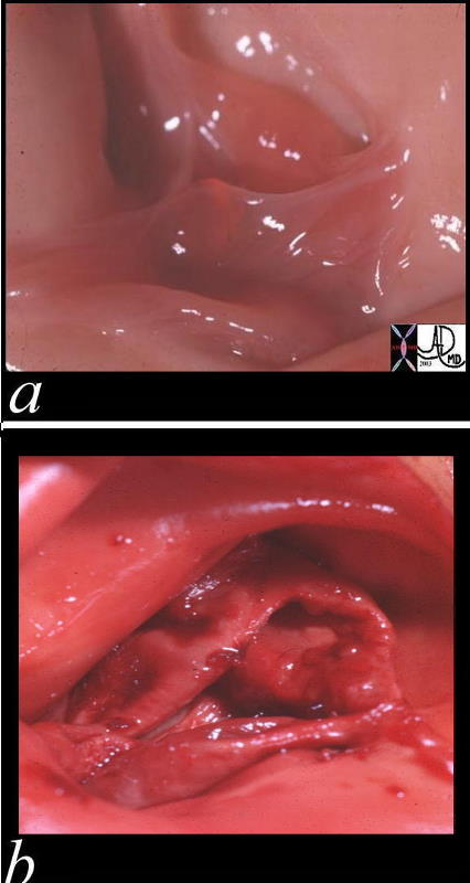
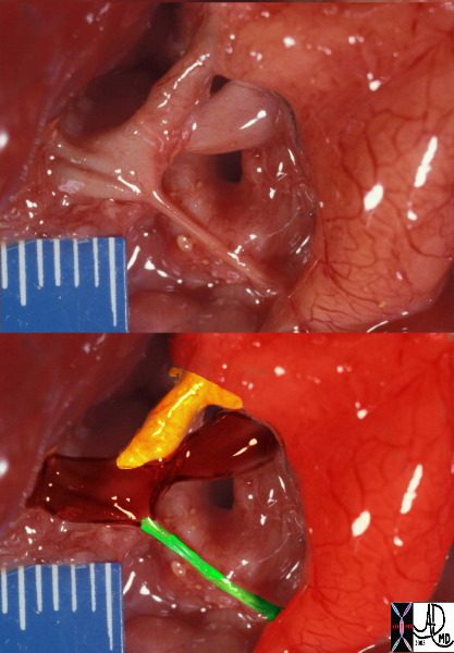
These two pathological specimens of the pulmonary valve show the delicate leaflets of the normal valve (a) in comparison to the thickened leaflets of the bicuspid pulmonary valve Courtesy Ashley Davidoff MD 00272c01.800
The lateral projection of this right ventricular angiogram reveals a case of severe pulmonary stenosis. The catheter enters the RV from the RA via the SVC. The RV inflow tract (purple) is hypoplastic. The vessels emanating and the-RV inflow are coronary arteries that are filling in retrograde fashion are due to the supra-systemic pressures in the RV indicating severe pulmonary stenosis with pressures in the RV that probably exceed 100mmhg. The infundibulum (right ventricular out flow tract (blue) is slightly narrow since it is hyperdynamic in an attempt to force the blood through the stenotic valve. The valve (green) is doming into the PA due the severe narrowing. The narrowing causes turbulence which causes the post stenotic dilatation.15036c01
Ashley Davidoff MD TheCommonVein.net
This is a post mortem specimen of a baby who died with pulmonary atresia. The atretic MPA overlayed in green in the lower image is connected to patrent branch pulmonary arteries in maroon, which are fed by a patent ductus arteriosus (yellow) arising from a anteriorly positioned left sided aorta, (red overlay) compatible with a diagnosis of transposition of the graet vessels – LTGA A.
Ashley Davidoff MD TheCommonVein.net 32627bC02
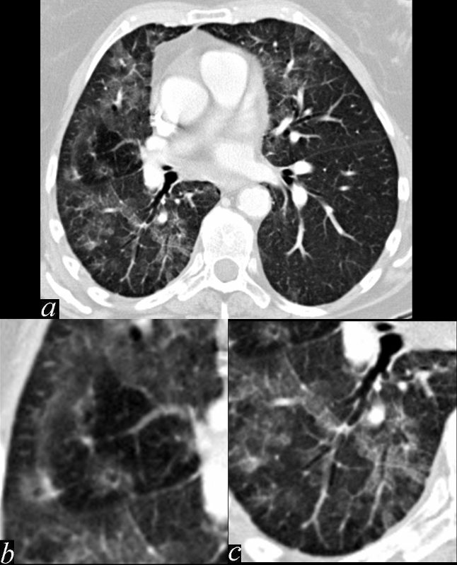
This series of images shows some subtle changes that reflect the local control of blood flow to a small segment of the right middle lobe. Note that in image a, there is a small area of increased lucency (blacker) in the right lung just lateral to the vessels of the right hilum. This region is highlighted in b. Note also that in b, the rapid diminution of the size of the blood vessel to that subsegment when compared to the size change of the vessels in the image in c. The lucent appearance of the lung suggests air trapping and the vasoconstriction reflects decreased perfusion – ie with decreased ventilation there is an associated consequent associated decrease in perfusion.
47170c01.800 broncho-centric inflammation lung bronchovascular bundle chest inflammation peribronchial halo air trapping mosaic perfusion ground glass changes alveolar change air bronchogram acute bronchovascular inflammation ddx allergic collagen vascular disease infection CTscan
Ashley Davidoff MD TheCommonVein.net