
Infection Inflammation Malignancy Mechanical/Atelectasis Trauma Metabolic Circulatory- Hemorrhage Immune
Infiltrative
Amyloidosis
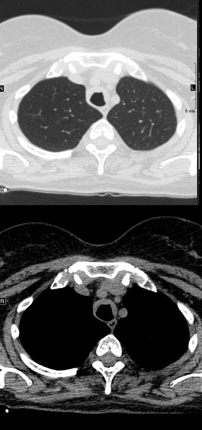
44-year-old female with immunoglobulin light chain (AL) amyloidosis of the trachea. The axial CT shows thickening of the trachea on its anterior and lateral walls, sparing the posterior membrane
Ashley Davidoff MD TheCommonVein.net 135866 249Lu
Idiopathic Iatrogenic
Idiopathic
Inherited
Mounier Kuhn
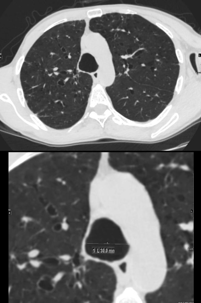
61 year old male with a history of treated mycobacterial infections and chronic cough
Axial CT at the level of the brachiocephalic vessels shows an enlarged trachea that measures 3cms which is abnormally enlarged. There are thin-walled cystic changes of the airways along the subsegmental arteries in the upper lobes likely reflecting bronchiectasis
Ashley Davidoff MD TheCommonVein.net 250Lu 135874ac
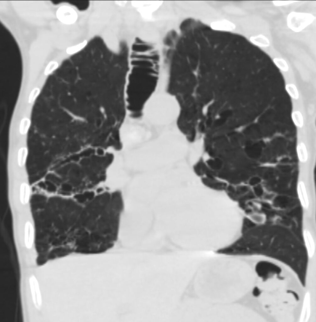
61 year old male with a history of treated mycobacterial infections and chronic cough
Coronal CT at the level of the trachea shows an enlarged trachea that measures 3cms which is abnormally enlarged. There are both thin-walled and mildly thickened cystic changes of the airways along the subsegmental bronchovascular bundle in the upper lobes and lower lobes reflecting bronchiectasis
Ashley Davidoff MD TheCommonVein.net 250Lu 135880
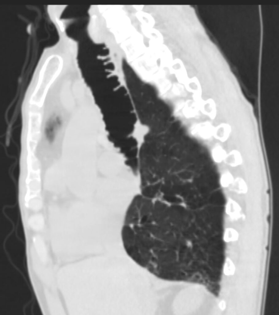
61 year old male with a history of treated mycobacterial infections and chronic cough
Sagittal CT at the level of the trachea shows an abnormally enlarged trachea. Mild thin walled bronchiectasis is also noted Flattened hemidiaphragm indicates hyperinflation
Ashley Davidoff MD TheCommonVein.net 250Lu 135884
Mechanical
Barium Swallow Aspiration
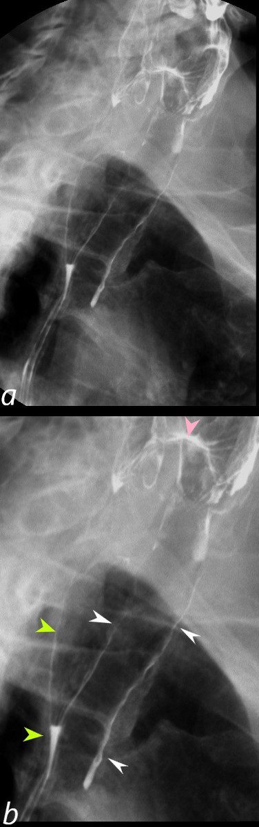
Barium swallow in the LPO projection shows abnormal accumulation of contrast along the walls of the trachea (b, white arrowheads) secondary to aspiration. Contrast also lines the wall of the posteriorly positioned esophagus (b, light green arrowheads) with a small air fluid level in the primary stripping wave of the esophagus (b, lower light green arrowhead). The contrast lined superior aspect of the epiglottis (pink arrowhead) is seen among the vallecula and pyriform sinuses.
Ashley Davidoff MD TheCommonVein.net 46505cL