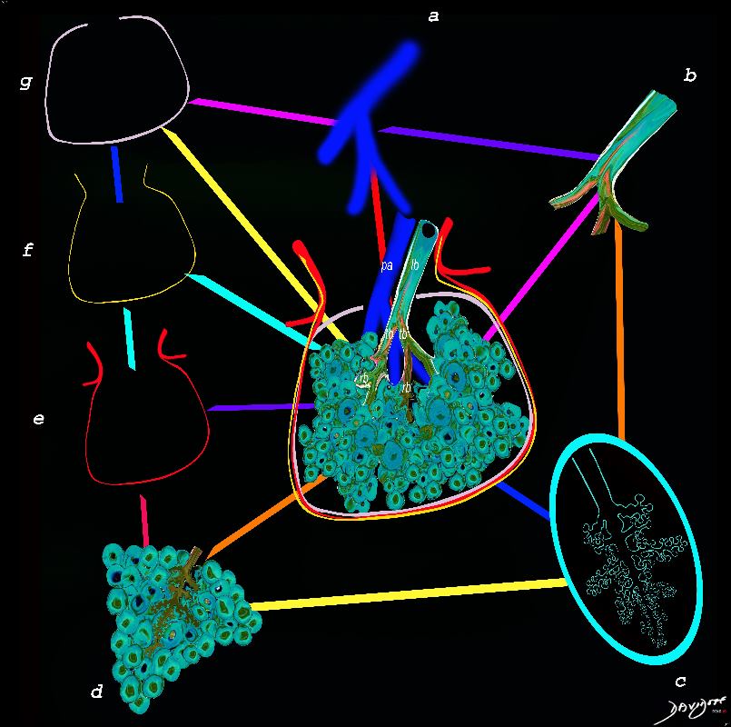
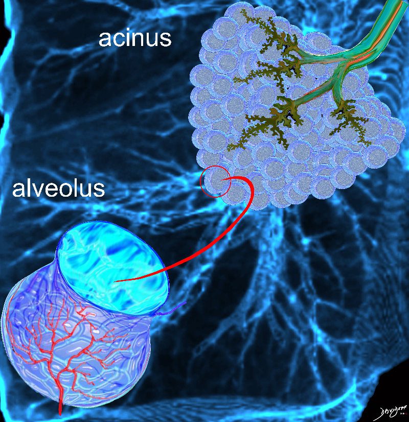
The secondary lobule is subtended by the lobular arteriole (a) and the lobular bronchiole (b) which which in turn branches into the respiratory bronchioles, alveolar ducts, and nd alveolar sacs (c) The acinus (d) consists of a respiratory bronchiole and its associated alveolar ducts, sacs, and alveoli and represents the functional unit of the lung.
The secondary lobule is drained by the pulmonary venule (e) which runs in the interlobular septum also containing the lymphatics (f). The whole unit is housed and surrounded by a connective tissue framework (g) . The latter 3 structures form the interlobular septum.
Ashley Davidoff MD TheCommonVein.net 0751 -lo resL
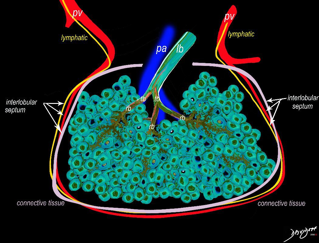
The secondary lobule is housed in a connective tissue framework in which run the lymphatic and venular tributaries . Together these 3 structures form the interlobular septum.
The lobar arteriole enters the framework, accompanied by the lobar bronchiole, and they all run together and form the interlobular septa. This structure measures between .5cms and 2cms and is visible on CT scan.
It is important in clinical radiology since many of the structures can be identified in health, and more particularly in disease, enabling the identification and characterization of many pathological processes.
Courtesy Ashley Davidoff MD The CommonVein.net
lungs-0036-low res
The arteries and airways pair up and travel together from the interlobular septa to the hilum. The pulmonary lobule, also called the secondary lobule is a structural unit surrounded by a membrane of connective tissue, and it is smaller than a subsegment of lung but larger than an acinus. This diagram shows two secondary lobules lying side by side. The pulmonary arteriole (royal blue) and bronchiole (pink) are shown together in the centre of the lobule (“centrilobular”), while the oxygenated pulmonary venules (red) and lymphatics (yellow) are peripheral and also form a formidable and almost inseparable pair.
42440b03
Ashley Davidoff MD
TheCommonVein.net

Ashley Davidoff MD TheCommonVein.net
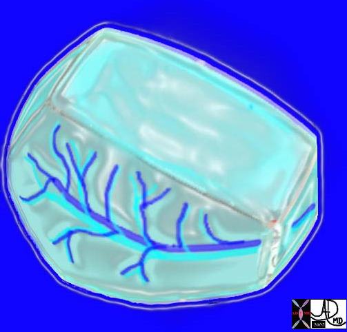
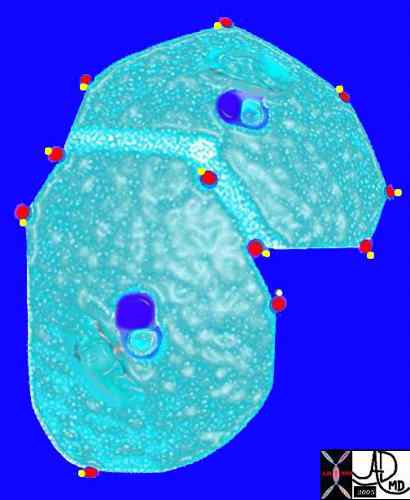
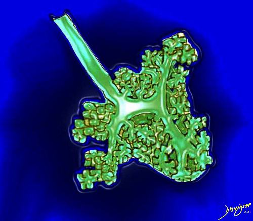
Here is a picture of the outside of the polyhedral pulmonary lobule from the side. It looked quite futuristic. Through the transparent side window we saw a couple similar to ourselves. From this vantage point the morphing did not look too different from what we had already been through – division after division – leaner and meaner. Ashley Davidoff MD. The Common Vein.net 42449b02
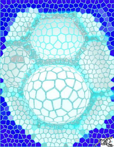
3D image of the polyhedral shapes secondary lobule with a centrilobular bronchovesicular bundle Ashley Davidoff MD. The Common Vein.net lungs-0003
The lobular (most distal of the subsegmental airways give rise to the terminal bronchiole which give rise to the membranous airways. These include in order, the respiratory bronchiole, alveolar ducts and alveolar sacs
Ashley Davidoff TheCommonvein.net lungs-0004
Ashley Davidoff MD TheCommonVein.net lungs-0008
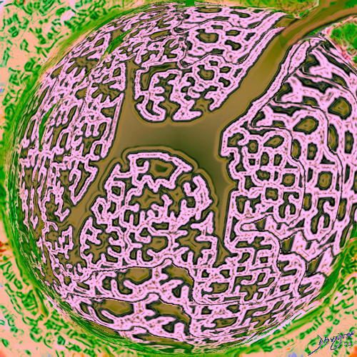
This picture shows us on the left with a white ring around us (we were the tallest) and the other couples who looked so much like us (also ringed). We called our tribe the “bronchovascular bundle” with the one part of the bundle being the progeny of the bronchus and the other the progeny of the pulmonary artery. In the distance at the periphery we could see the pairs from the other friendly tribe – the red pulmonary vein with its smaller yellow buddy the lymphatic. Behind them we could see the transparent window membrane through which we had peaked earlier. Oh my goodness!!! Look what has happened to my body!!!!!!!…… Ashley Davidoff MD. The Common Vein.net 42447b03b01
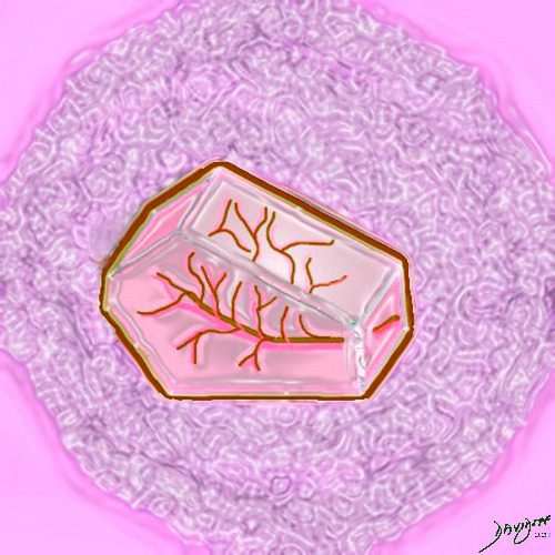
This picture was taken just before the real drama started. The image gives a sense of what was to come. You can see here in the house of the lobule that we were all dividing into smaller parts and were getting smaller and the picture was quite colorful and rosy. I fully expected to have intimate contact with the arteriole… but it did not happen as I expected…… Ashley Davidoff MD. The Common Vein.net 42447b05b02
Courtesy Ashley Davidoff MD
lungs-0028-low res
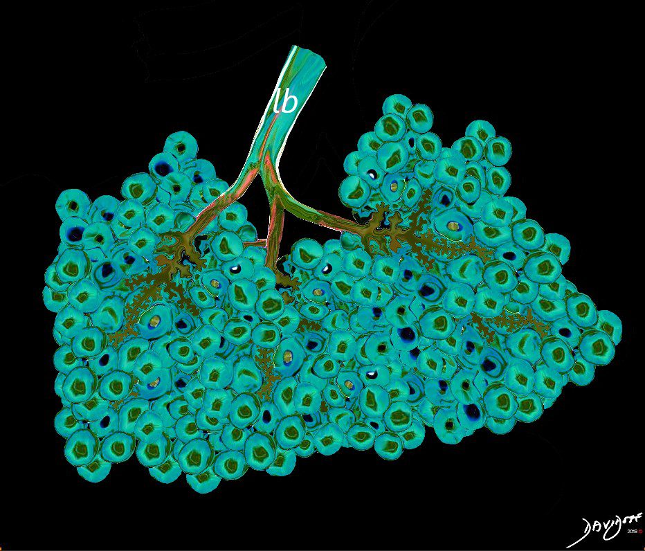
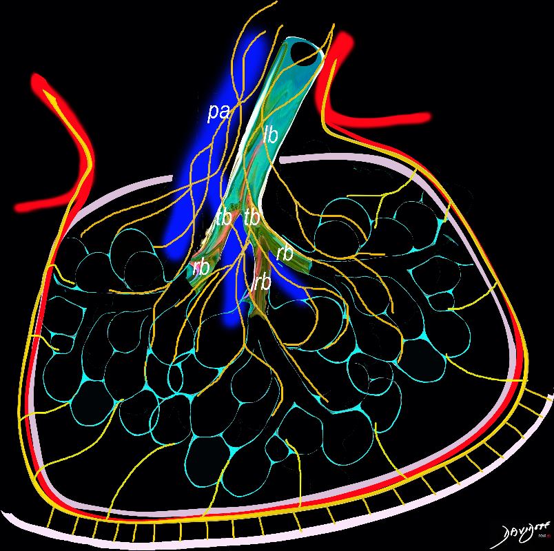
Between 10 and 30 acini combine to form a secondary lobule which is between .5- 2 cms in diameter. It is subtended by a single lobar bronchiole (lb), and is accompanied by arterioles, venules, lymphatics and connective tissue. It is important in clinical radiology since many of the structures can be identified in health, and more particularly in disease, enabling the identification and characterization of many disease processes.
Courtesy Ashley Davidoff MD
lungs-0035-low res
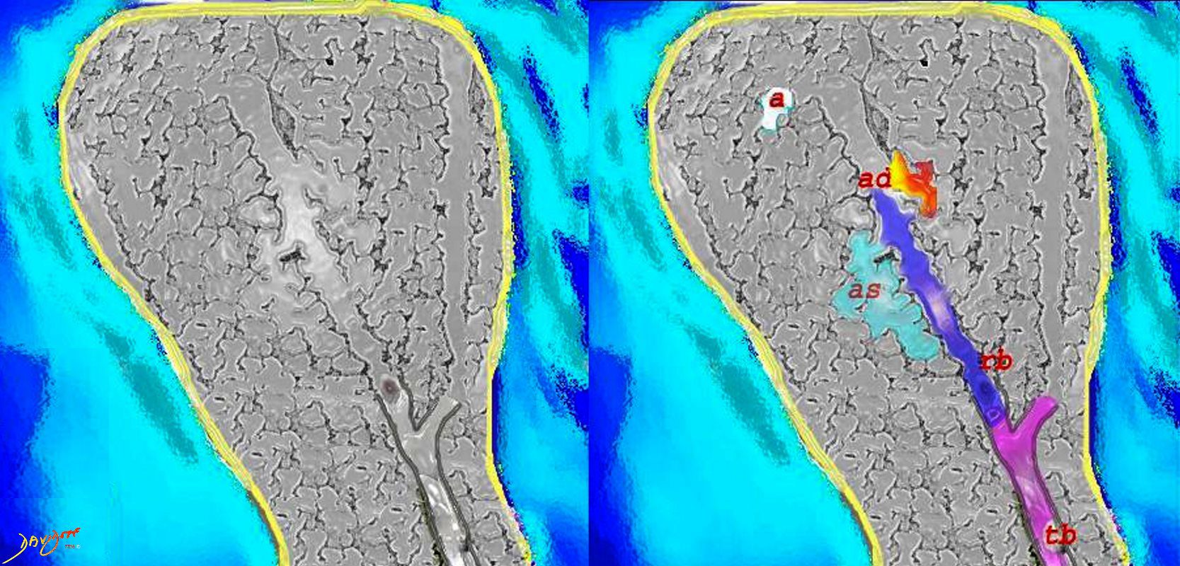
The subsegmental medium sized airways give rise to the terminal bronchiole (tb) which gives rise to the membranous airways. These include in order, the respiratory bronchiole (rb), alveolar duct (ad) and alveolar sac (as)
Ashley Davidoff TheCommonvein.net lungs-0007
Ashley Davidoff MD TheCommonvein.net lungs-0056
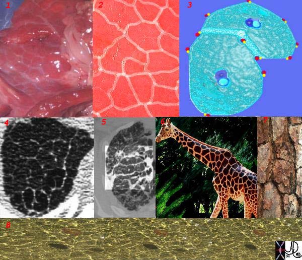
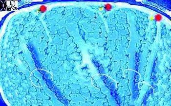
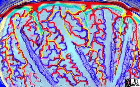
This is a series of images demonstrating the shape of the secondary lobule. The first image (1) is a post mortem specimen with congested lungs showing the interlobular septa, while the next (2), is an overlay of the septa in white showing their polygonal shape. The next drawing reveals side-by-side secondary lobules with central bronchovascular bundles and peripheral lympho-vascular bundles. Image 4 is a CT image through the apex of the lung showing thickened secondary lobules in a patient with mild emphysema, and 5 shows marked thickening of the interlobular septa in a patient with end stage sarcoidosis. 6,7,8 show the shape of the secondary lobules in the skin of a giraffe, the bark of a pine, and the ripples of the water respectively.
Ashley Davidoff MD TheCommonVein.net 31866collage
Lymphatics and the Secondary Lobule
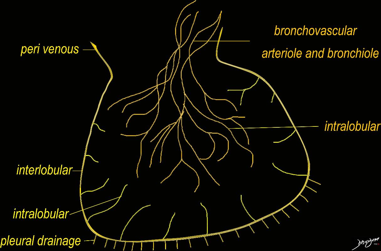
The diagram shows the 2 systems of lymphatic drainage at the level of the secondary lobule. The superficial system drains some of the interstitium of the secondary lobule, runs in the interlobular septa and drains all the pleura. Thee pathway to the lymph nodes in the mediastinum is via the pulmonary veins. The deeper system drains the interstitium in the interalveolar septa, and then they travel along the bronchovascular bundle accompanying the bronchi and pulmonary artery and into the lymph nodes of the hila and mediastinum
Ashley Davidoff MD TheCommonVein.net lungs-0767