
There is a hint of the reversed S of Golden
Ashley Davidoff MD TheCommonVein.net RnD image
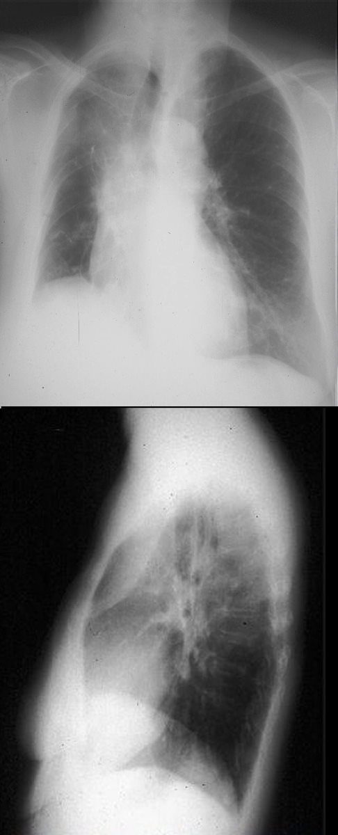
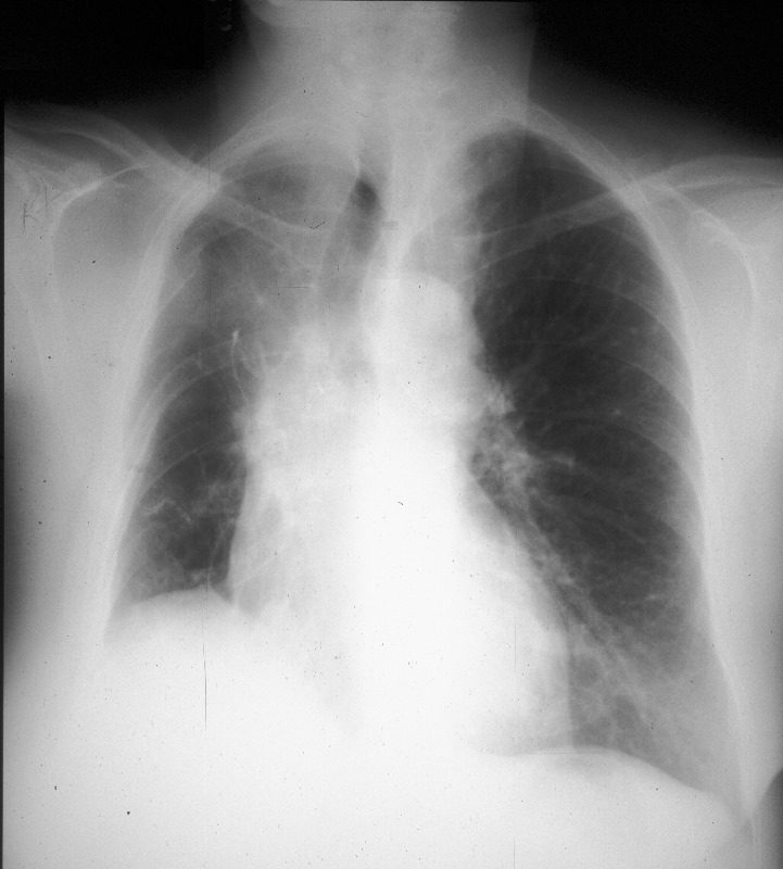
Frontal and lateral views of the chest show signs of volume loss characterized by elevation of the right hemidiaphragm (black arrowhead), rightward tracheal and mediastinal shift and elevation of the minor fissure contributing to the reverse S sign of Golden. There is a vague infiltrate in the right upper lobe correlating with an anterior pie shaped density on the lateral examination, consistent with collapse of the anterior segment of the RUL. This combination of images is consistent with a malignant mass in the hilum causing obstruction of the right mainstem bronchus.
Ashley Davidoff MD TheCommonVein.net 237Lu 32292c01
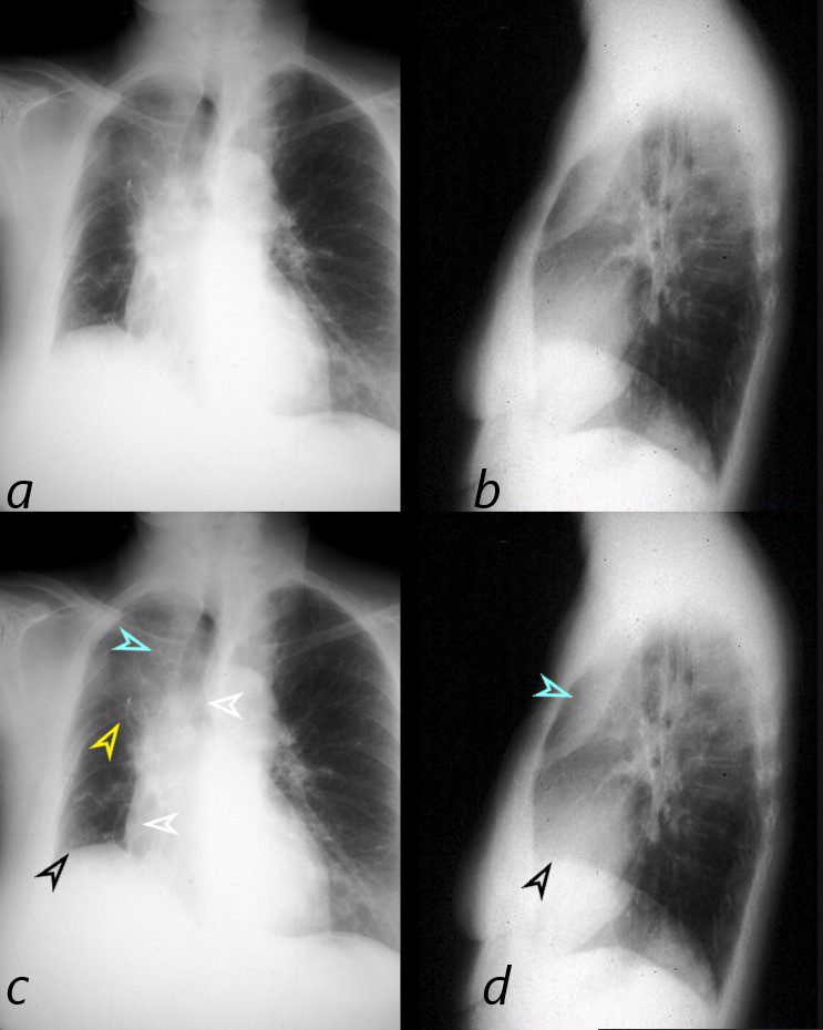
This combination of images shows the manifestations of a malignant mass in the hilum causing compression of the right mainstem bronchus. The PA CXR shows signs of volume loss (atelectasis characterized by elevation of the right hemidiaphragm (black arrowhead), rightward tracheal and mediastinal shift (white arrowheads) and elevation of the minor fissure contributing to the reverse S sign of Golden. There is a vague infiltrate in the right upper lobe correlating with an anterior pie shaped density of the lateral (blue arrowheads), consistent with collapse of the anterior segment of the RUL
32292cL01
Ashley Davidoff MD TheCommonVein.net
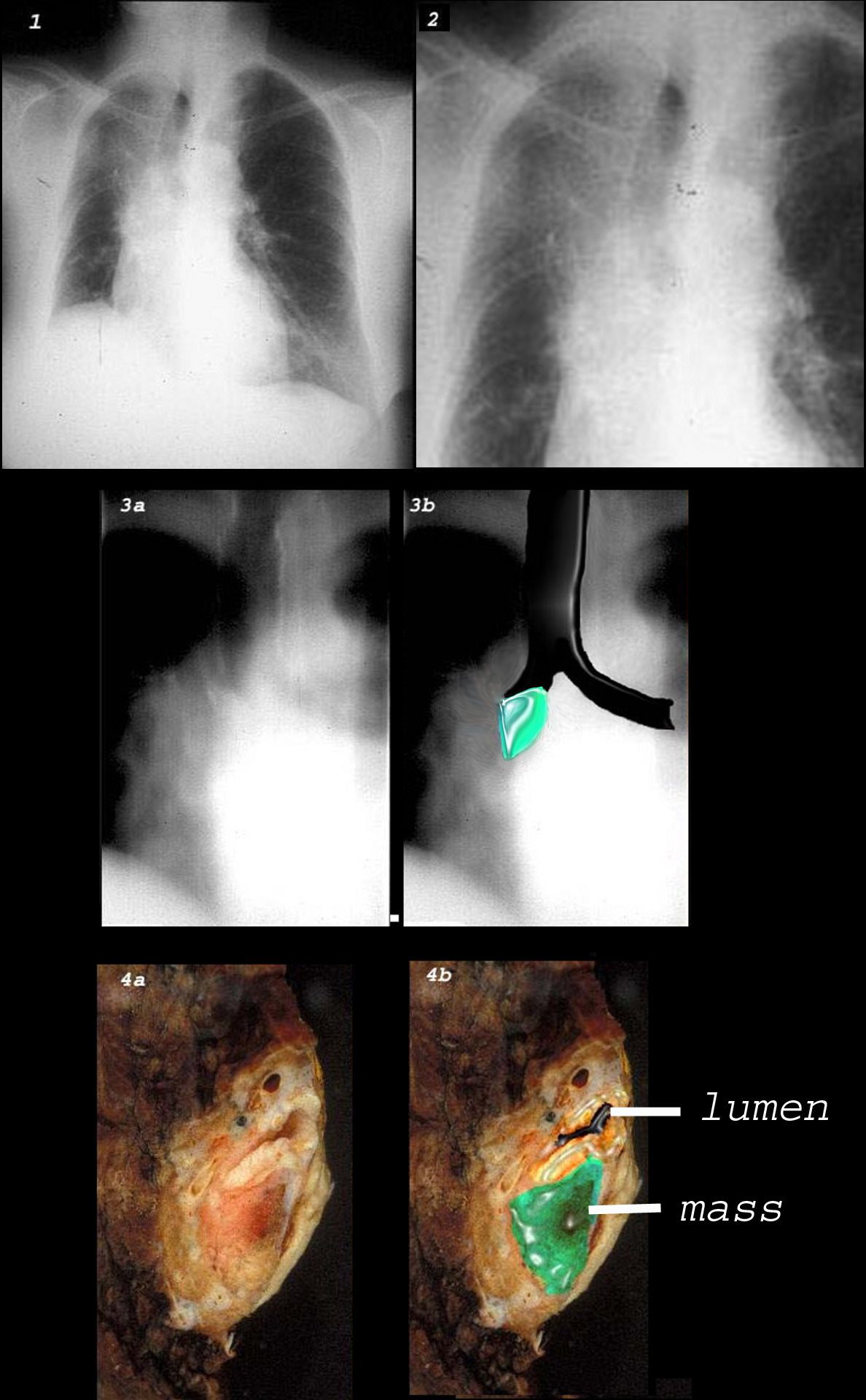
This combination of images shows the manifestations of a malignant mass in the hilum causing compression of the right mainstem bronchus. There is elevation of the right hilum on the CXR, associated with collapse of the anterior segment of the RUL seen as a vague density in the P-A . The tomogram (3a) shows an abrupt cut off of the right mainstem bronchus while the overlay in 3b shows the occlusion of the right mainstem bronchus, the implied tumor overlaid in green. Images 4a and 4b are the correlative gross pathology images showing the tumor in green pushing and occluding the right mainstem
Ashley Davidoff MD TheCommonVein.net 32292cw