
Infection
TB
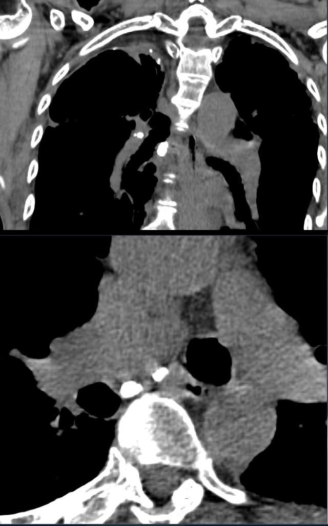
80- year-old non-smoker with childhood history of treated TB, presents with a chronic cough
CT scan in the coronal (upper image) and axial (lower image) planes show calcified hilar and mediastinal nodes consistent with chronic granulomatous disease
Ashley Davidoff MD TheCommonVein.net 292Lu 136629c
Inflammation
Malignancy
Mechanical
Atelectasis
Trauma
Metabolic
Circulatory-
Hemorrhage
Immune
Infiltrative
Amyloid
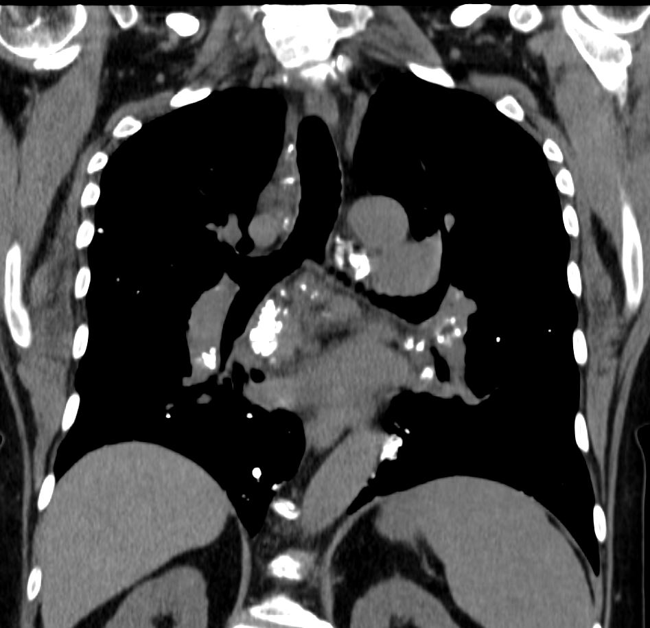
CT scan in the coronal plane of a 60-year-old female with known diagnosis of AL amyloidosis shows multiple heterogeneously calcified lymph nodes in the mediastinum and hila
Ashley Davidoff MD TheCommonVein.net 266Lu 136191
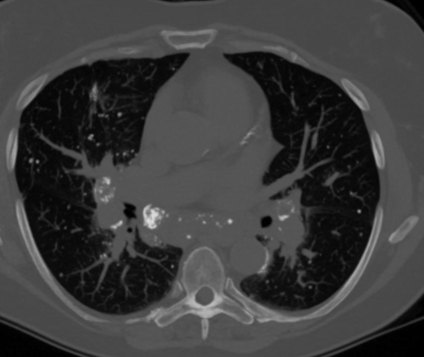
CT scan in the axial plane of a 60-year-old female with known diagnosis of AL amyloidosis shows multiple heterogeneously calcified lymph nodes in the mediastinum and hila
Ashley Davidoff MD TheCommonVein.net 266Lu 136188
Idiopathic Iatrogenic