
Allergic Bronchopulmonary Aspergillosis Finger in Glove and Low Density Lymph Nodes
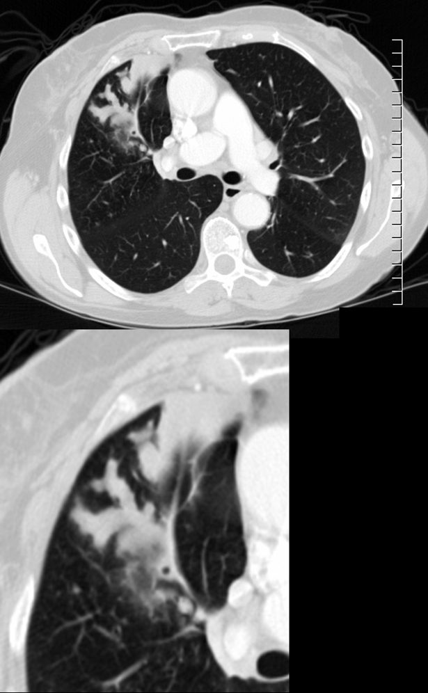
CT scan with contrast shows finger in glove appearance of the anterior segmental airways of the right upper lobe ) with a focal region of subsegmental atelectasis medially. The finger in glove sign results thick, mucus plugs within the bronchi due to the exaggerated inflammatory and immune response caused by Aspergillus fumigatus leading to airway inflammation, mucus production, bronchial wall thickening, and bronchiectasis.
Ashley Davidoff MD The CommonVein.net 294Lu 117966c
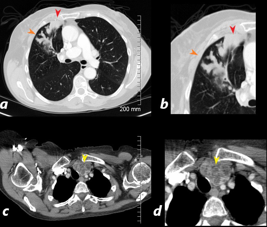
CT scan with contrast shows finger in glove appearance of the anterior segmental airways of the right upper lobe (orange arrowheads a, b) with a focal region of subsegmental atelectasis (red arrow head a,b).
The lower panel shows a cluster of low density lymph nodes in the anterior mediastinum in the retro-clavicular region (yellow arrowheads c,d) .
Ashley Davidoff MD The CommonVein.net 294LU 117972b
56-year-old male presents with chronic cough dyspnea and weight loss. CT scan in coronal projection shows a subcarinal esophageal mass which was diagnosed as a leiomyoma,
Ashley Davidoff MD TheCommonVein.net 136220
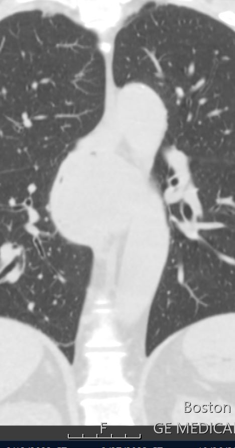
19 year old female with cystic fibrosis and bronchiectasis
19 year old female with cystic fibrosis and bronchiectasis
CT scan through the upper lung fields shows mucin filled subsegmental bronchi of the right upper lobe with morphology reminiscent of the “finger in glove” sign
Courtesy Priscilla Slanetz MD MPH TheCommonVein.net
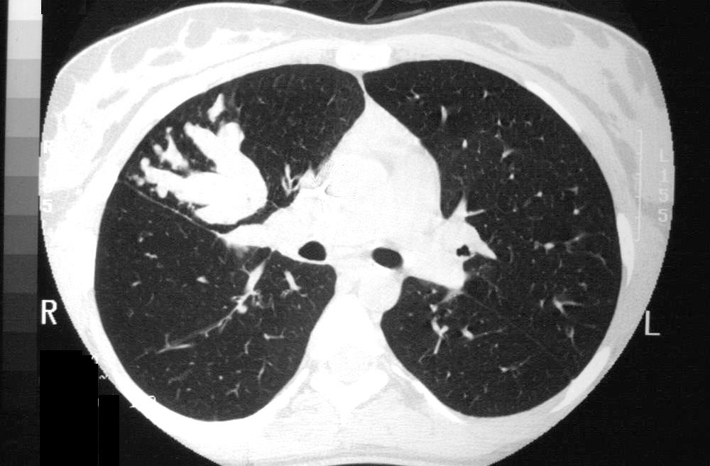
77 Female Asthma and APBA

CT – with Noted Tubular Structures in the Upper Lobes
77 year old female with history of asthma, allergic bronchopulmonary aspergillosis (ABPA) and COPD
CXR shows prominent bronchovascular bundles in the upper lung fields (green arrowheads a, and b) . CT shows fluid filled bronchiectatic airways (green arrowheads in image d, which is a magnified image of c) reminiscent of the finger in glove appearance of ABPA)
Ashley Davidoff TheCommonVein.net
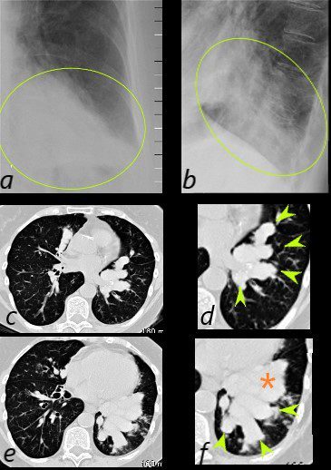
77 year old female with history of asthma, allergic bronchopulmonary aspergillosis (ABPA) and COPD
CXR shows LLL infiltrate in the PA (green oval in a) and lateral views (green oval in d and f) which reflect mucus filled bronchiectatic airways magnified image s of the CT scan of the LLL ) reminiscent of the finger in glove appearance of ABPA. There is a LL infiltrate possibly atelectatic (orange asterisk)
Ashley Davidoff TheCommonVein.net
Squamous Cell Carcinoma Masquerading as ABPA
56-year-old male presents with chronic cough dyspnea and weight loss. CT scan in coronal projection shows an appearance reminiscent of finger in glove in the right lower lobe. There s segmental and subsegmental thickening of the airways in the upper lobes, and paraseptal emphysema. Micronodules in the upper lobes suggest smoker’s bronchiolitis. The subcarinal esophageal mass was diagnosed as a leiomyoma, Pathology of the right lower process was a squamous cell carcinoma
Ashley Davidoff MD TheCommonVein.net 136219
56-year-old male presents with chronic cough dyspnea and weight loss. CT scan in axial projection shows an appearance reminiscent of finger in glove in the right lower lobe. There s a para-fissural soft tissue mass that seems “soft” since it does not displace nor deform the fissure.. Pathology of the right lower process was a squamous cell carcinoma
Ashley Davidoff MD TheCommonVein.net 136221
56-year-old male presents with chronic cough dyspnea and weight loss. CT scan in axial projection shows an appearance reminiscent of finger in glove in the right lower lobe. Pathology of the right lower process was a squamous cell carcinoma
Ashley Davidoff MD TheCommonVein.net 136223