
28-year-old immigrant with cough
CXR – Reactivation TB Cavitating Pneumonia – Left Upper Lobe
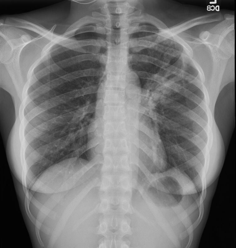
Frontal CXR of a 28-year-old immigrant with cough shows a cavitating pneumonia in the left upper lobe (magnified in the lower image)
Lab tests confirmed the diagnosis of TB and the patient was treated with RISE a 4-month treatment regimen of rifapentine-moxifloxacin for mycobacterium tuberculosis.
Ashley Davidoff MD TheCommonVein.net 255Lu 136071
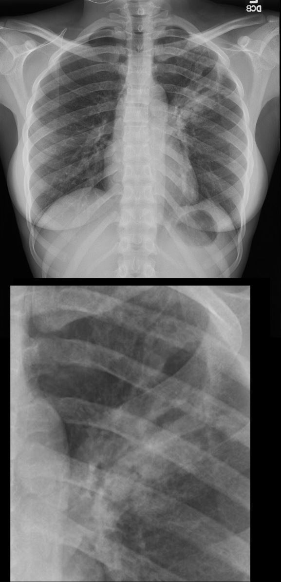
Frontal CXR of a 28-year-old immigrant with cough shows a cavitating pneumonia in the left upper lobe (magnified in the lower image)
Lab tests confirmed the diagnosis of TB and the patient was treated with RISE a 4-month treatment regimen of rifapentine-moxifloxacin for mycobacterium tuberculosis.
Ashley Davidoff MD TheCommonVein.net 255Lu 136071c
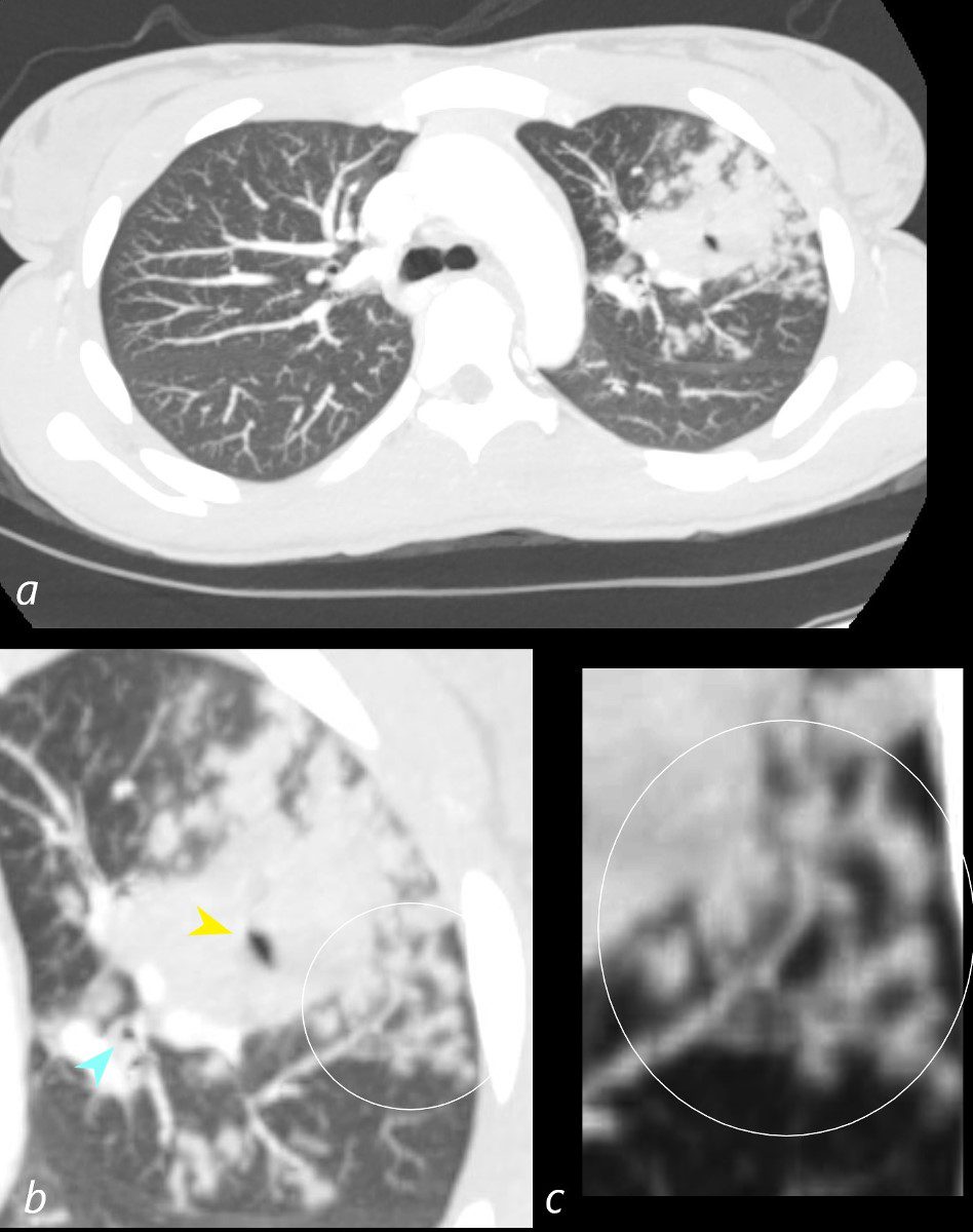
CT –TB Pneumonia
Cluster of Subsegmental Consolidations –
suggestion of Tree in Bud Changes
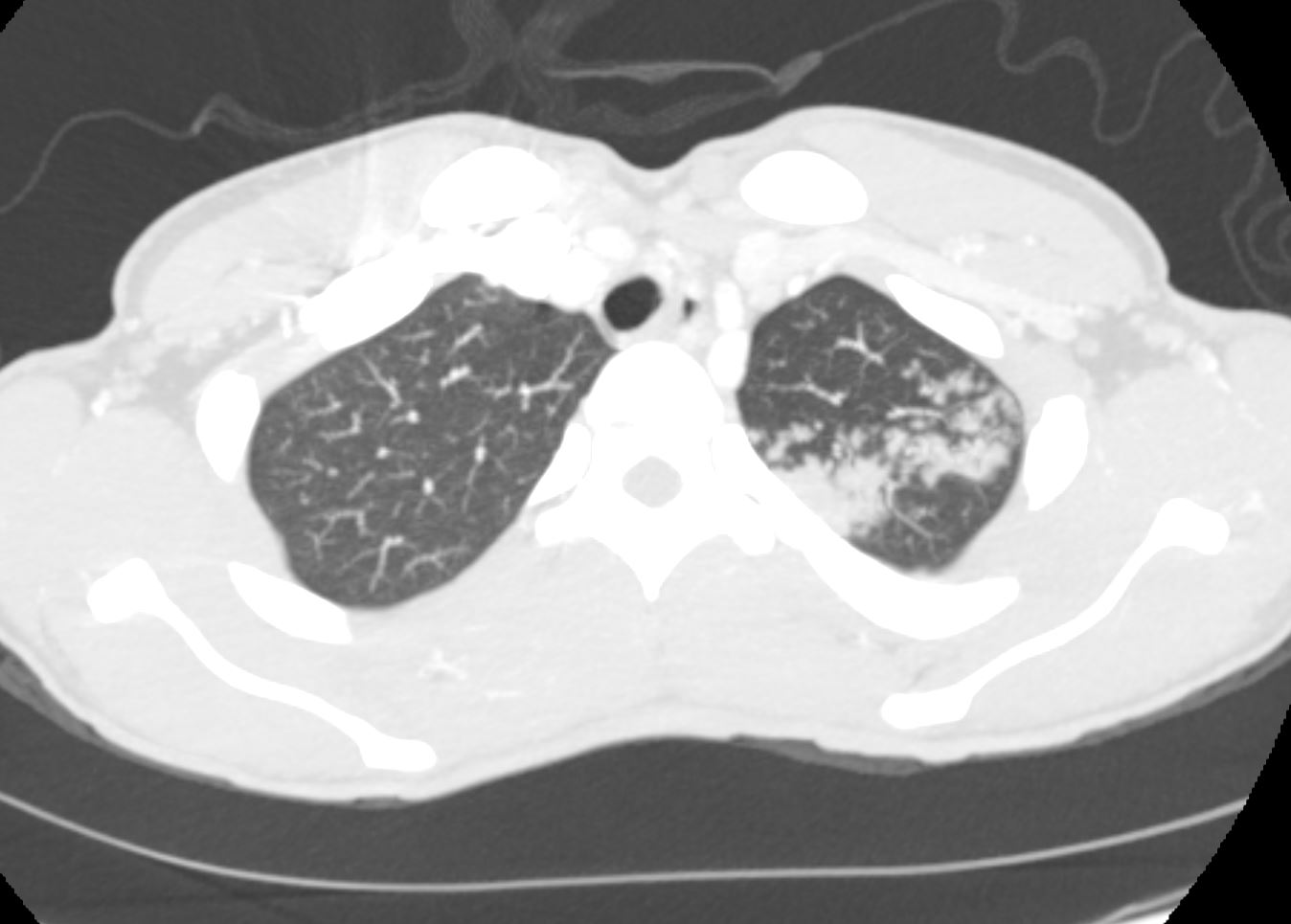
CT scan in the axial plane of the left upper lobe a 28-year-old immigrant with cough shows a a cluster of subsegmental consolidations with suggestion of expanded tree in bud changes . Lab tests confirmed the diagnosis of TB and the patient was treated with RISE a 4-month treatment regimen of rifapentine-moxifloxacin for mycobacterium tuberculosis.
Ashley Davidoff MD TheCommonVein.net 255Lu 136073
CT –TB Pneumonia – Left Upper Lobe Cavitation
CT scan in the axial plane of the left upper lobe of a 28-year-old immigrant with cough shows a thick walled cavitating mass subtended by a subsegmental thick-walled airway. Lab tests confirmed the diagnosis of TB and the patient was treated with RISE, a 4-month treatment regimen of rifapentine-moxifloxacin for mycobacterium tuberculosis.
Ashley Davidoff MD TheCommonVein.net 255Lu 136074c
Cavitating Infiltrate Left Upper Lobe Connecting with Airways
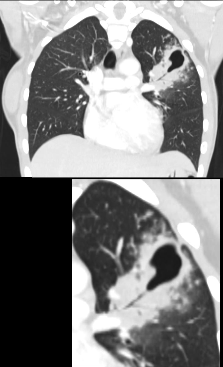
CT scan in the coronal plane of the left upper lobe of a 28-year-old immigrant with cough shows a thick walled cavitating mass subtended by a subsegmental thick-walled airway. Lab tests confirmed the diagnosis of TB and the patient was treated with RISE, a 4-month treatment regimen of rifapentine-moxifloxacin for mycobacterium tuberculosis.
Ashley Davidoff MD TheCommonVein.net 255Lu 136081c
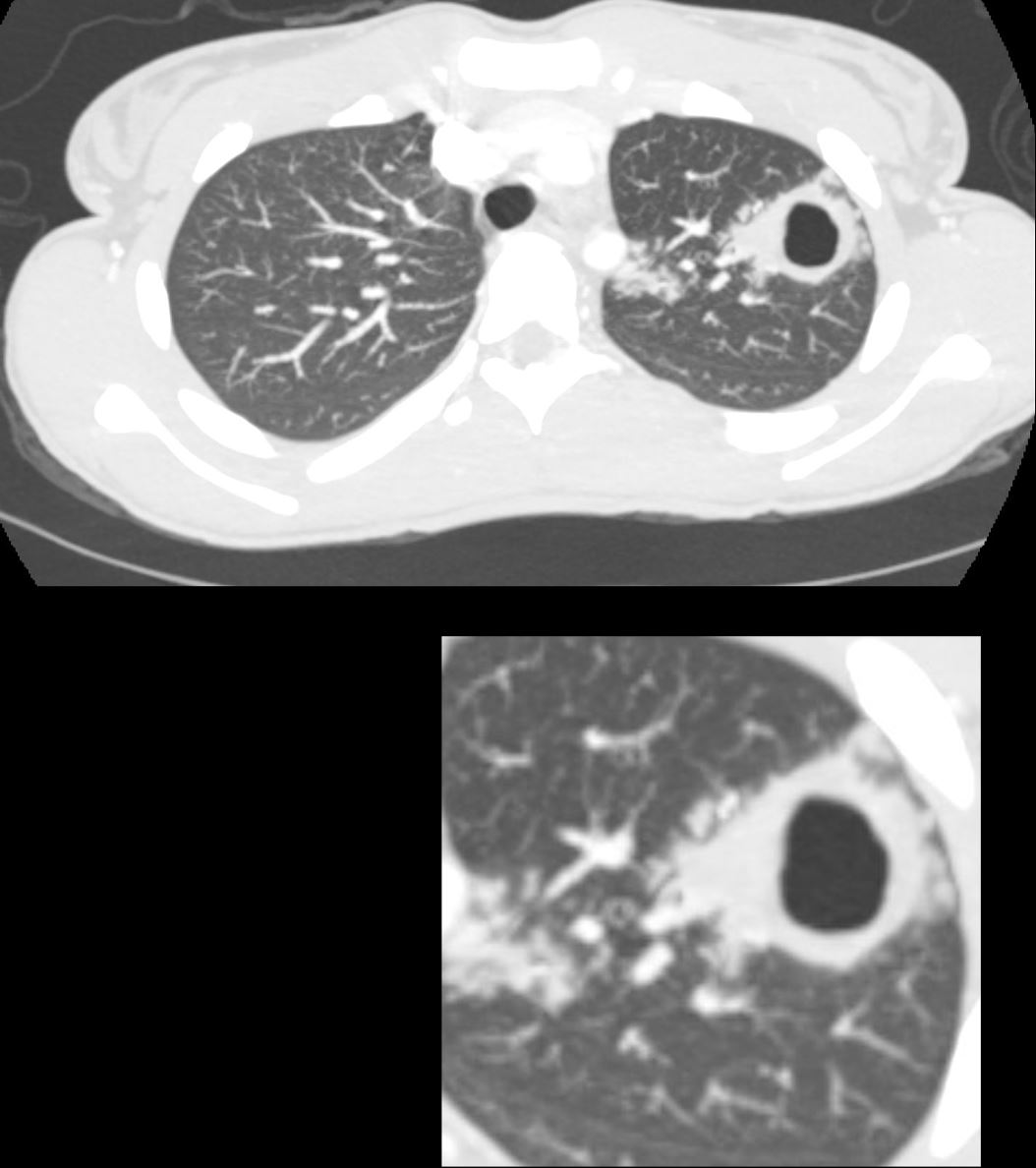
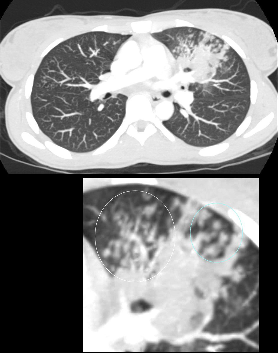
TB Pneumonia – Endobronchial Spread
Left Upper Lobe Cavitation and Tree in Bud Disease
CT scan in the axial plane of the left upper lobe of a 28-year-old immigrant with cough shows a focal subsegmental consolidation with focal cavitation (yellow arrowhead) subtended by a thick-walled subsegmental airway. There are extensive tree in bud changes ringed in white (b and c) indicating transbronchial spread. Lab tests confirmed a diagnosis of TB and the patient was treated with RISE, a 4-month treatment regimen of rifapentine-moxifloxacin for mycobacterium tuberculosis.
Ashley Davidoff MD TheCommonVein.net 255Lu 136075cL
Left Upper Lobe Airway Disease
Segmental Subsegmental and Small Airway Involvement
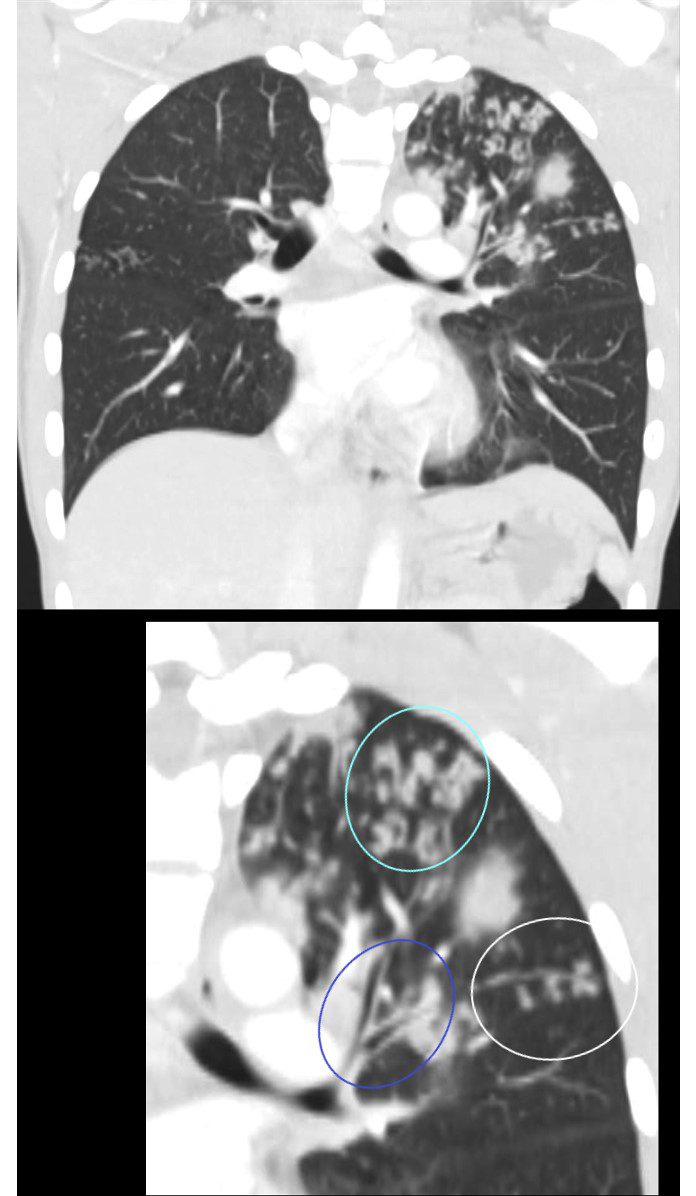
CT scan in the coronal plane of the left upper lobe of a 28-year-old immigrant with cough shows a thickening of the walls of the segmental, (blue circle) and subsegmental airway disease (teal circle ) as well as small airways disease characterised by tree in bud changes (ringed in whit)e These findings indicate transbronchial spread.
Lab tests confirmed a diagnosis of TB and the patient was treated with RISE, a 4-month treatment regimen of rifapentine-moxifloxacin for mycobacterium tuberculosis.
Ashley Davidoff MD TheCommonVein.net 255Lu 136079cL
CT scan in the axial plane of the left upper lobe of a 28-year-old immigrant with cough shows a focal subsegmental consolidation. There are extensive tree in bud changes ringed in white indicating transbronchial spread. Prominent centrilobular nodules indicating small airway impaction are noted as well (ringed in blue lower panel)
Lab tests confirmed a diagnosis of TB and the patient was treated with RISE, a 4-month treatment regimen of rifapentine-moxifloxacin for mycobacterium tuberculosis.
Ashley Davidoff MD TheCommonVein.net 255Lu 136077cL
Reactivation TB Pneumonia –
Cavitating Infiltrate Left Upper Lobe Pre and Post Treatment
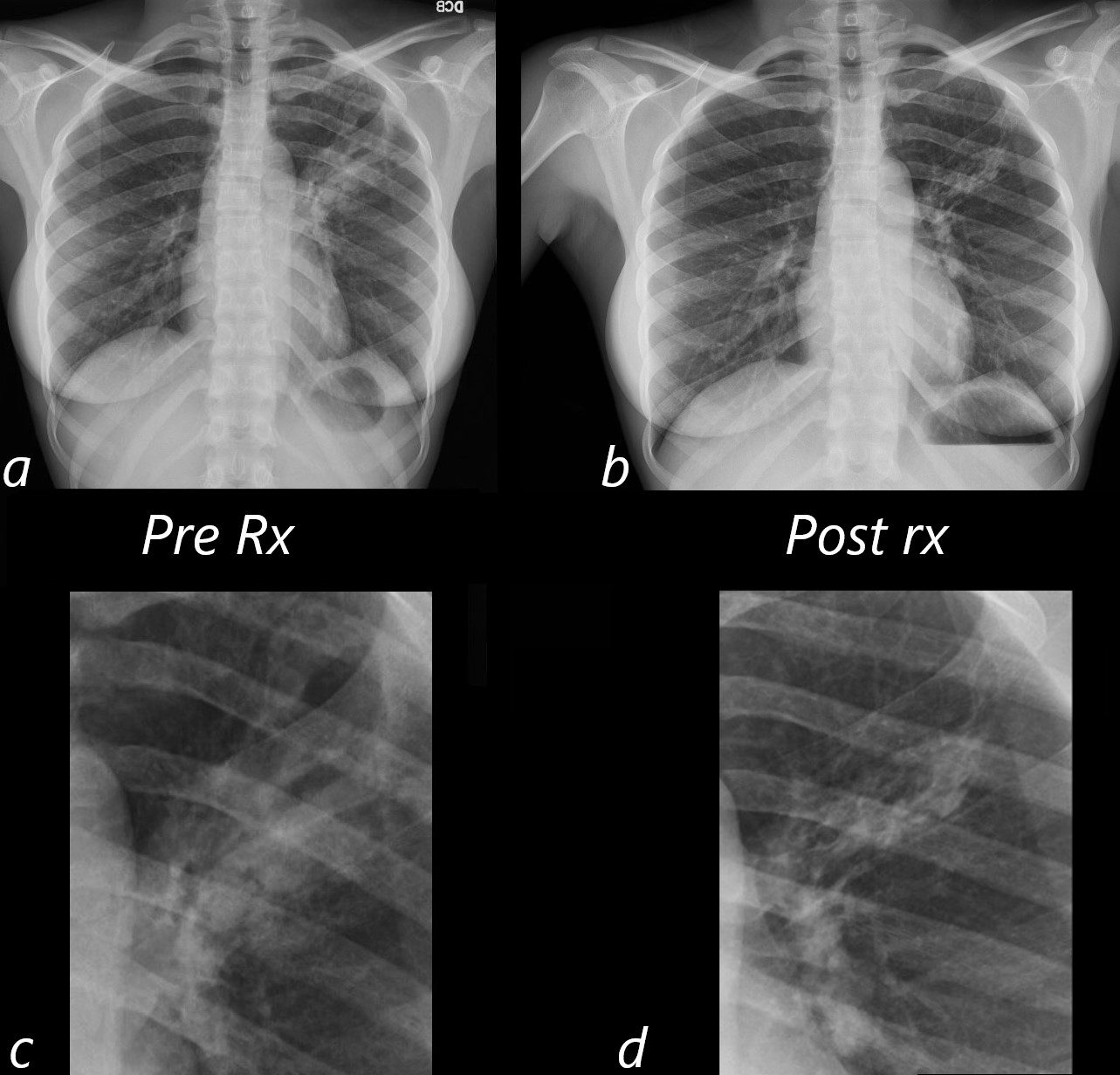
Frontal CXR at time of presentation of a 28-year-old immigrant with cough shows a cavitating pneumonia in the left upper lobe (a, magnified in c). Lab tests confirmed the diagnosis of TB and the patient was treated with RISE, a 4-month treatment regimen of rifapentine-moxifloxacin for mycobacterium tuberculosis.
Follow up frontal CXR shows significant improvement of the left upper lobe pneumonia (b and magnified in d)
Ashley Davidoff MD TheCommonVein.net 255Lu 136082c02L