
Upper Lobes
Anterior and Medial Upper Lobes

CXR shows right upper lobe (RUL) atelectasis. Final diagnosis was a central RUL proximal squamous cell carcinoma with extensive filling of the distal bronchi-ectatic segmental and subsegmental airways
Ashley Davidoff TheCommonVein.net

Ashley Davidoff TheCommonVein.net
Lingula Superior and Anterior Migration
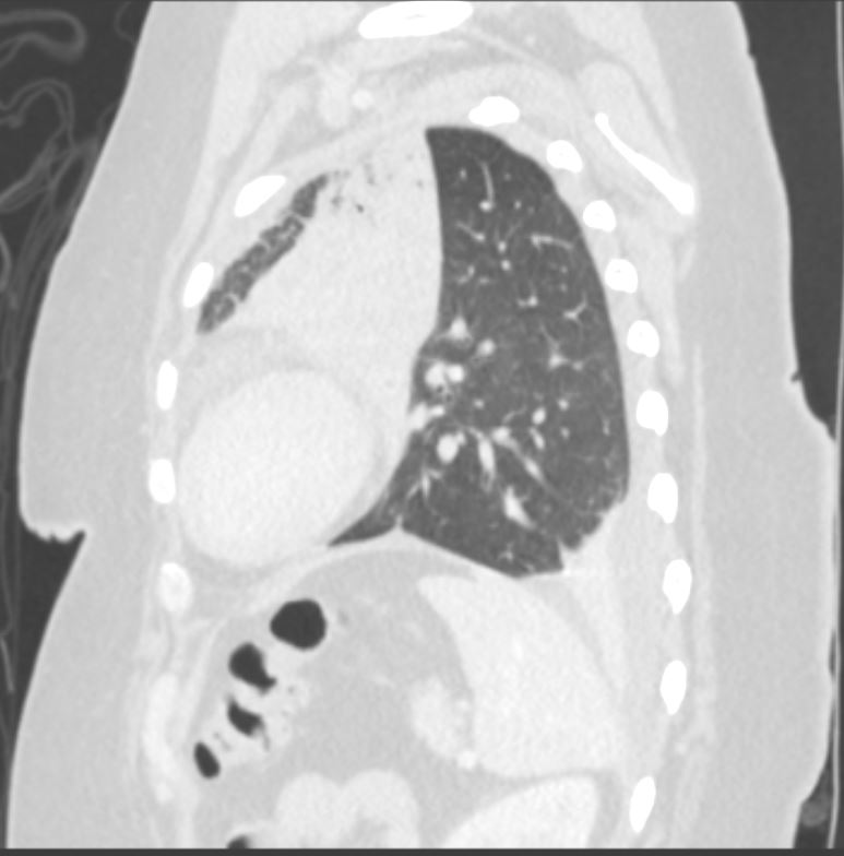
58-year-old female presents with a cough. CT in the sagittal plane shows post obstructive atelectasis of the lingula, hyperinflation of the left lower lobe, superior and anterior migration of the left major fissure, and a small portion of aerated left upper lobe anteriorly. There is a loculated effusion with subsegmental compressive atelectasis of the left lower lobe.
Pathology revealed findings consistent with a carcinoid tumor in the left mainstem bronchus
Ashley Davidoff MD TheCommonVein.net 257Lu 136121
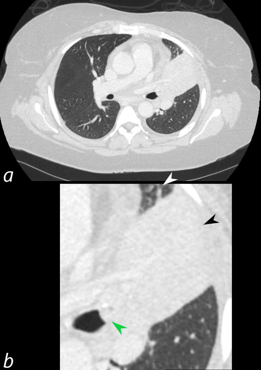
58-year-old female presents with a cough. CT in the axial plane shows an obstructing lesion in the left mainstem bronchus of the lung (green arrowhead) with post obstructive atelectasis of the lingula (black arrowhead) and a small portion of aerated left upper lobe anteriorly (white arrowhead). The major fissure is displaced anteriorly.
Pathology revealed findings consistent with a carcinoid tumor of the left bronchus.
Ashley Davidoff MD TheCommonVein.net 257Lu 136110cL
Lower Lobes Posterior and Medial Lower Lobes
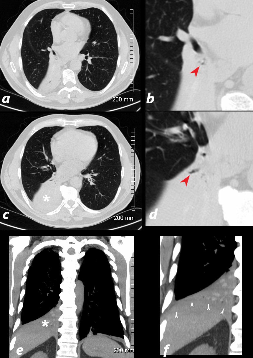
77 year old male presents chest discomfort
CT scan without contrast shows atelectasis of the right lower lobe )asterisk c and r) and also seen axial projection (a) magnified in (b) and in (c) magnified in {d) Red arrowheads in b and d show airways filled with material. Aspergillus was isolated at bronchoscopy. Coronal imaging (e magnified in f) show silhouetting of the right hemidiaphragm by the atelectatic lung (white arrowheads
Ashley Davidoff TheCommonVein.net 117786cL
Middle Lobe
Right Middle Lobe
Segmental Atelectasis
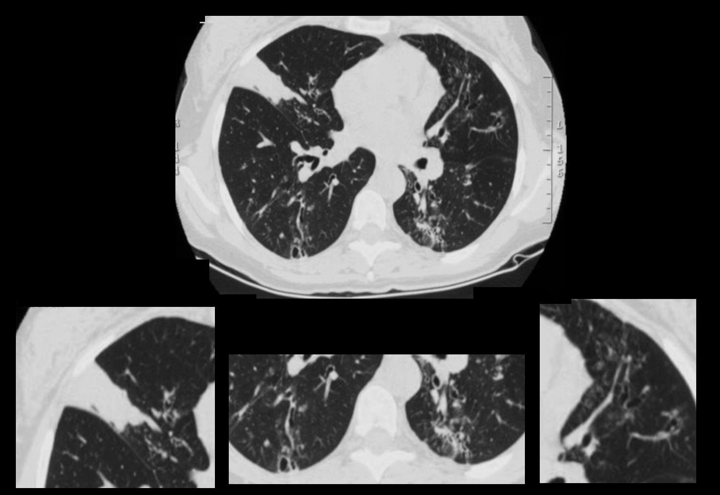
CT Allergic Bronchopulmonary Aspergillosis (ABPA)
48 year old female with a history of asthma presents with productive cough. CT scan 18 months prior confirms atelectasis in the middle lobe (upper panel and right lower panel) . There is diffuse mild multicentric foci of bronchial wall thickening in the segmental and subsegmental airways of the middle lobe, lingula and the lower lobes bilaterally (upper panel magnified in lower 3 panels).
Ashley Davidoff MD TheCommonVein.net
Right Lower Lobe Left Upper Lobe Left Lower Lobe