Infection Inflammation Malignancy Mechanical/Atelectasis Trauma Metabolic Circulatory- Hemorrhage Immune Infiltrative Idiopathic Iatrogenic Idiopathic
Infection
Transbronchial Spread of TB with Cavitation
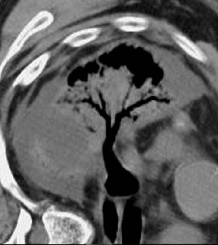
CT scan in the coronal plane of the left upper lobe of a 28-year-old immigrant with cough shows a thick walled cavitating mass subtended by a subsegmental thick-walled airway. Lab tests confirmed the diagnosis of TB and the patient was treated with RISE, a 4-month treatment regimen of rifapentine-moxifloxacin for mycobacterium tuberculosis.
Ashley Davidoff MD TheCommonVein.net 255Lu 136081c
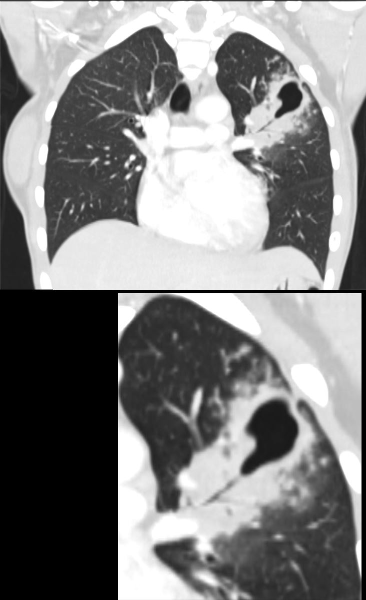
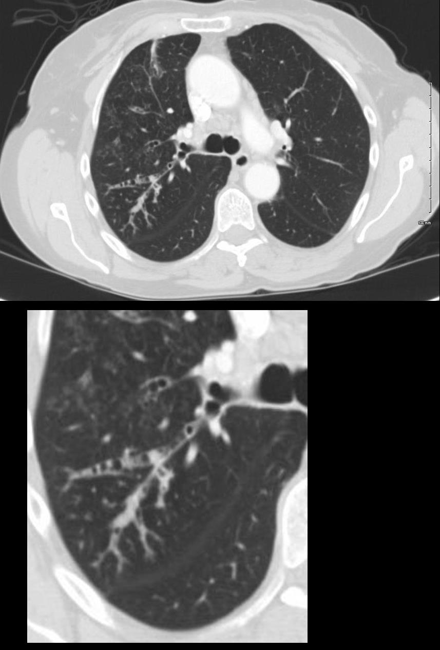
CT scan in the axial plane of the left upper lobe of a 28-year-old immigrant with cough shows a focal subsegmental consolidation with focal cavitation (yellow arrowhead) subtended by a thick-walled subsegmental airway. There are extensive tree in bud changes ringed in white (b and c) indicating transbronchial spread. Lab tests confirmed a diagnosis of TB and the patient was treated with RISE, a 4-month treatment regimen of rifapentine-moxifloxacin for mycobacterium tuberculosis.
Ashley Davidoff MD TheCommonVein.net 255Lu 136075cL
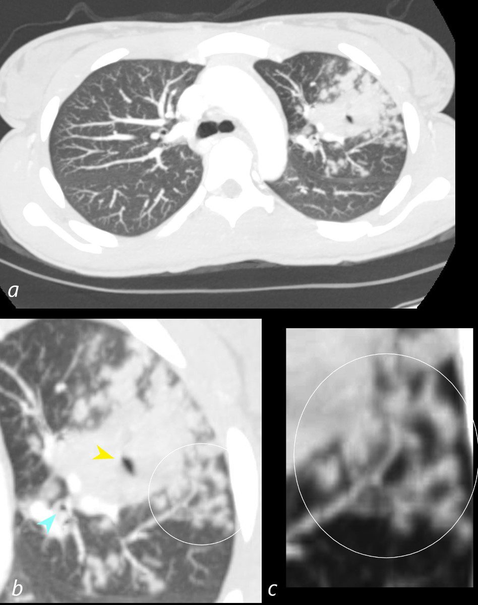
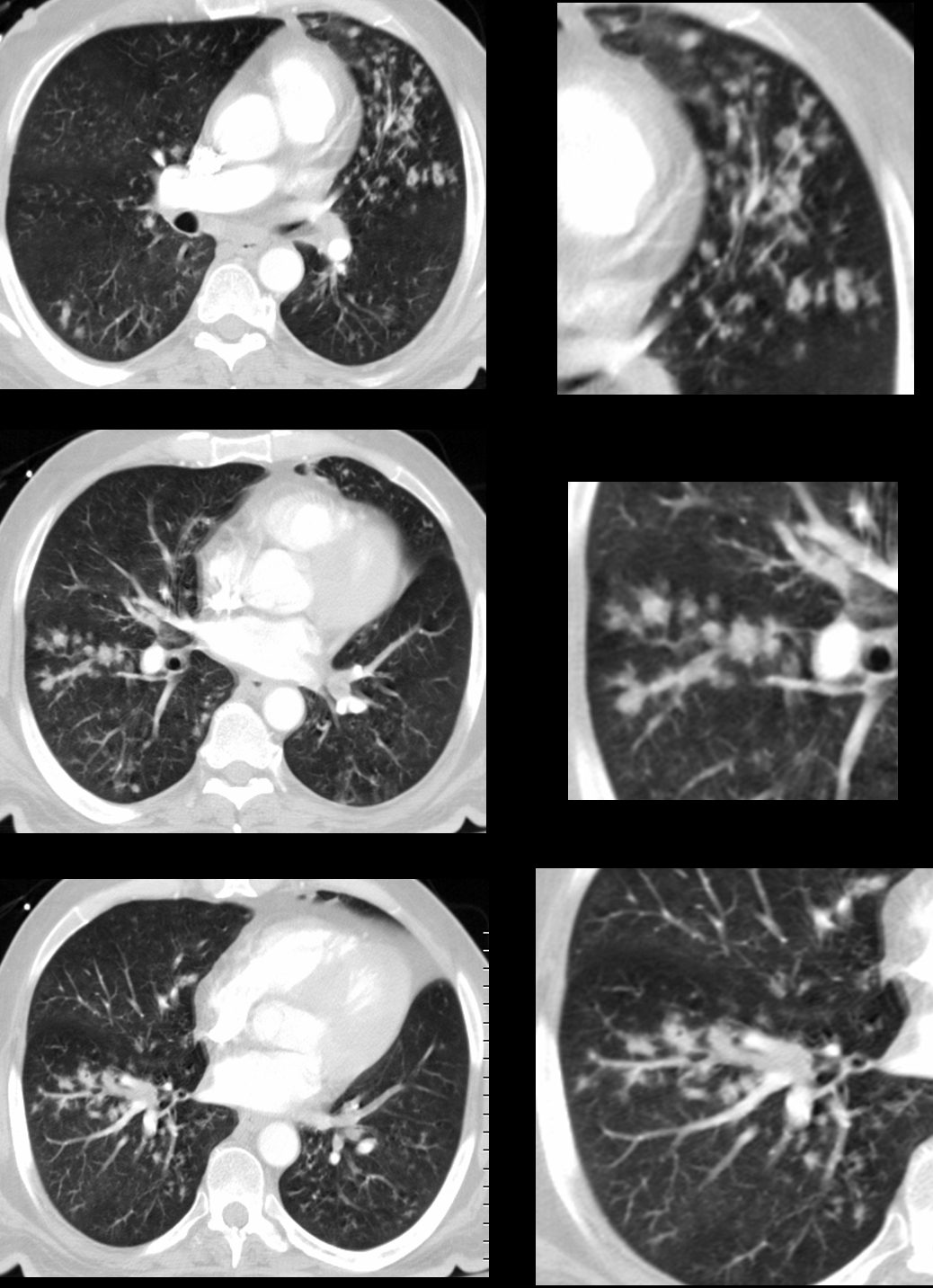
39-year-old immigrant Vietnamese male presents night sweats fever and cough. CT in the axial plane through the mid chest shows innumerable micronodules resulting from transbronchial spread with resultant tree in bud pattern scattered through the right lung (magnified in the right lower image). There are minimal similar changes in the lingula (magnified left lower image)..
Ashley Davidoff MD TheCommonvein.net 135789c 006Lu
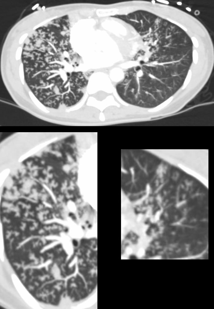
Small Airway Disease and Tree in Bud Changes -Right Upper Lobe Active TB
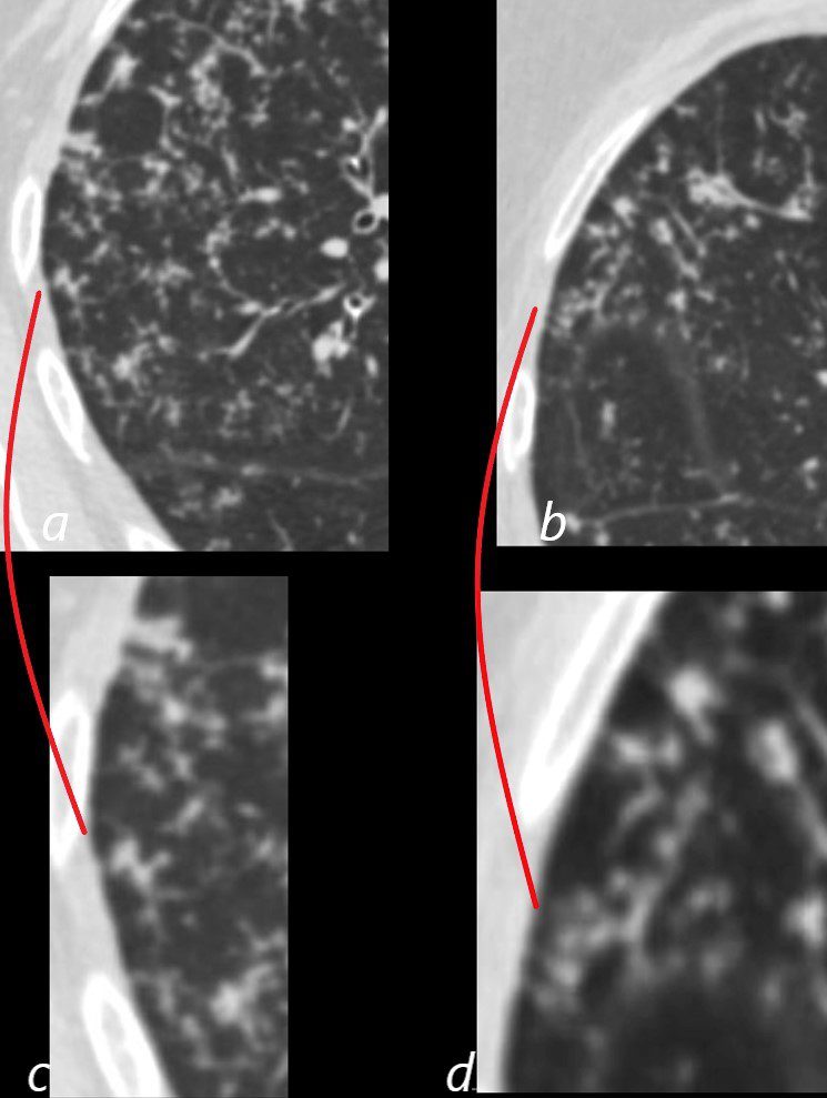
CT in the axial plane shows extensive small airway disease dominant in the right upper lobe characterized by innumerable, ground glass micronodules, and tree in bud changes. (a magnified in c and b magnified in d)
Ashley Davidoff MD TheCommonVein.net 135827aL 192Lu
Inflammation
ABPA Early
ABPA Current
CT Scan RUL
72 year old female with asthma with cough and chronic dyspnea.
CT in the axial plane shows thickening of the segmental and subsegmental airways of the posterior segment of the right upper lobe with mucoid impaction and tree in bud formation
Ashley Davidoff MD TheCommonVein.net 294Lu 117967c
ABPA Advanced
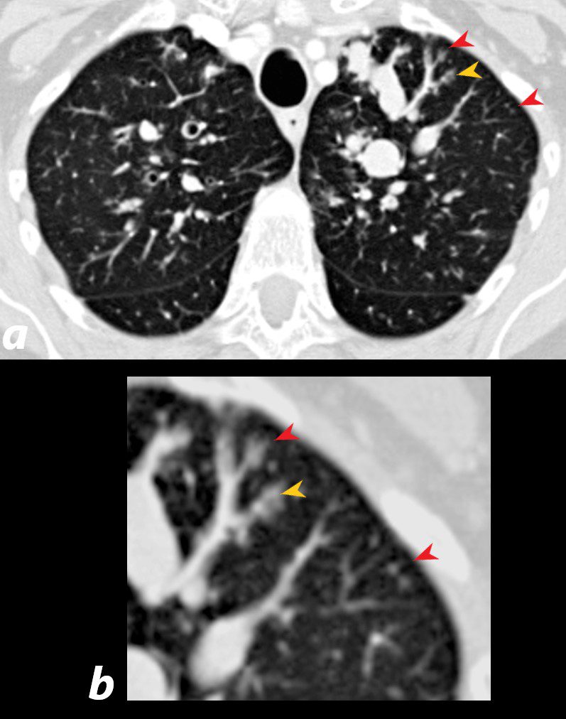
60 year old male with history of asthma, allergic bronchopulmonary aspergillosis (ABPA)
CT scan shows left upper lobe bronchiectasis and soft tissue/fluid impaction in the anterior segmental and subsegmental airways associated with tree in bud appearance (yellow arrowhead) and centrilobular nodules (red arrowhead) reminiscent of associated small airway disease
Ashley Davidoff TheCommonVein.net
ABPA
43 year old man with known aspergillus infection. Note the thickening of the walls of the segmental subsegmental and small airways with extensive tree in bud changes and bronchial wall thickening. There are centrilobular nodules indicating the small airway disease
Ashley Davidoff MD TheCommonVein.net
117816c