

Ashley Davidoff MD TheCommonvein.net lungs-0774
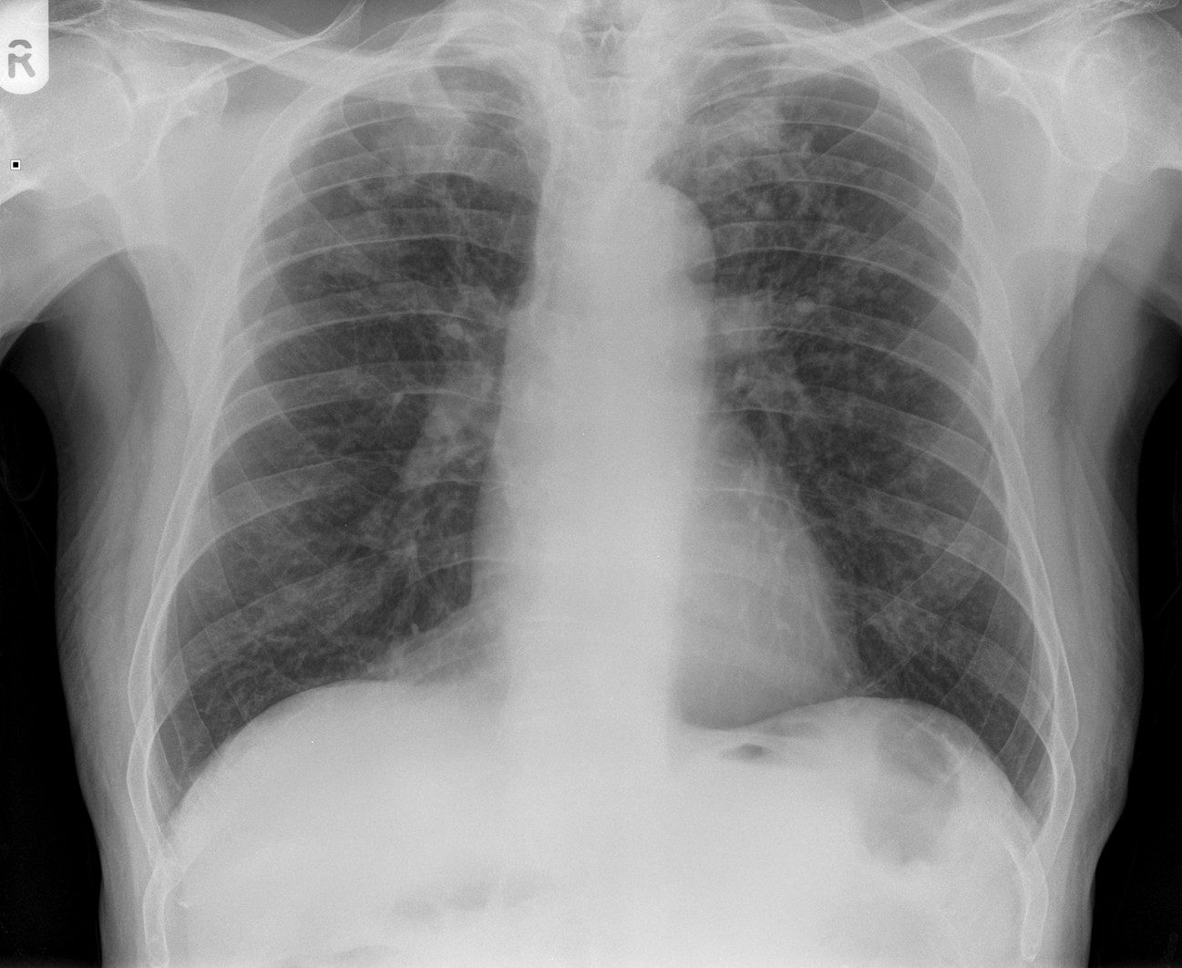
CXR (PA view) shows interstitial reticulonodular and coalescing opacities in the lungs bilaterally consistent with a diagnosis of classic complicated silicosis. Differential diagnosis includes coal worker’s pneumoconiosis and talcosis.
Case courtesy of Dr Ian Bickle, Radiopaedia.org, rID: 33227
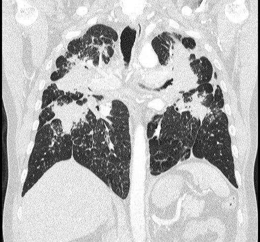
Coronal CT at the level of bronchi shows soft-tissue masses with irregular borders and significant bullous disease in bilateral lungs consistent with a diagnosis of classic complicated silicosis. Differential diagnosis includes coal worker’s pneumoconiosis and talcosis.
Case courtesy of Dr Michael P Hartung, Radiopaedia.org, rID: 71691
- The upper lung zones are particularly vulnerable to silica dust because they have a
- larger surface area and
- better ventilation
- more particles are likely to enter the upper lung zones and become
- trapped in the alveoli,
- larger lymphatic drainage in the upper lung zones
- allows silica particles to more easily enter the
- lymphatic system and
- spread to lymph node enlargement and
- induce autoimmune reactions.