
Calcified Broncholith
TB
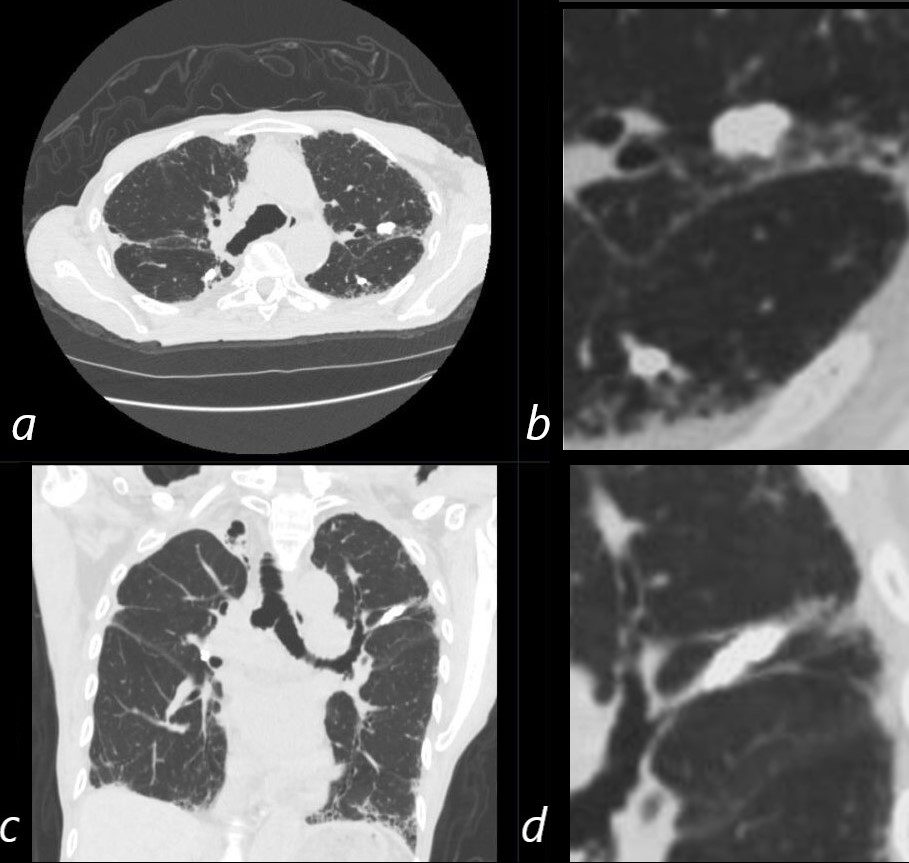
CT in axial and coronal projections of an 80- year-old non-smoker with childhood history of treated TB, shows multiple calcifications with tubular morphology (c and d) cionsistent with broncholiths. There is interstitial coarsening of the lung markings
Ashley Davidoff MD TheCommonVein.net 292Lu 136626c01
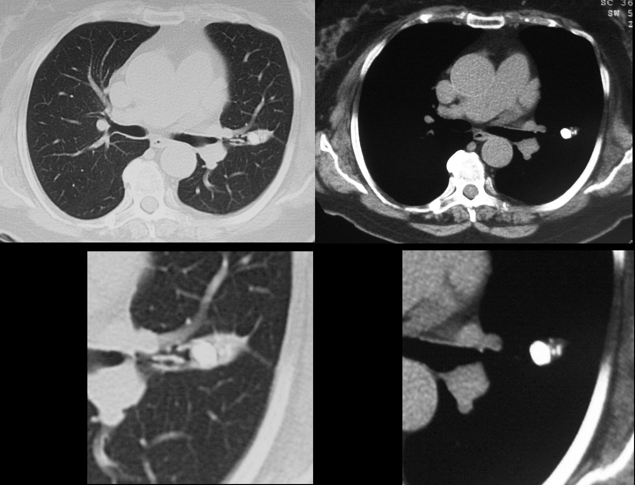
60 year old female presents with a chronic cough. CT in the axial plane shows a large calcified broncholith without downstream atelectasis
Ashley Davidoff MD TheCommonVein.net 31720c
Calcified Broncholiths and Chronic Atelectasis of the
Lateral Segment of Middle
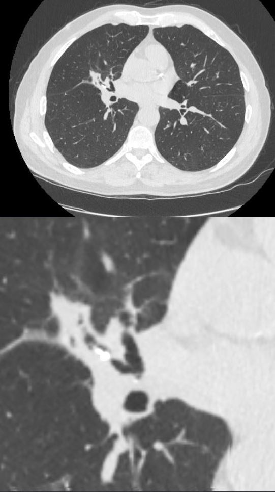
70 year old male with positive QuantiFERON test presents for evaluation of active TB CT in the axial plane shows a bilobed calcified broncholith in the lateral segment of the middle lobe with post obstructive atelectasis.
Ashley Davidoff MD TheCommonVein.net 136585b
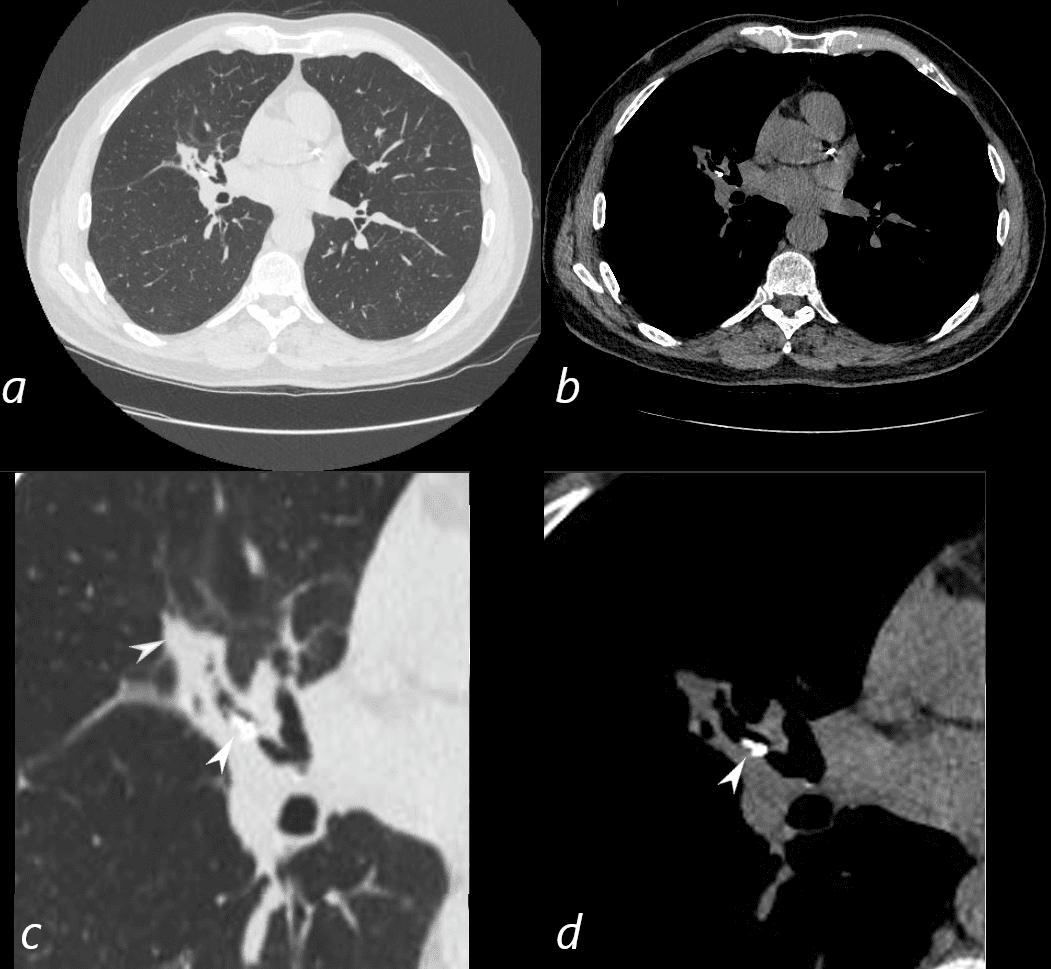
70 year old male with positive QuantiFERON test presents for evaluation of active TB CT in the axial plane shows a bilobed calcified broncholith in the lateral segment of the middle lobe (c, d white arrowheads) with post obstructive atelectasis (c, blue arrowhead)
Ashley Davidoff MD TheCommonVein.net 136585cL
Calcified Broncholith with Chronic Discoid Atelectasis of the Medial Segment of the Right Middle Lobe (RML)
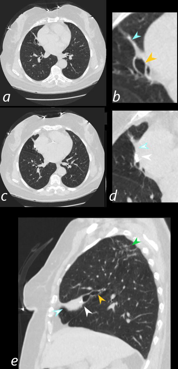
73 year old female with history of TB . CT in the axial plane shows a focus of bronchiectasis in the medial segment of the RML (blue arrowhead b, d, e) caused by a calcified broncholith in the medial segment of the middle lobe (white arrowhead d and e) with upstream bronchiectasis (orange arrowhead b and e). Linear nodular changed noted in the right upper lobe (green arrowhead, e) consistent with prior TB infection
Ashley Davidoff MD TheCommonVein.net 136532cL