
CXR Lung Abscess with a Cavity and an Air-Fluid Level
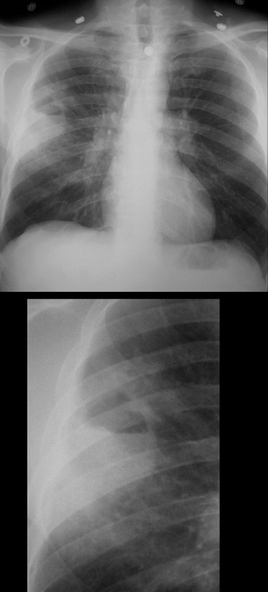
45 year old male presents with a fever
Frontal CXR shows a cavity in the right mid lung field with an air fluid level consistent with a lung abscess
Ashley Davidoff MD TheCommonvein.net
CT Right Upper Lobe Pneumonia with Abscess Formation
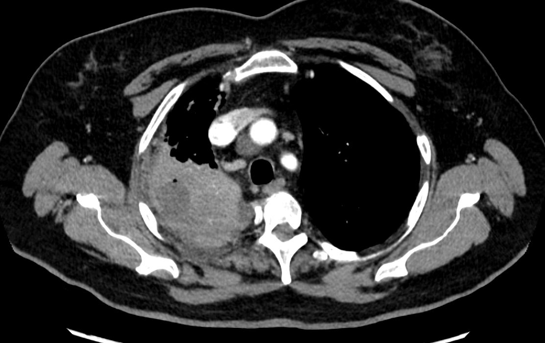
CT scan in the axial plane of a 63-year-old female with cough fever and leukocytosis, shows a right upper lobe consolidation with a 2.8cms fluid collection and a dependent bubble of air consistent with a diagnosis of a lung abscess secondary to pneumonia
Ashley Davidoff MD TheCommonVein.net 136170
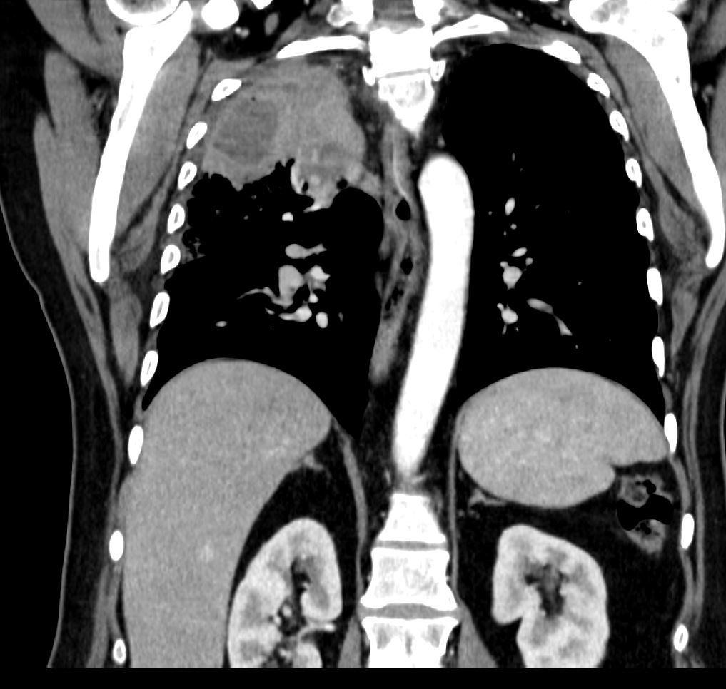
CT scan in the coronal plane of a 63-year-old female with cough fever and leukocytosis, shows a right upper lobe consolidation with a 2.8cms fluid collection and a bubble of air consistent with a diagnosis of a lung abscess secondary to pneumonia
Ashley Davidoff MD TheCommonVein.net 136171
CXR Lung Abscess with Air Fluid Level
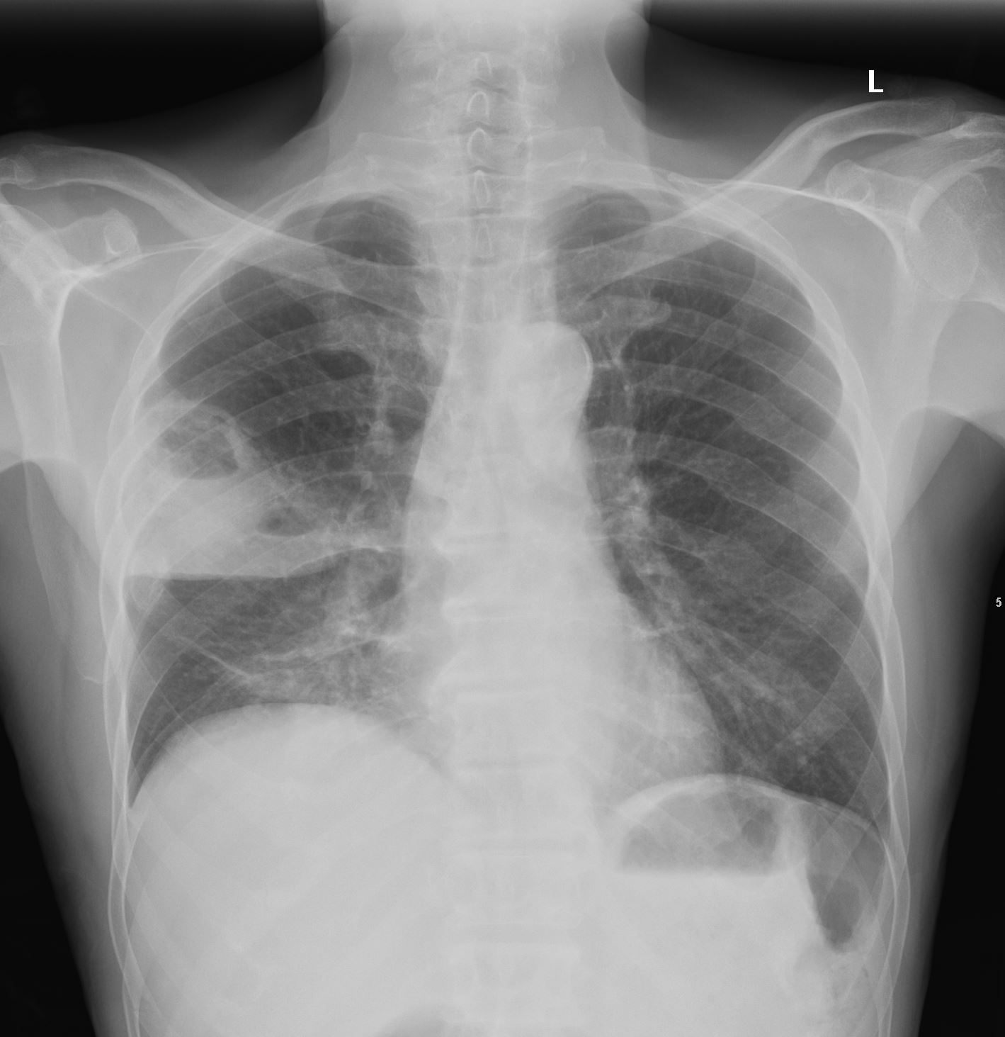
61-year-old – male presented with 3-week cough productive of phlegm and blood, right-sided chest pain, and 20-lb weight loss over the past 4 months.
CXR showed consolidative opacity in R upper lobe with two intrinsic air fluid levels concerning for abscess.
Ashley Davidoff MD TheCommonVein.net 110 LU 136164
CT chest showed a cavitating right upper lobe abscess, with a region of consolidation and a smaller region of ground glass opacity
Ashley Davidoff MD TheCommonVein.net 110 LU 136165
CXR Frontal and Lateral –
Left Hilar Carcinoid Tumor s/p Stent and
Lung Abscess
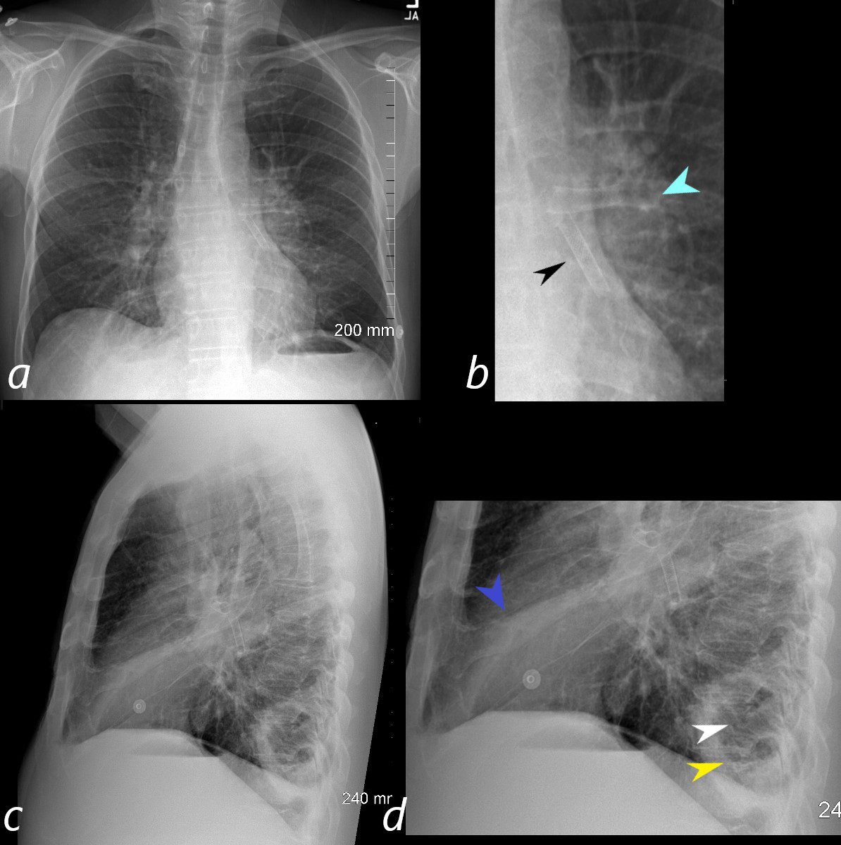
56-year-old male with known central carcinoid tumor causing lingula atelectasis, s/p placement of a stent. There is fullness to the left hilum (b, teal arrowhead) with ill-defined abutting infiltrate. A stent in the lingula airway is present (b, black arrowhead)
The lateral examination shows a segmental lingula atelectasis (d, blue arrowhead) and a large cavitating abscess (d, white arrowhead), and air-fluid level (d yellow arrowhead).
Ashley Davidoff MD TheCommonVein.net 261Lu 118379cL
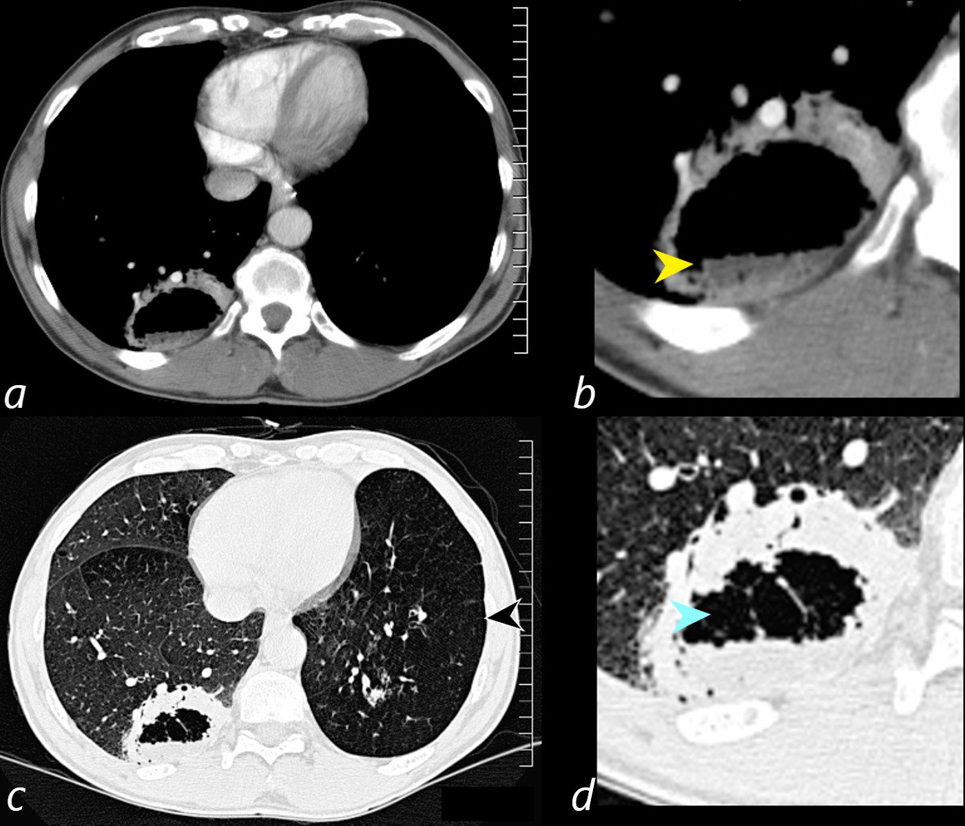
Axial CT scan at the level of the lung bases in a 56-year-old male with an obstructing carcinoid tumor of the lingula shows a cavitating abscess cavity(d, blue arrowhead) with an air fluid level in the right lower lobe (b yellow arrowhead).
The left lower lobe is relatively lucent, reflecting compensatory hyperinflation secondary to the lingula atelectasis (c, black arrowhead)
Ashley Davidoff MD TheCommonVein.net 261Lu 118383cL
Cavitating Mass Left Upper Lobe – Culture Confirms Abscess
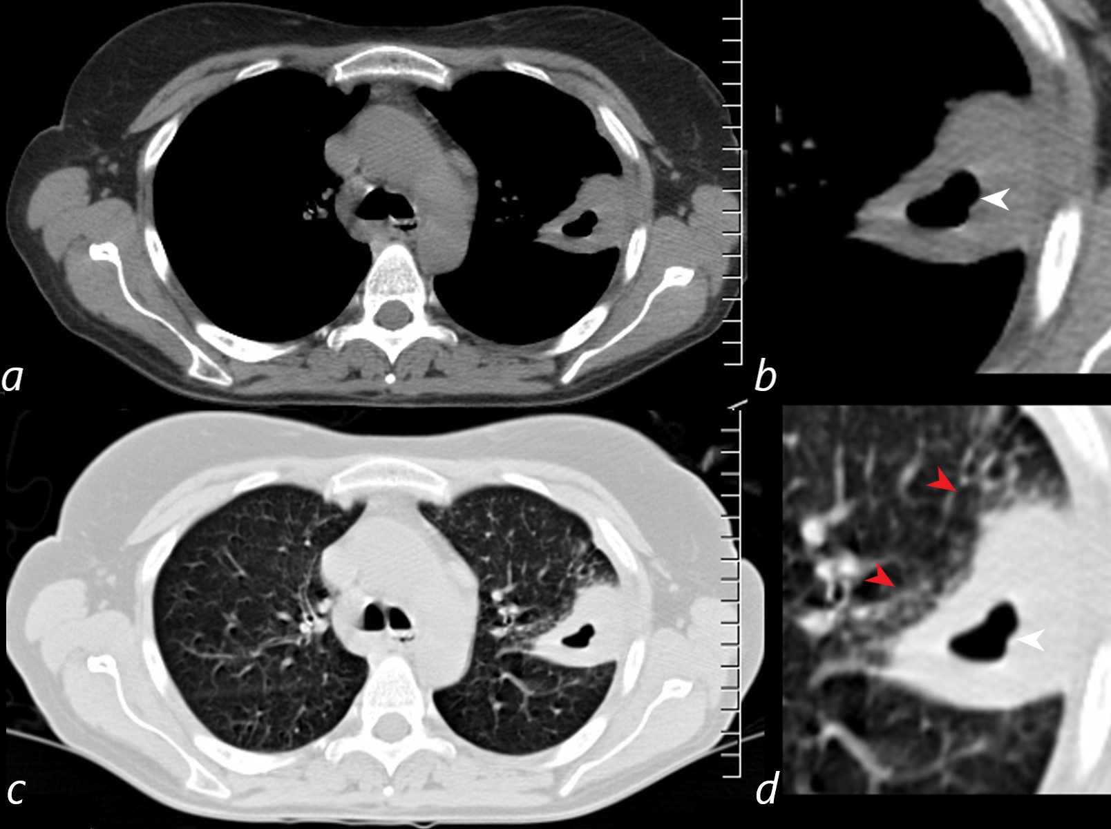
72-year-old female presents with cough, fever and leukocytosis
The CT confirms a peripheral subsegmental consolidation in the posterior segment of the LUL with cavitation (b and d white arrowheads. There is surrounding ground glass change reflecting surrounding edema (d, red arrowheads). Cultures confirmed bacterial abscess
Ashley Davidoff MD TheCommonVein.net 261Lu 118357cL
72-year-old female presents with cough, fever and leukocytosis
The CT reveals a peripheral subsegmental consolidation in the posterior segment of the LUL with cavitation Biopsy and cultures confirmed bacterial abscess
Ashley Davidoff MD TheCommonVein.net 261Lu 118359
CT –Right Upper Lobe Pneumonia with
Multicentric Abscess Formation
54-year-old female presents with a cough, fever and leukocytosis. CT in the axial plane shows a consolidation in the right upper lobe, with multiple abscesses with enhancing walls, consistent with lung abscesses.
Ashley Davidoff MD TheCommonVein.net 19598
Cavitating Pneumonia with Abscess Formation
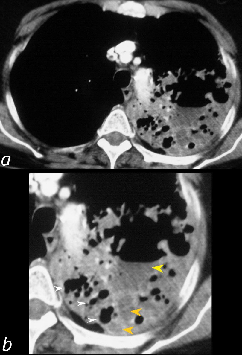
70-year-old female presents with a cough, fever and leukocytosis. CT in the axial plane shows a cavitating pneumonia (b, white arrowheads) with large abscess and air-fluid level (b, yellow arrowheads), and multiple small intraparenchymal abscesses (b, orange arrowheads)
Ashley Davidoff MD TheCommonVein.net 31591cL
Aspiration Pneumonitis Complicated by Abscess Formation
Aspiration Pneumonia
76-year-old female presents with a cough, fever and leukocytosis. CT in the axial plane shows ground glass pneumonitis involving the left lower lobe associated with a small left effusion. There is a moderate right effusion with compressive atelectasis.
Ashley Davidoff MD TheCommonVein.net 261Lu 46128a003
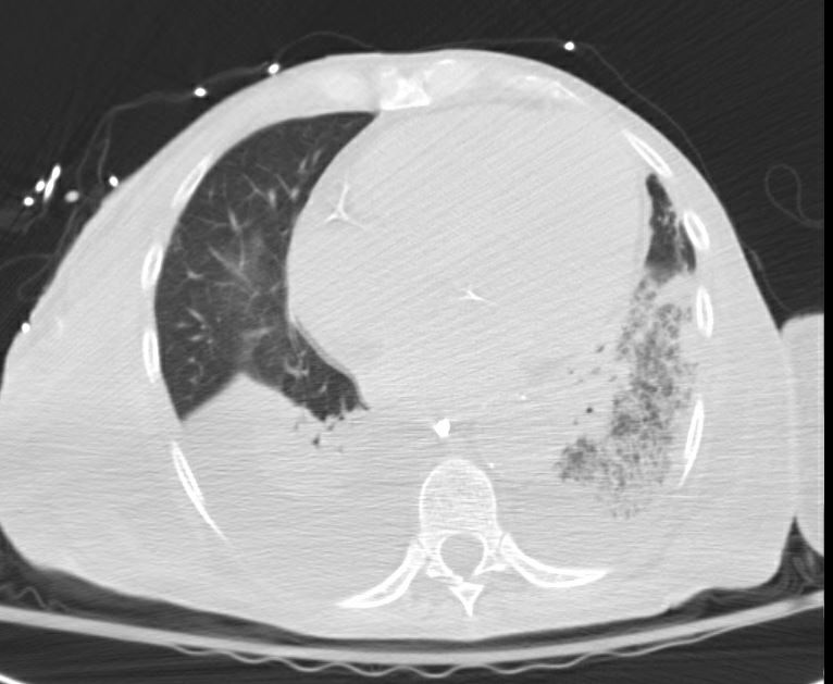
Left Lower Lobe Lung Abscess with Drainage Catheter
76-year-old female presents 2 months after an episode of aspiration pneumonia with ongoing sepsis, now on dialysis. Axial CT shows s thick walled left lower lung abscess with indwelling drainage catheter, and associated parapneumonic effusion.
Ashley Davidoff MD TheCommonVein.net 261Lu 46130
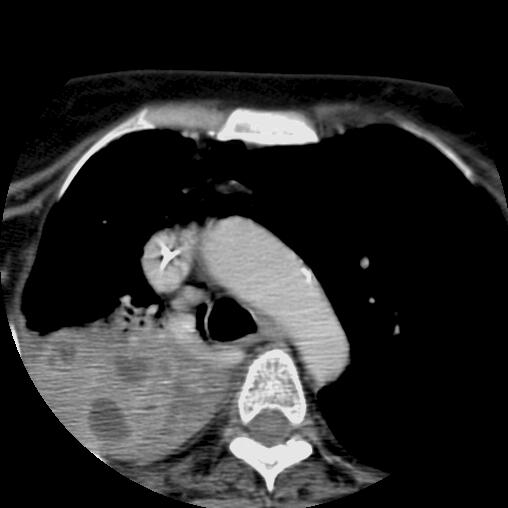
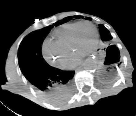
Emphysema, Infected Bullous Lung Disease, Biapical Abscesses
65-year-old male with emphysema of the lungs presents with a cough, fever and leukocytosis. CT in the axial plane shows extensive apical bullous lung disease. There is a large right upper lobe bulla with an air fluid level and a smaller left upper lobe bulla with an air fluid level.
Ashley Davidoff MD TheCommonVein.net 259Lu 117471
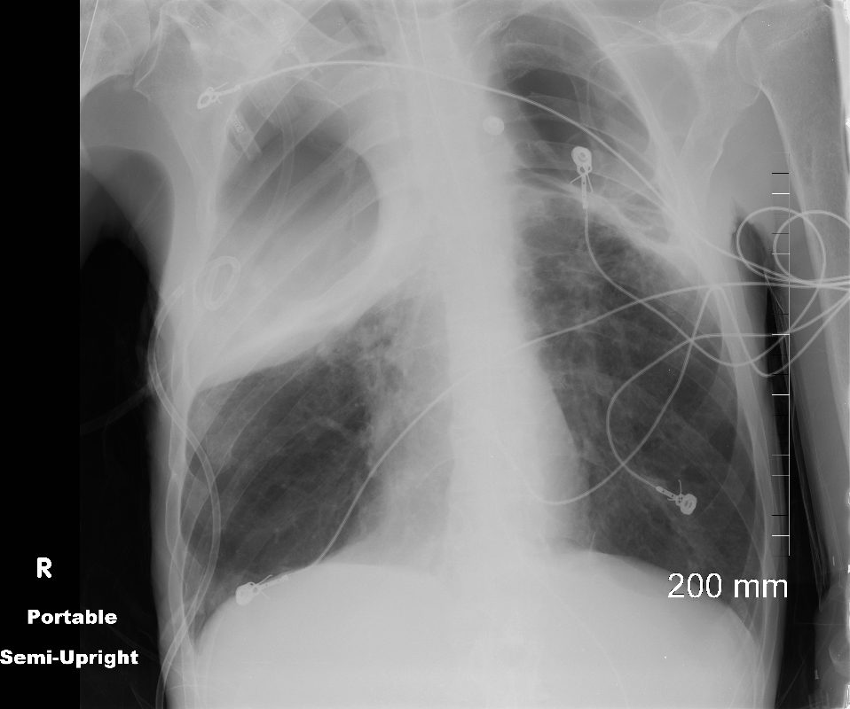
65-year-old male with emphysema of the lungs presents with a cough, fever and leukocytosis.
Frontal CXR in semi-upright position, shows a pigtail catheter in the large right apical abscess within a giant apical bulla with persistent fluid as evidenced by ground glass curtain of fluid bilaterally. Hyperinflation with resultant small heart is noted.
Ashley Davidoff MD TheCommonVein.net 259Lu 117505.8
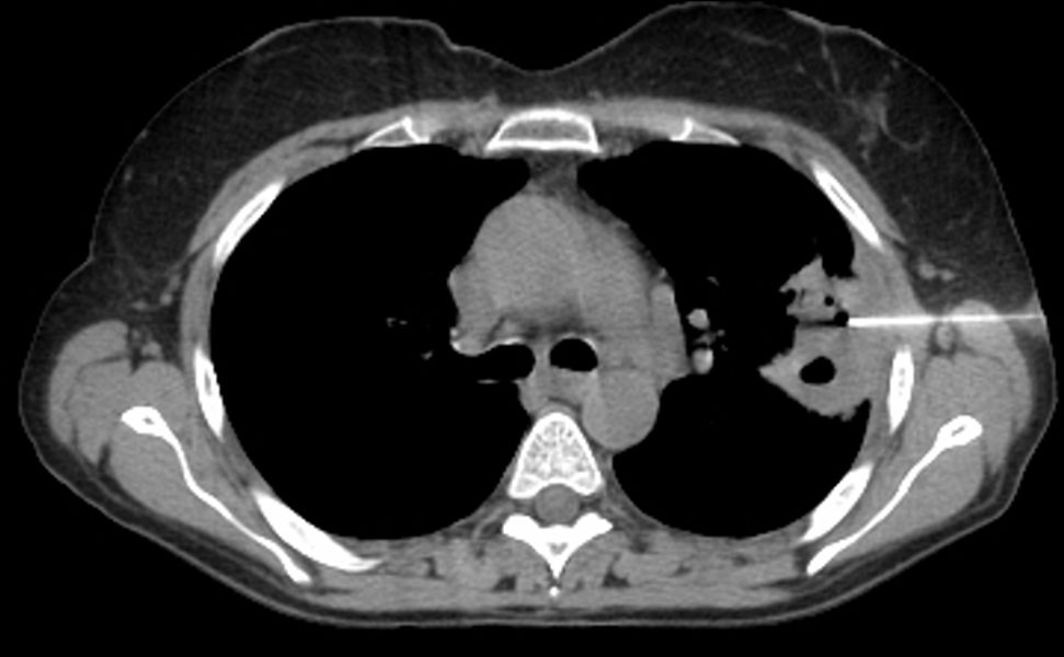
Post Partum Cardiomyopathy Necrotizing Pulmonary Infarction s/p Pulmonary Embolus (PE) New Abscess in Region of Prior Infarction
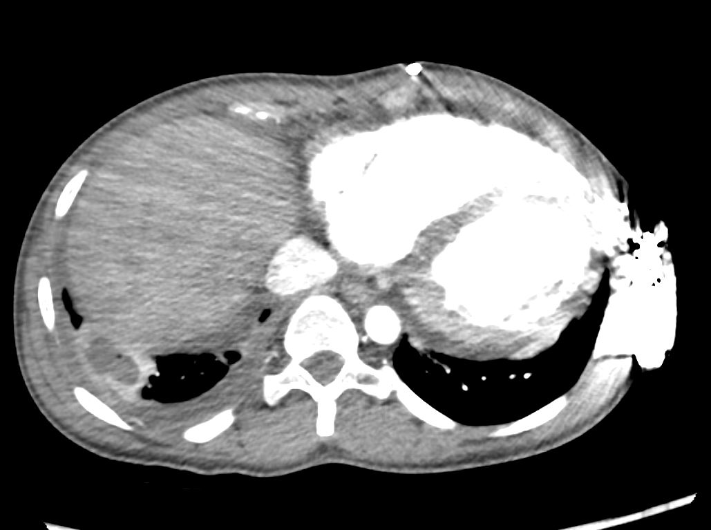
35-year-old female with an 8-year history of post- partum cardiomyopathy presents with a history of ongoing chest pain and fever 1 week following new cavitation of a pulmonary infarction. CT of the chest with contrast in an axial projection, at the level of the heart shows an enlarged right ventricle, evolution of the posterolateral infarction into an abscess with a bubble of air, enhancing wall, a small right pleural effusion with suggestion of pleural enhancement in the left lower lobe.
Ashley Davidoff MD TheCommonVein.net 258Lu 136174
Lung Abscess LIP HIV AIDS Lymphoma
27 year old male with a history of perinatal HIV with intermittent highly active antiretroviral therapy (HAART) compliance with a CD4 count of < 50 with biopsy confirmed B cell lymphoma of the liver, s/p CHOP therapy , chronic esophageal strictures s/p dilatations, esophageal candidiasis, LIP, bronchiectasis pancreatitis, and portal vein and splenic vein thrombosis.
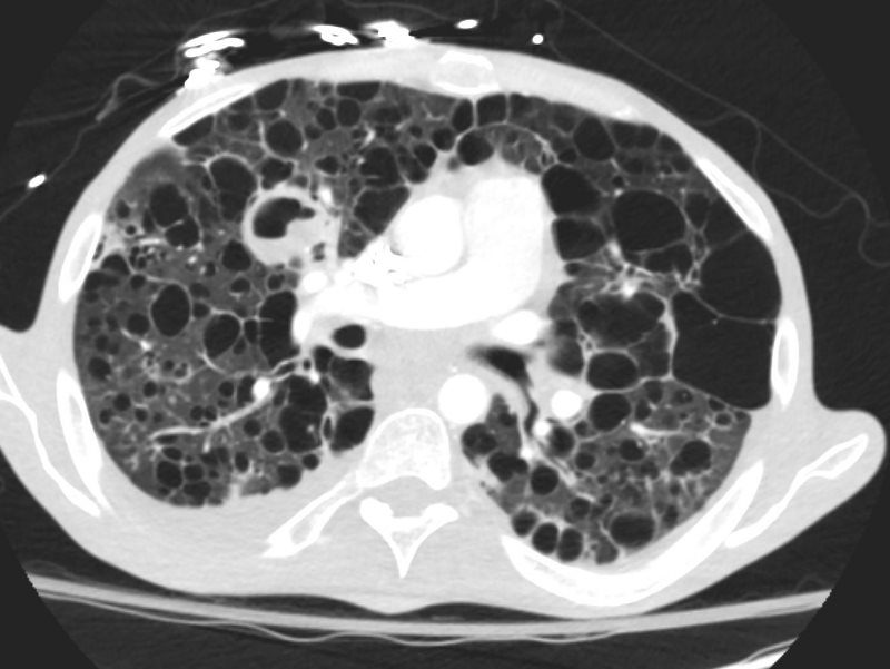
Initial Chest X-ray shows a diffuse reticular pattern with cystic changes dominant at the bases.
CT at that time confirmed the presence of diffuse cystic changes with the largest cysts at the lung bases. Ascites and splenomegaly are also present
He presented one month later with fever and neutropenia.
CT showed an abscess cavity in the right upper lobe in the right upper lobe, thickened distal esophagus with edematous wall, atrophic gastritis and ascites. Bronchovascular thickening along a bronchiectatic segment in the right upper lobe was present in the last CT
Ashley Davidoff MD TheCommonVein.net 017Lu 132031

LIP HIV AIDS and LYMPHOMA
PULMONARY 27 year old male with a history of perinatal HIV with intermittent highly active antiretroviral therapy (HAART) compliance with a CD4 count of < 50 with biopsy confirmed B cell lymphoma of the liver, s/p CHOP therapy , chronic esophageal strictures s/p dilatations, esophageal candidiasis, LIP, bronchiectasis pancreatitis, and portal vein and splenic vein thrombosis.
Initial Chest X-ray shows a diffuse reticular pattern with cystic changes dominant at the bases.
CT at that time confirmed the presence of diffuse cystic changes with the largest cysts at the lung bases. Ascites and splenomegaly are also present
He presented one month later with fever and neutropenia.
CT showed an abscess cavity in the right upper lobe in the right upper lobe, thickened distal esophagus with edematous wall, atrophic gastritis and ascites. Bronchovascular thickening along a bronchiectatic segment in the right upper lobe was present in the last CT
Ashley Davidoff MD TheCommonVein.net 017Lu 132030
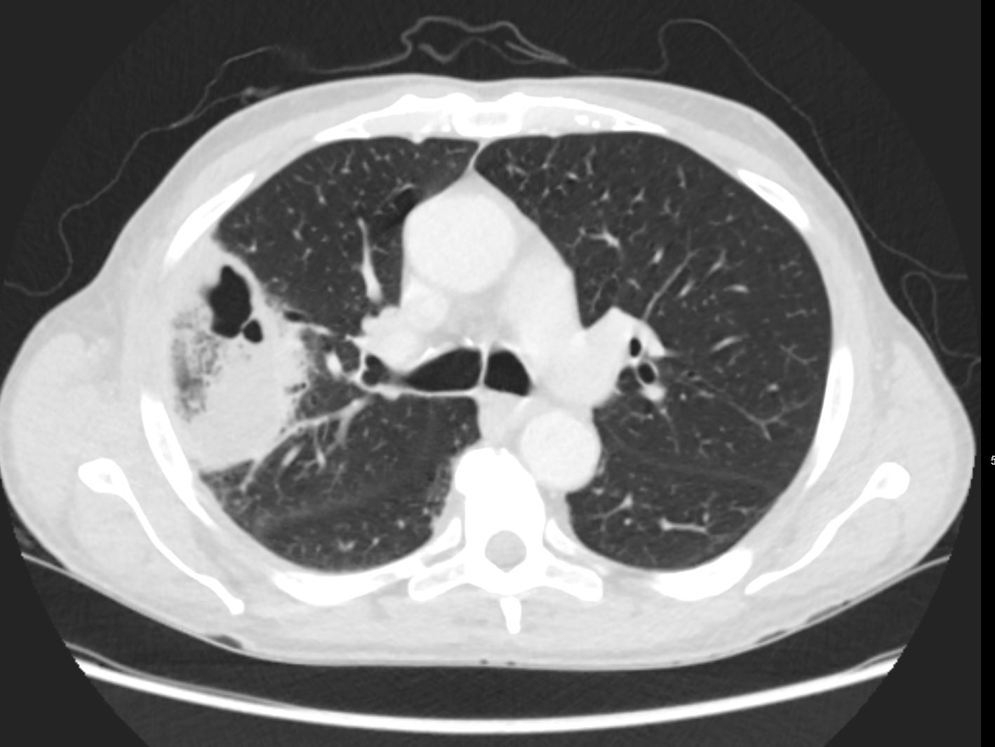
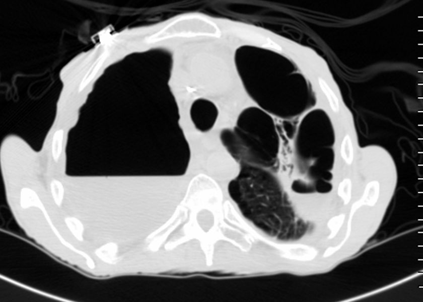
Aspergilloma in the Left Apex and
Consolidation and Abscess in the Right Apex
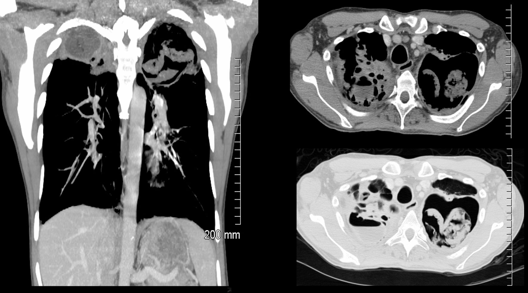
CT of a 54 year old male shows a large left apical cavity with aspergilloma. These findings are consistent with chronic pulmonary aspergillosis In the apex of the right lung, there is pneumonic consolidation and abscess formation . Note the air fluid level in the right apex on axial soft tissue and lung windows.
Ashley Davidoff TheCommonvein.net 225Lu 134202