
Where is Waldo or Sherlock
Where Is Waldo
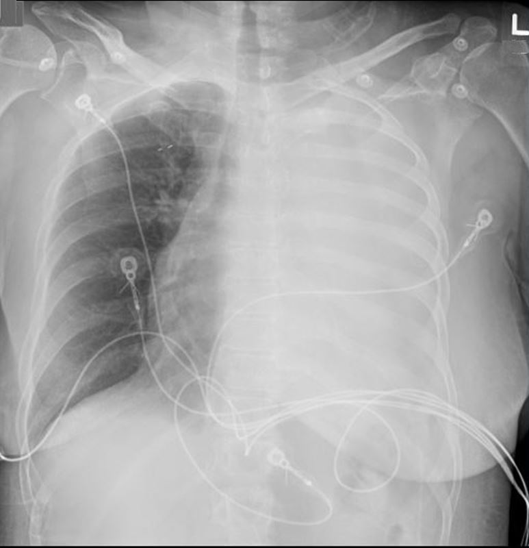
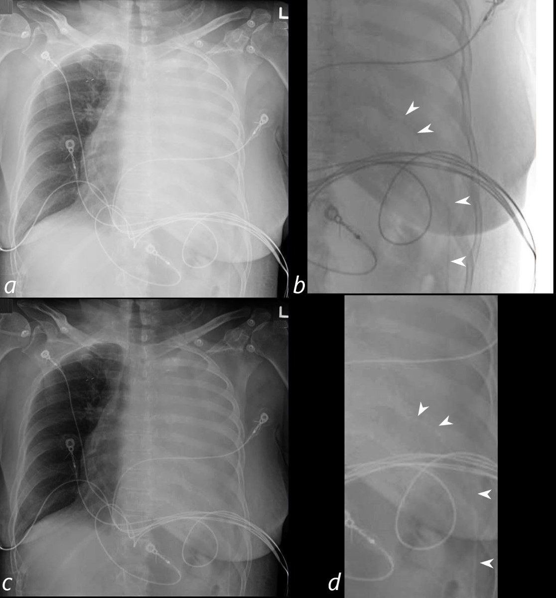
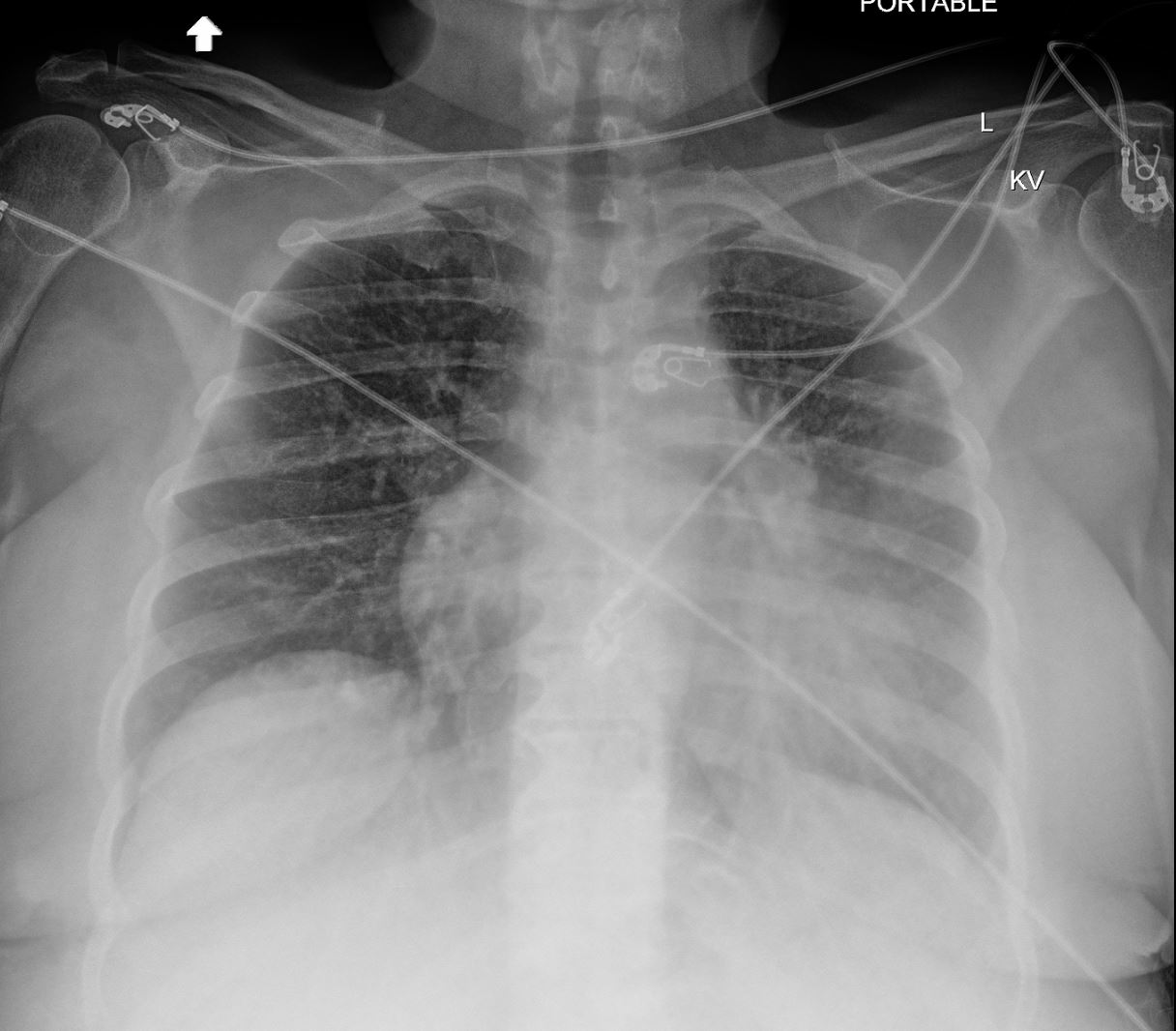
Pericardial Drain
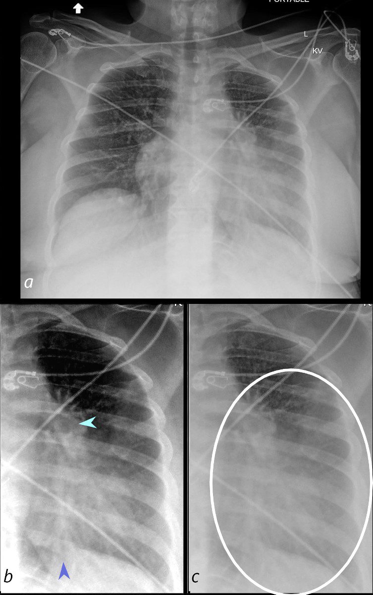
62-year-old female presents with acute dyspnea and chest pain
Frontal CXR shows a “white out” of the left hemithorax
She was subsequently diagnosed with a small cell lung carcinoma that was obstructing the left main stem bronchus
Ashley Davidoff MD TheCommonVein.net 298Lu 136714
Frontal CXR shows a “white out” of the left hemithorax
Echocardiogram revealed a moderate effusion with elevated right sided pressures, but without “frank” tamponade. It was elected to perform a pericardiocentesis. The frontal CXR faintly reveals the pericardial catheter entering from the LUQ and overlies the expected location of the cardiac shadow.
Ashley Davidoff MD TheCommonVein.net 298Lu 136715cL
Is There a Right Lower Lobe Pneumonia?
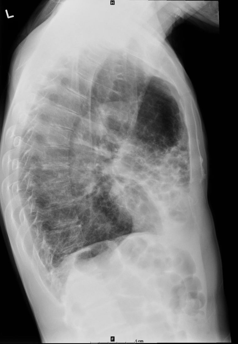
33-year-old female presents with a cough. Chest X-ray in the frontal view shows a region of increased density in the medial right lower lung field. The cardio mediastinal shadow is shifted to the left. These findings are consistent with pectus excavatum.
Courtesy Ashley Davidoff MD TheCommonVein.net 136533a
33-year-old female presents with a cough. Chest X-ray in the frontal view shows a region of increased density in the medial right lower lung field. The cardio mediastinal shadow is shifted to the left. On the lateral view a moderate sized pectus excavatum causes a decrease in the A_P diameter of the chest, compresses the lung accounting for the increased density and causes the cardio-mediastinal shadow to shift leftward.
Courtesy Ashley Davidoff MD TheCommonVein.net 136533b
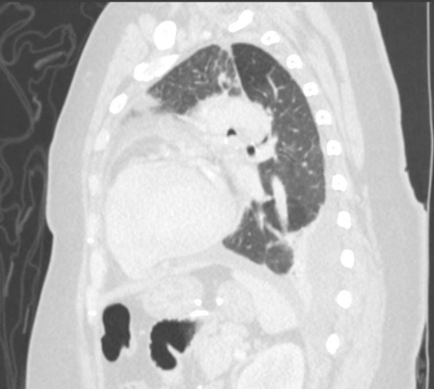
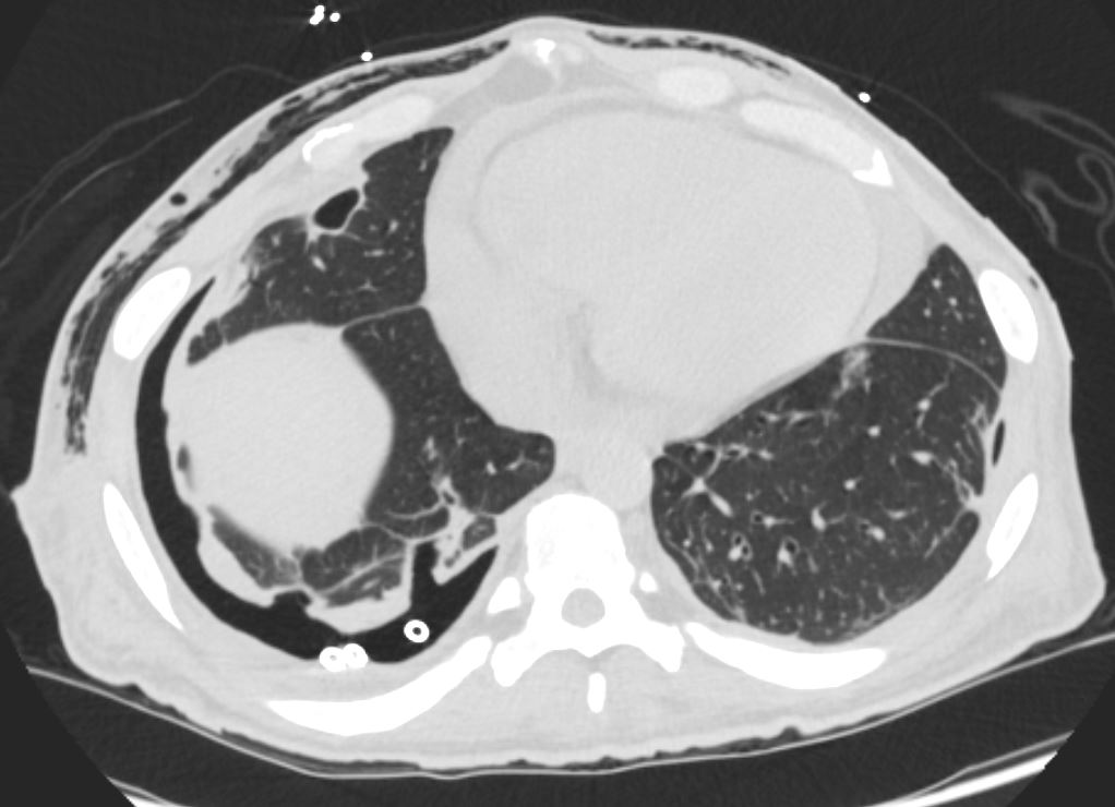
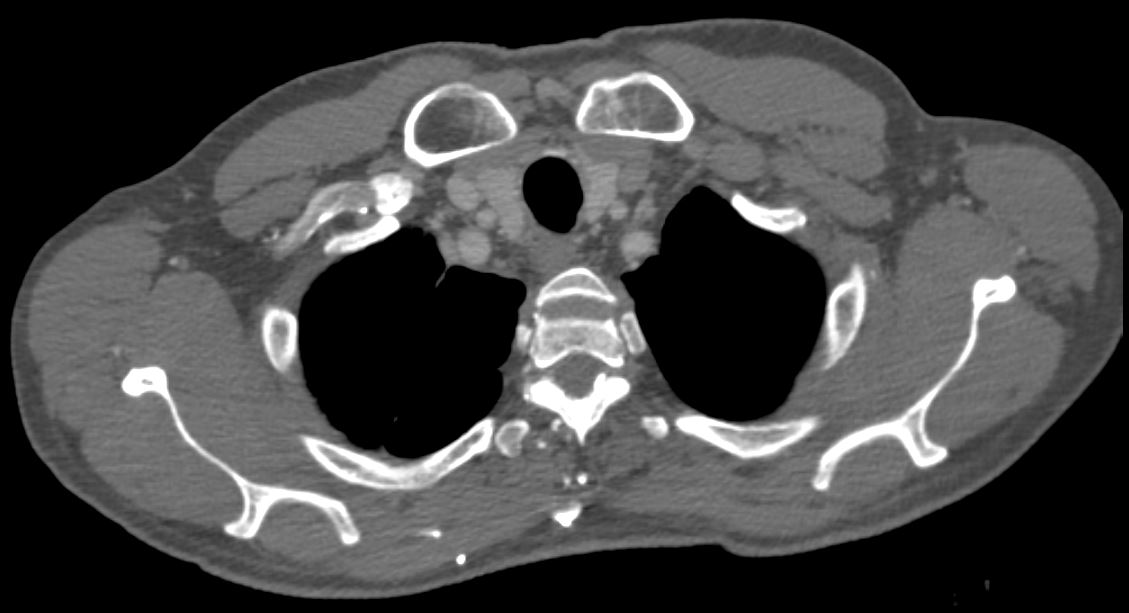
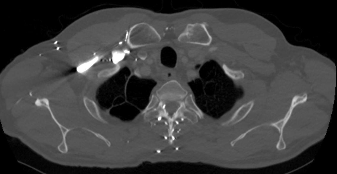
Cardiomyopathy with Pulmonary Emboli to Right Lower Lobe External Defibrillator
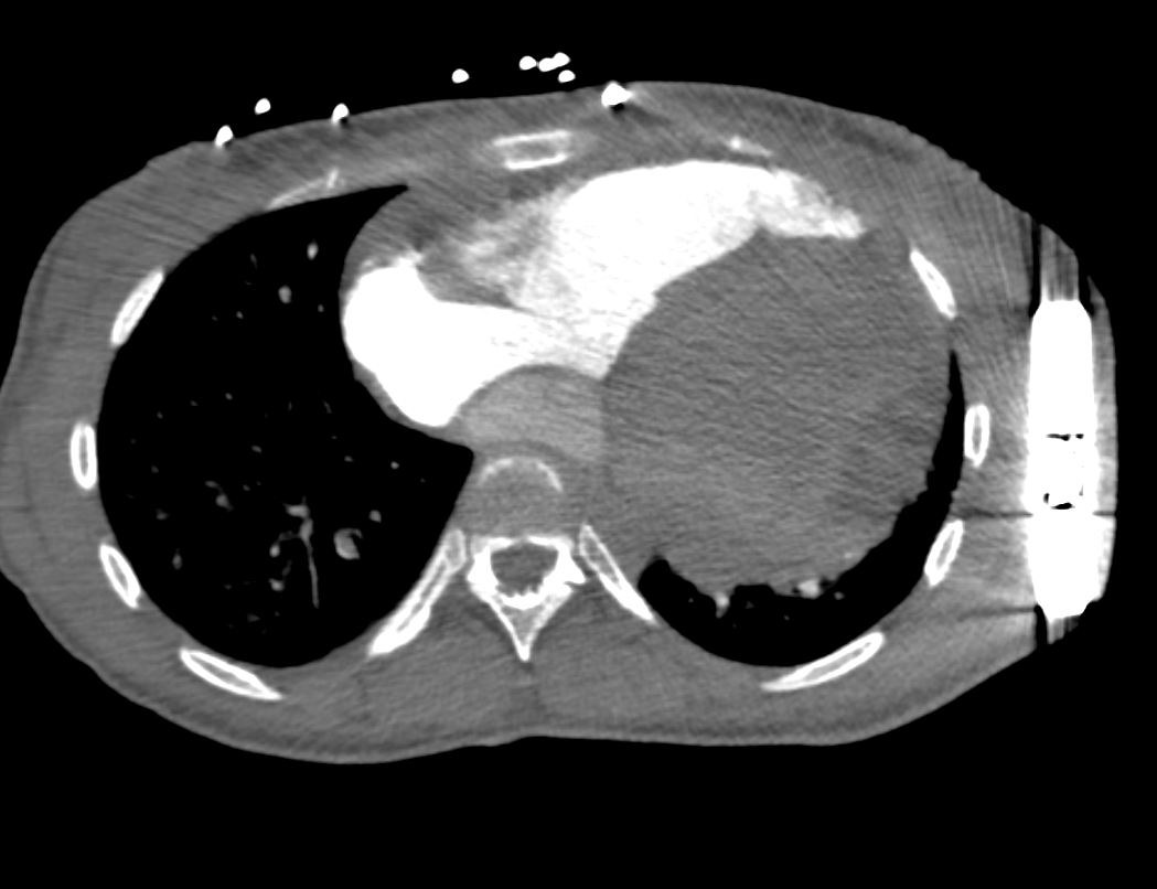
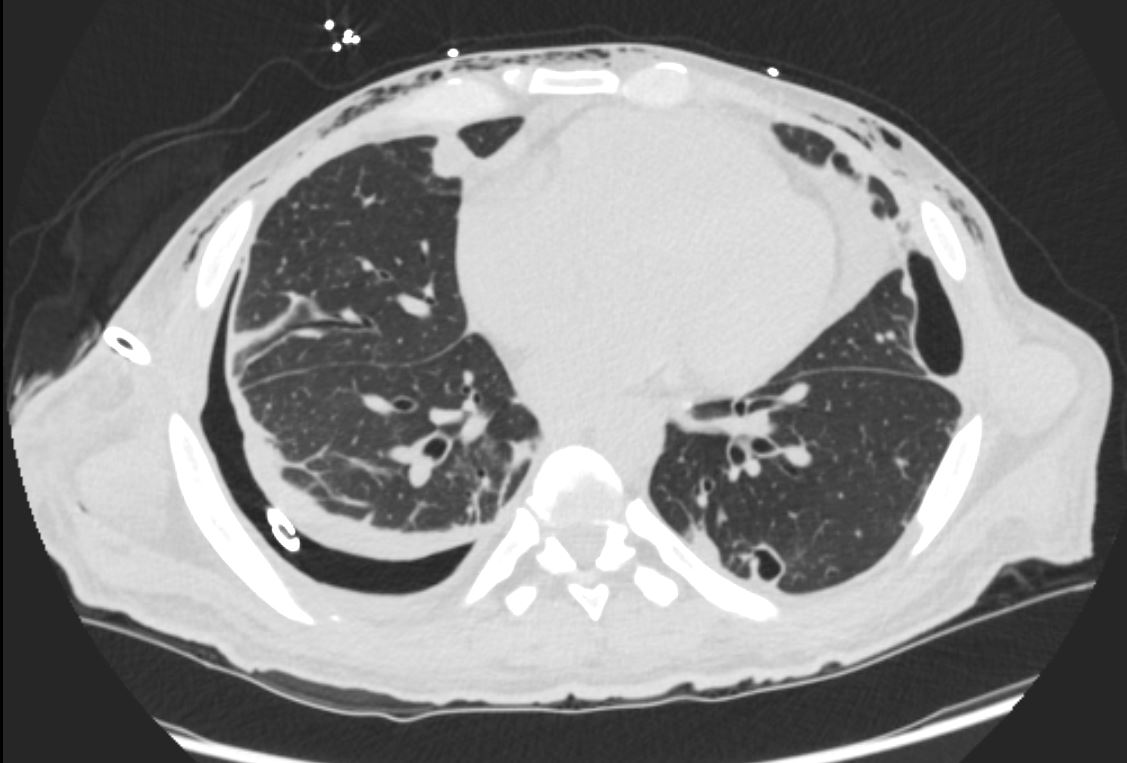
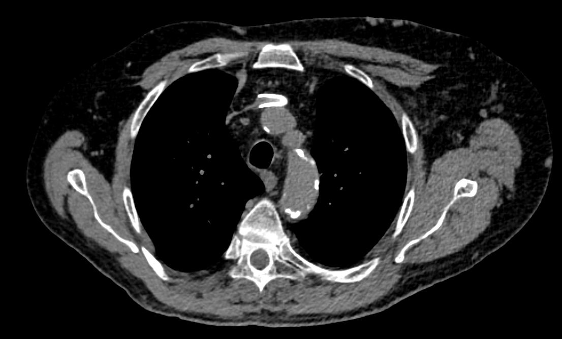
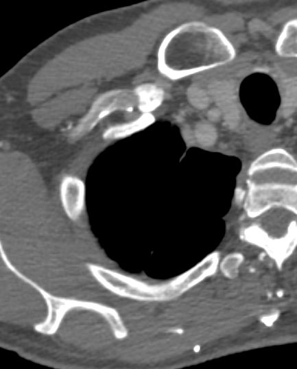
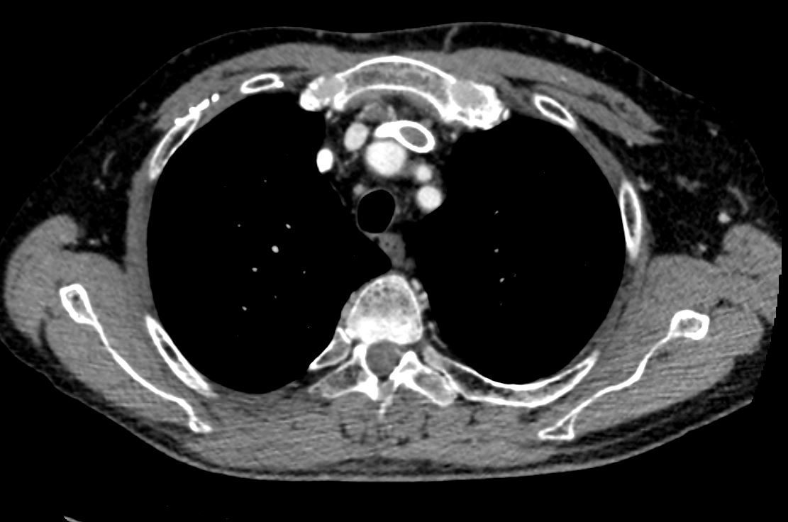
35-year-old female with an 8-year history of post- partum cardiomyopathy presents with a history of chest pain. CT of chest with contrast in an axial projection, at the level of the heart, shows an enlarged left ventricle. The right lower lobe segmental arteries show filling defects and absence of contrast compared to the left lower lobe arteries. An external defibrillator is present.
Ashley Davidoff MD TheCommonVein.net 253Lu 136165
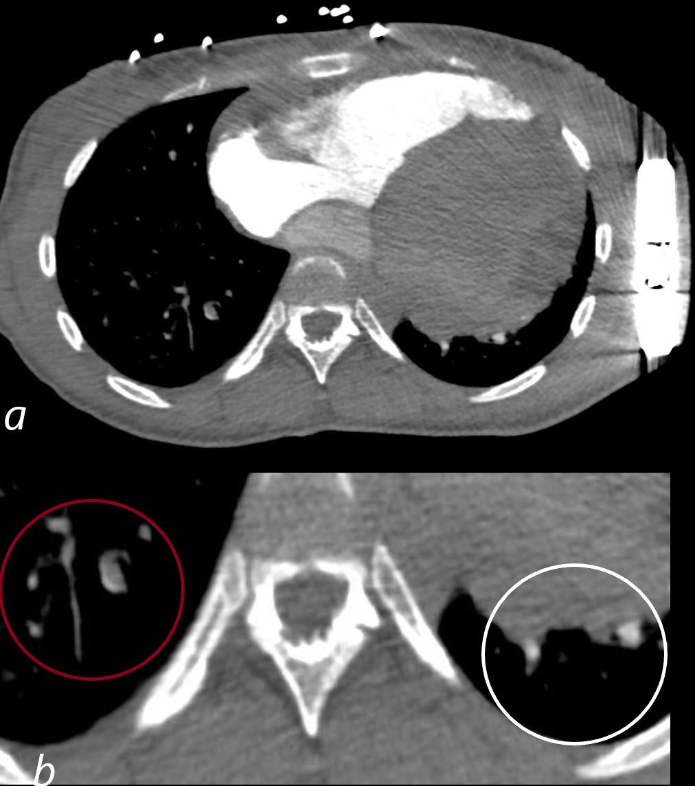
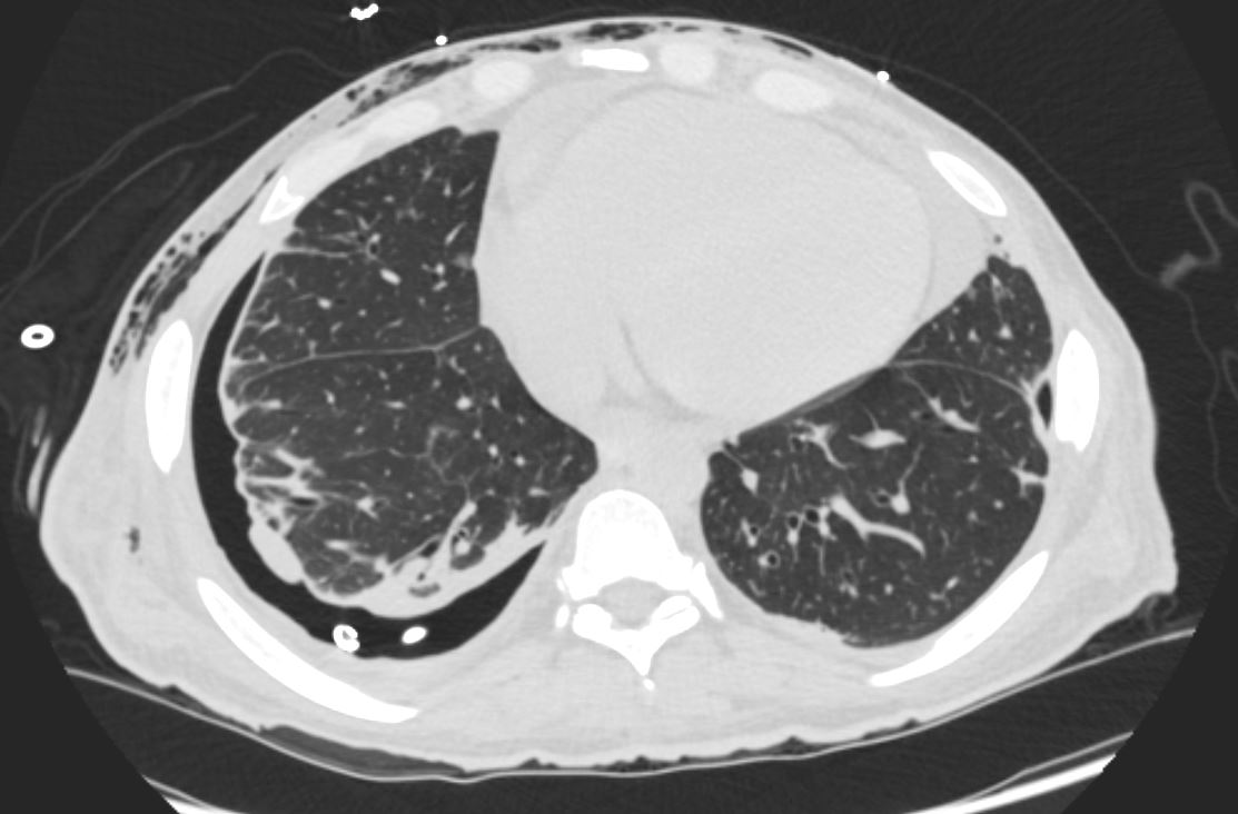
35-year-old female with an 8-year history of post- partum cardiomyopathy presents with a history of chest pain. CT of the chest with contrast in an axial projection, at the level of the heart, shows an enlarged left ventricle. The right lower lobe segmental arteries show filling defects and absence of contrast (maroon circle in b), compared to the left lower lobe arteries (white circle b). An external defibrillator is present.
Ashley Davidoff MD TheCommonVein.net 253Lu 136165cL
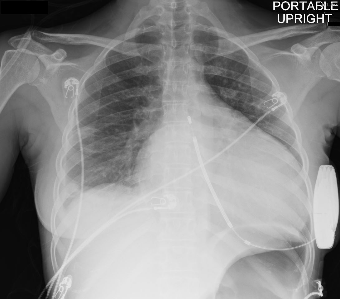
35-year-old female with a 8 year history of post- partum cardiomyopathy presents with of chest pain. Frontal CXR shows global cardiomegaly, blunting of the right costophrenic angle with a suggestion of a subsegmental infiltrate in the right costophrenic angle, and a region of linear atelectasis in the right mid lung field. A small loculated right effusion is present. An external defibrillator is noted. No definite CHF
Ashley Davidoff MD TheCommonVein.net 253Lu 136164
58-year-old female presents with a cough Frontal CXR shows silhouetting of the left heart border with hazy or veiling opacity extending out from the left hilum and fading out inferiorly . The left hilum is pulled superiorly, resulting in an almost horizontal course of the left main bronchus and vertical orientation of the left lower lobe bronchus
Ashley Davidoff MD TheCommonVein.net 257Lu 136109
58-year-old female presents with a cough Frontal CXR shows silhouetting of the left heart border with hazy or veiling opacity extending out from the left hilum and fading out inferiorly (white circle c). The left hilum is pulled superiorly (teal arrowhead b) , resulting in an almost horizontal course of the left main bronchus and vertical orientation of the left lower lobe bronchovascular bundle (dark blue arrowhead b)
Ashley Davidoff MD TheCommonVein.net 257Lu 136109cL01
58-year-old female presents with a cough. CT in the sagittal plane shows a nodule in the left mainstem bronchus of the lung with post obstructive atelectasis of the lingula, a hyperinflated portion of the apical segment of the left lower lobe, superior and anterior migration of the left major fissure and a small portion of aerated left upper lobe anteriorly that appears congested . In the left lower lobe, there is a loculated effusion with compressive atelectasis.
Pathology revealed findings consistent with a carcinoid tumor in the left mainstem bronchus
Ashley Davidoff MD TheCommonVein.net 257Lu 136119
Aspirate Occluding the Right Lower Lobe Bronchus
CT of a 72-year-old male with acute dyspnea shows a focal accumulation of low-density aspirate in the right lower lobe. Distal to the obstruction the posterior segmental and medial segmental airways are patent, but associated atelectasis is noted in those segments of the right lower lobe. The esophagus is displaced to the right, and appears to contain some aerated content. There is atelectasis of the medial and posterior segments of the right lower lobe secondary to the aspiration
Ashley Davidoff MD TheCommonVein.net 136038
Mounier Kuhn syndrome
61 year old male with a history of treated mycobacterial infections and chronic cough
Lateral view shows an enlarged trachea and thick walled cystic changes overlying the heart consistent with known bronchiectasis. There is evidence of hyperinflation
Ashley Davidoff MD TheCommonVein.net 250Lu 135872a
61 year old male with a history of treated mycobacterial infections and chronic cough
Lateral view shows an enlarged trachea and thick walled cystic changes overlying the heart consistent with known bronchiectasis. There is evidence of hyperinflation
Lateral view (a magnified in b, and shows an enlarged trachea (white arrowheads) and thick walled cystic changes overlying the heart consistent with known bronchiectasis
Ashley Davidoff MD TheCommonVein.net 250Lu 135872ac01L
bronchopleural fistula 6a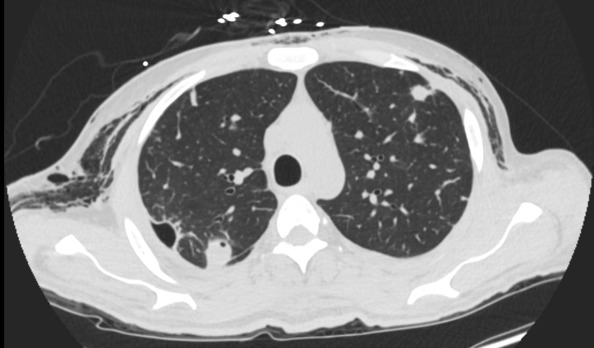
heart lung endocarditis empyema loculated pneumothorax bronchopleural fistulaRnD detectives 05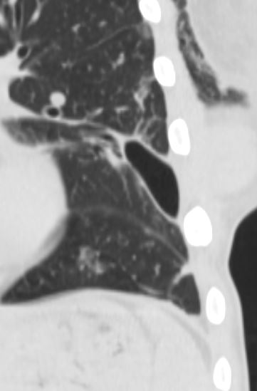
lung cancer mucinous adenocarcinoma spiculated nodule feeding vessel airway cavitation RND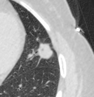 lung cherry picking testicular metastases with pleural defect and subcutaneous ephysema RnD
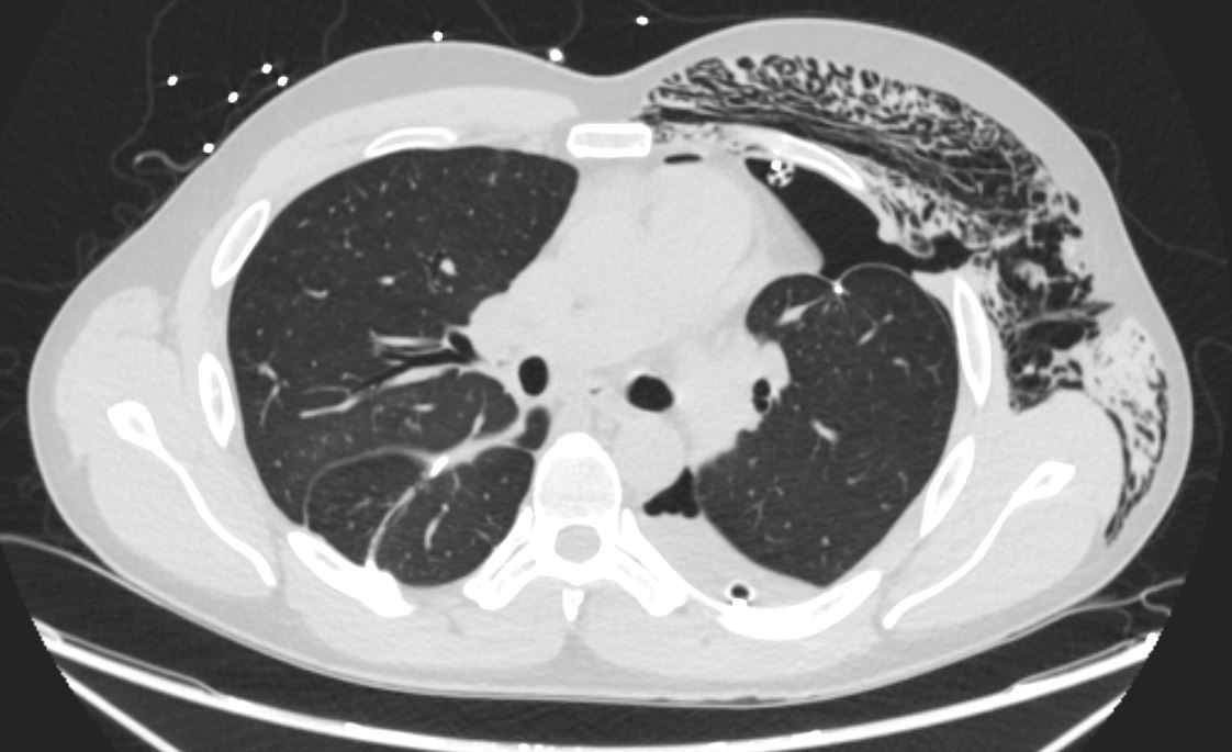
lung consolidation and ground glass thickened interlobular septa RnD IF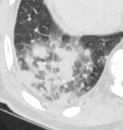
lungs heart endocarditis feeding vessel sign cavitation wedge shaped infarction RnD IF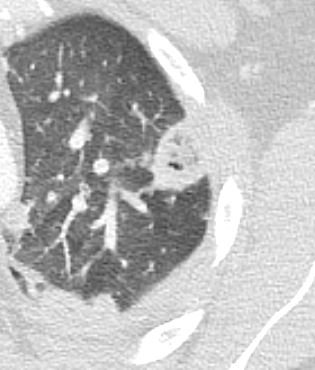
pleura lung thickened split pleura question TB split pleura sign RnD IF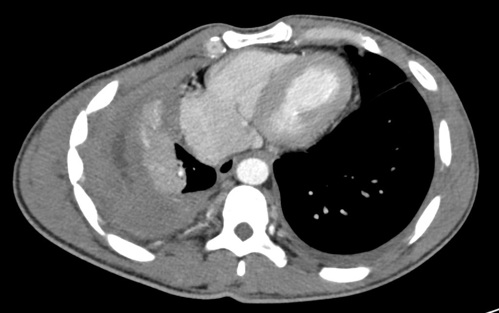
vein stent brachiocephalic collateral left chest RnD detectives 002
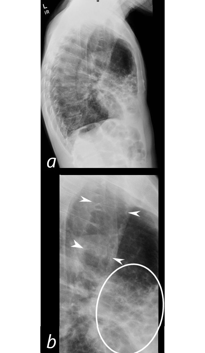
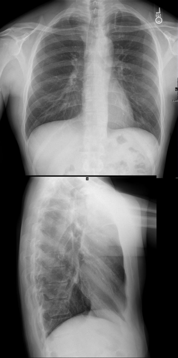
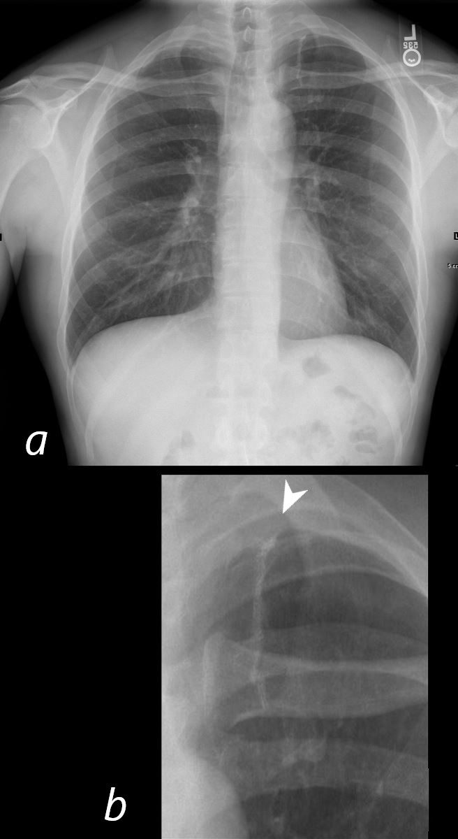
Spontaneous Pneumothorax
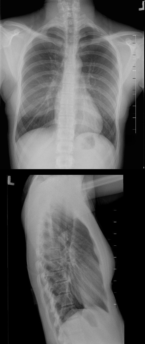
20-year-old female presents with acute left sided chest pain. She has a narrow A-P diameter exemplified in the lateral projection (below) and the asthenic build raises the suspicion for spontaneous pneumothorax. Frontal CXR shows a small subtle pneumothorax characterised by a thin pleural line and relative lucency of the left apex compared to the right
Ashley Davidoff MD TheCommonVein.net 117246c
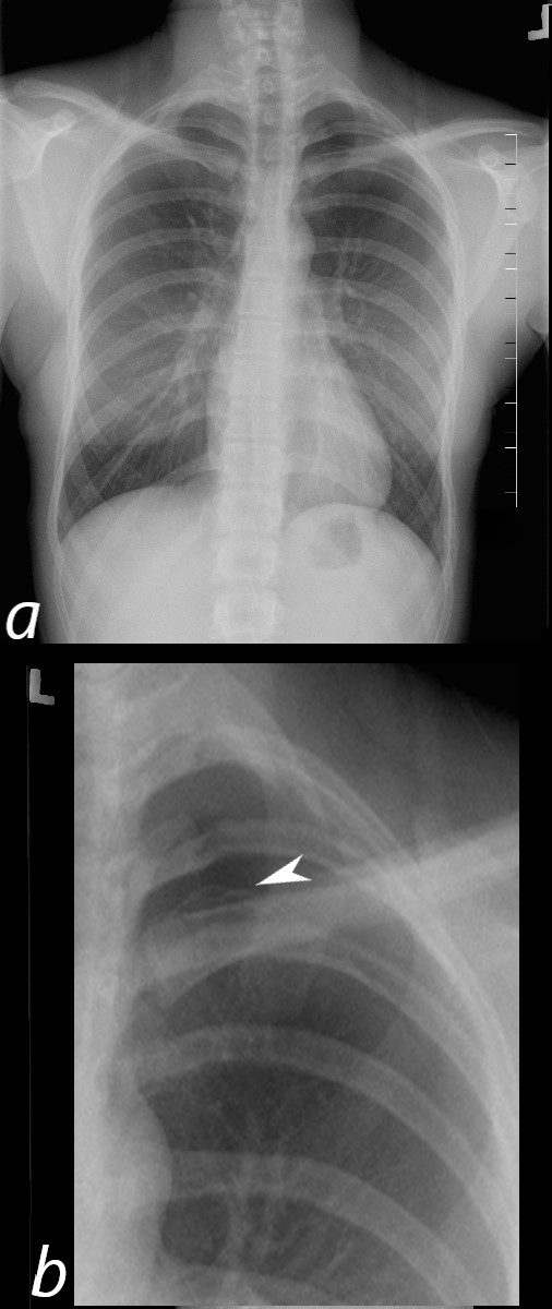
20-year-old female presents with acute left sided chest pain. She has asthenic build which raises the suspicion for a spontaneous pneumothorax. Frontal CXR shows a small subtle pneumothorax characterised by a thin pleural line (b, white arrowhead) and relative lucency of the left apex
Ashley Davidoff MD TheCommonVein.net 117246c01
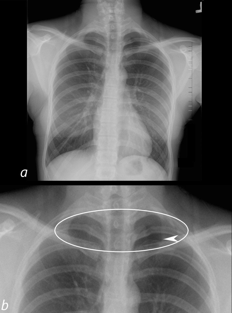
20-year-old female presents with acute left sided chest pain. She has asthenic build which raises the suspicion for a spontaneous pneumothorax. Frontal CXR shows a small subtle pneumothorax characterised by a thin pleural line (b, white arrowhead) and relative lucency of the left apex compared to the right (b, ringed)
Ashley Davidoff MD TheCommonVein.net 117246c02
CXR Surgical Repair Left Apical Bulla
28-year-old male presents for follow up post bullectomy after having a spontaneous pneumothorax. He has a narrow A-P diameter and an asthenic build and the surgical sutures are noted in the left apex of the lung
Ashley Davidoff MD TheCommonVein.net 136231c
28-year-old male presents for follow up post bullectomy after having a spontaneous pneumothorax. He has a narrow A-P diameter and an asthenic build and the surgical sutures are noted in the left apex of the lung (b, arrowhead)
Ashley Davidoff MD TheCommonVein.net 136231c