
35 year old female IVDU with HIV presents with
Fever and Dyspnea
CXR Left Lower Lobe Pneumonia
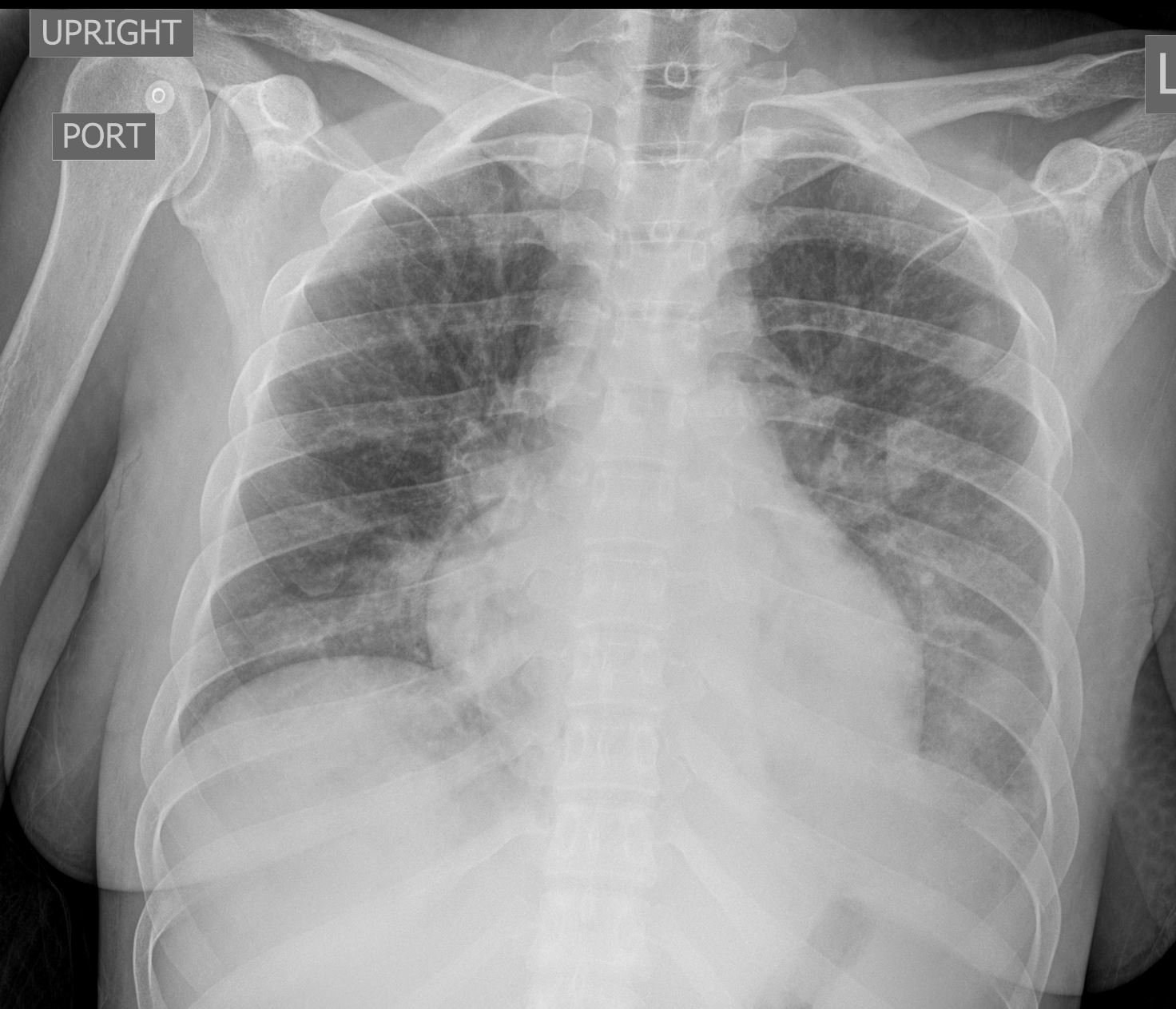
35-year-old woman, presents with dyspnea and fever. Frontal CXR shows a retrocardiac infiltrate, with suggestion of air bronchograms and silhouetting of the diaphragm medially. There are ground glass changes in the remaining left lower lobe peripherally and a suggestion of a nodular infiltrate in the left upper lung zone. The are no findings to suggest volume loss. The CXR is most compatible with an acute pneumonia when taken in the clinical context
Courtesy Ashley Davidoff MD TheCommonVein.net 289Lu 136561
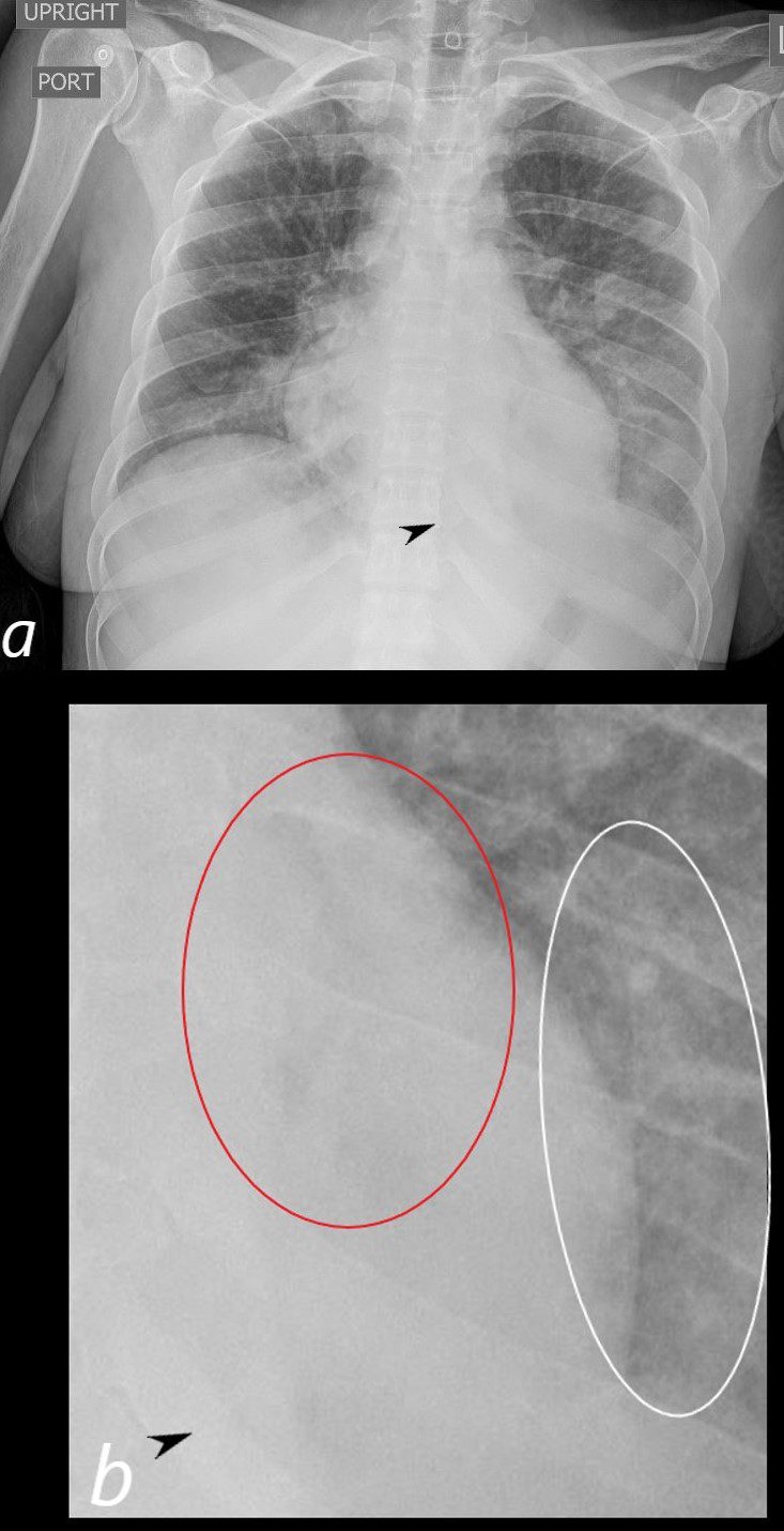
35-year-old woman, presents with dyspnea and fever. Frontal CXR (a, and magnified in b) shows a retrocardiac infiltrate, (b red ring) with suggestion of air bronchograms and silhouetting of the diaphragm medially (a, b black arrowhead). There are ground glass changes in the remaining left lower lobe peripherally (b white ring) and a suggestion of a nodular infiltrate in the left upper lung zone. The are no findings to suggest volume loss. The CXR is most compatible with an acute pneumonia when taken in the clinical context
Courtesy Ashley Davidoff MD TheCommonVein.net 289Lu 136561cL
CT – Left Lower Lobe Bronchopneumonia
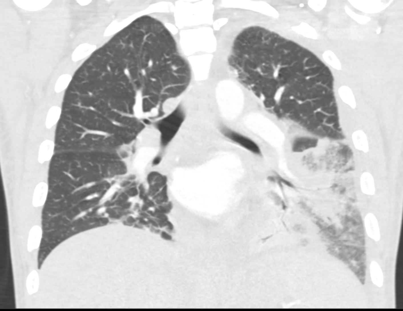
35-year-old woman, presents with dyspnea and fever. Coronal CT shows a bronchocentric pneumonic infiltrate in the superior segment of the left lower lobe and extending into the medial basal segment, and silhouetting the medial aspect of the diaphragm. There is an adjacent region of ground glass infiltrate. These findings are consistent with a bronchopneumonia in the left lower lobe.
Courtesy Ashley Davidoff MD TheCommonVein.net 289Lu 136560
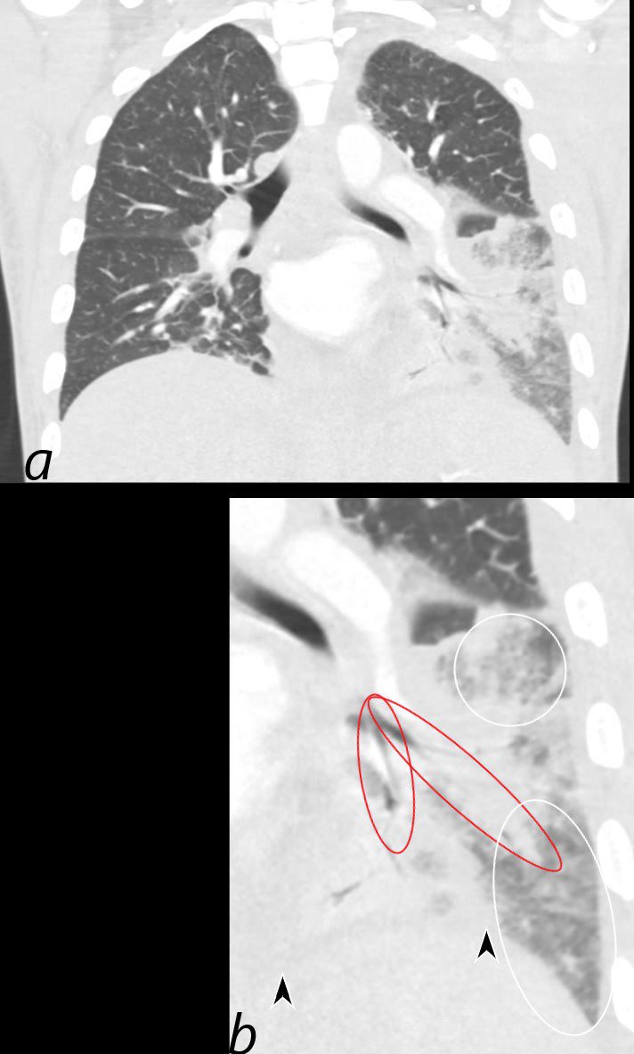
35-year-old woman, presents with dyspnea and fever. Coronal CT shows a bronchocentric pneumonic infiltrate in the superior segment of the left lower lobe (red rings) and extending into the medial basal segment, resulting in silhouetting of the medial aspect of the left hemidiaphragm (between the black arrowheads). There is an adjacent region of ground glass infiltrate. (b, white rings) These findings are consistent with a bronchopneumonia in the left lower lobe.
Courtesy Ashley Davidoff MD TheCommonVein.net 289Lu 136560cL
CXR and CT Left Lower Lobe Bronchopneumonia
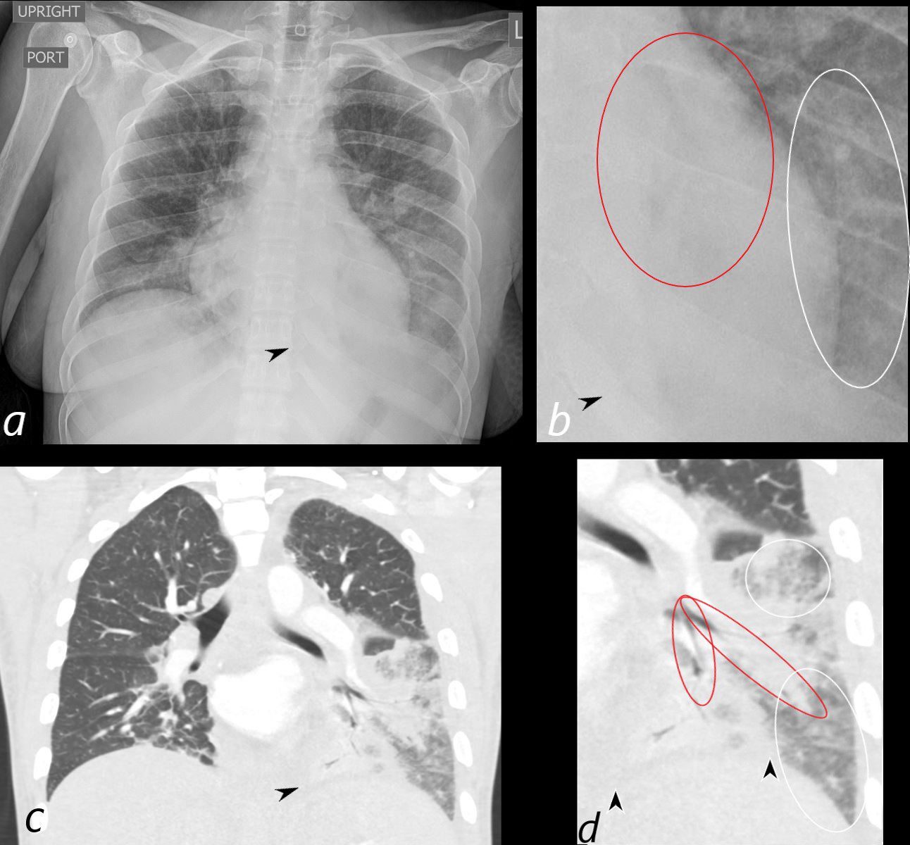
35-year-old woman, presents with dyspnea and fever. Frontal CXR (a, and magnified in b) shows a retrocardiac infiltrate, with suggestion of air bronchograms, ground glass changes (b, white ring) and silhouetting of the diaphragm medially (a, b, black arrowheads)
Coronal CT shows a bronchocentric pneumonic infiltrate in the superior segment of the left lower lobe and extending into the medial basal segment, (red rings), consistent with a bronchopneumonia. There is an adjacent region of ground glass infiltrate (d, white rings)
Courtesy Ashley Davidoff MD TheCommonVein.net 289Lu 136562cL