
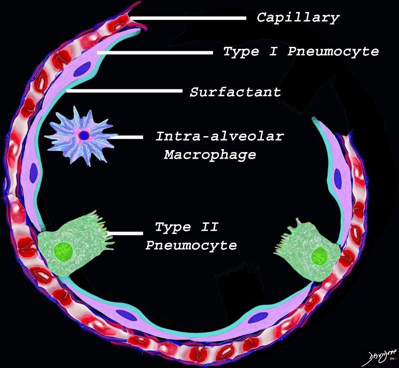
The diagram shows the lining of the normal alveolus composed of type 1 pneumocyte squamous in nature and the cuboidal cell (type pneumocyte) which rest on a lamina propria, and basement membrane (not shown) shared with the inner endothelial layer of the capillary. Intra-alveolar macrophage lies within the alveolar lumen
Ashley Davidoff TheCommonVein.net lungs-0689
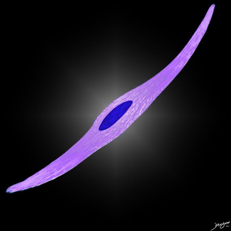
– flattened for gas exchange, forms a part of the Blood-Gas Barrier
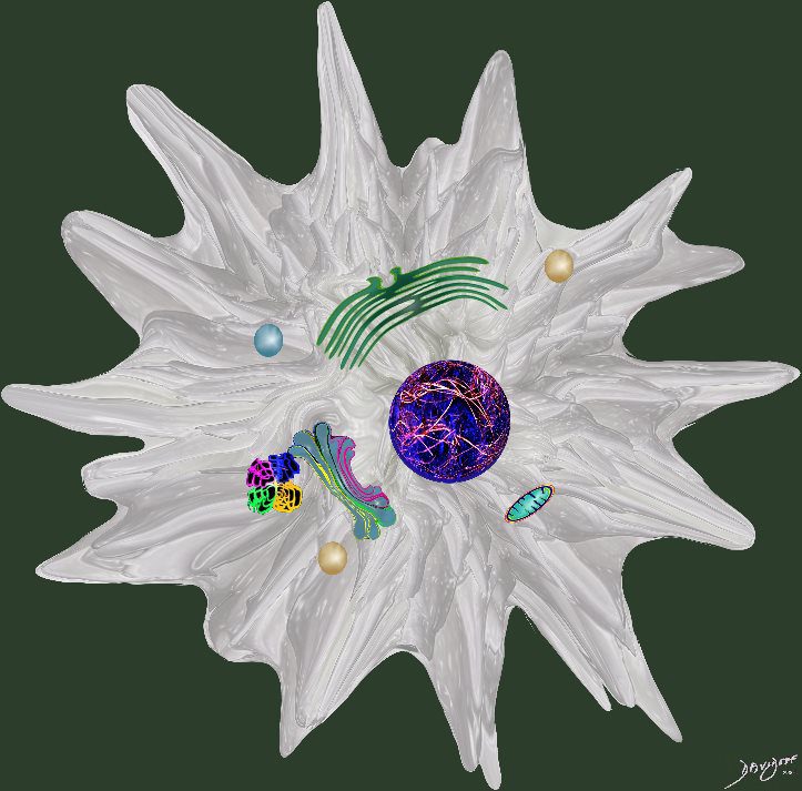
nd sometimes vacuolated cytoplasm
It produces the phospholipid – part of the surfactant that reduces surface tension and allows the alveoli to remain open
#cells
Ashley Davidoff
TheCommonVein.net lungs-0061

It produces the phospholipid – part of the surfactant that reduces surface tension and allows the alveoli to remain open
#cells
Ashley Davidoff
TheCommonVein.net lungs-0062
Ashley Davidoff MD The4CommonVein.net cells-0072
Ashley Davidoff MD
TheCommonVein.net
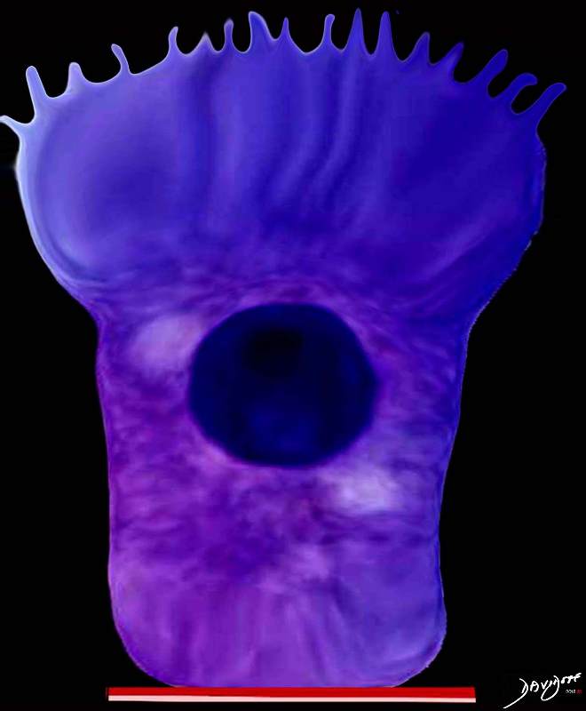
The club cell aka bronchiolar exocrine cells formerly known as the Clara cell is a low columnar cell with short microvilli and are most abundant in the bronchioles
Ashley Davidoff MD TheCommonVein.net lungs-0743
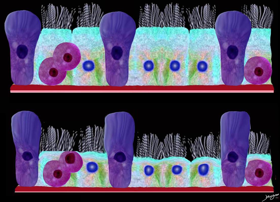
This upper diagram shows the ciliated columnar epithelium present throughout the 20- 25 generations of branching, until the airways start to transition their function from a transport system to a gas exchange system at the respiratory bronchiole level The ciliated columnar epithelium becomes a ciliated cuboidal epithelium. There are no goblet cells in the bronchioles In addition to the ciliated cells there are 2 other types of cells including the club cell (purple) and the neuroendocrine cell. The club cells Purple with dome shaped superior aspects – formerly Clara Cell) have many functions. The neuroendocrine cell (NE) (dark pink and round ) can be seen as a single cell (NE) and sometimes seen in a cluster, known as a neuroendocrine body (NEB) . The cells rest on a basement membrane, with prominent muscle layer (maroon) as well as elastic tissue (pink). There is no cartilage
Ashley Davidoff MD TheCommonVein.net
lungs-0741
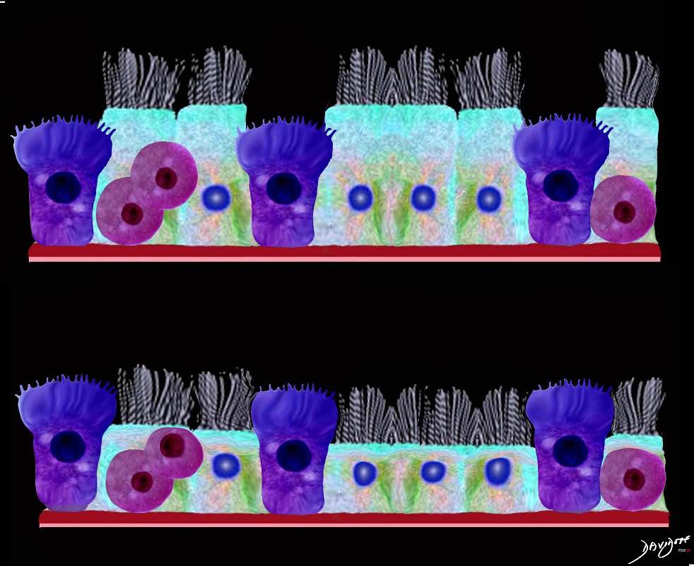
This upper diagram shows the ciliated columnar epithelium present throughout the 20- 25 generations of branching, until the airways start to transition their function from a transport system to a gas exchange system at the respiratory bronchiole level The ciliated columnar epithelium becomes a ciliated cuboidal epithelium. There are no goblet cells in the bronchioles In addition to the ciliated cells there are 2 other types of cells including the club cell (purple) and the neuroendocrine cell. The club cells Purple with dome shaped superior aspects – formerly Clara Cell) have many functions. The neuroendocrine cell (NE) (dark pink and round ) can be seen as a single cell (NE) and sometimes seen in a cluster, known as a neuroendocrine body (NEB) . The cells rest on a basement membrane, with prominent muscle layer (maroon) as well as elastic tissue (pink). There is no cartilage
Ashley Davidoff MD TheCommonVein.net
lungs-0741n

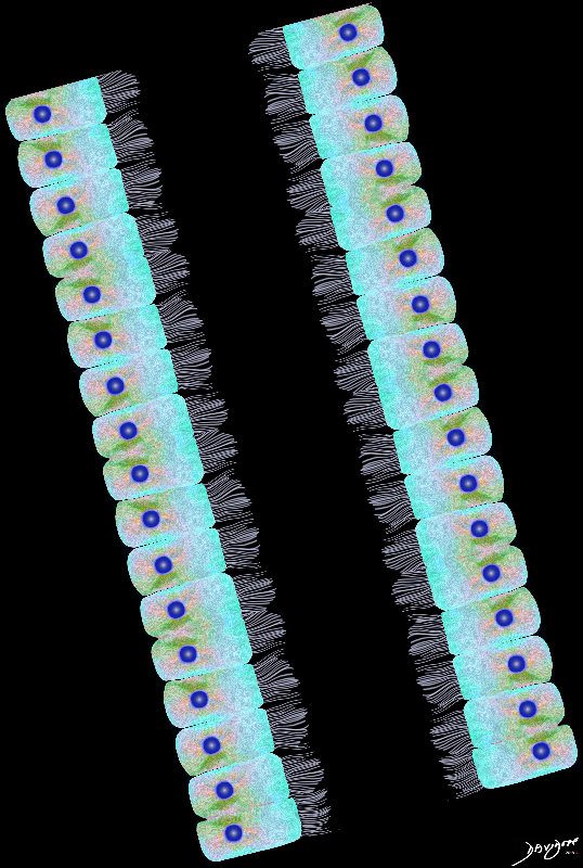
Airways are lined by a pseudostratified ciliated columnar epithelium interspersed with mucus secreting goblet cells
Ashley Davidoff
TheCommonVein.net lungs-00674b01-lo res
Ashley Davidoff
TheCommonVein.net
lungs-00675-lo-res
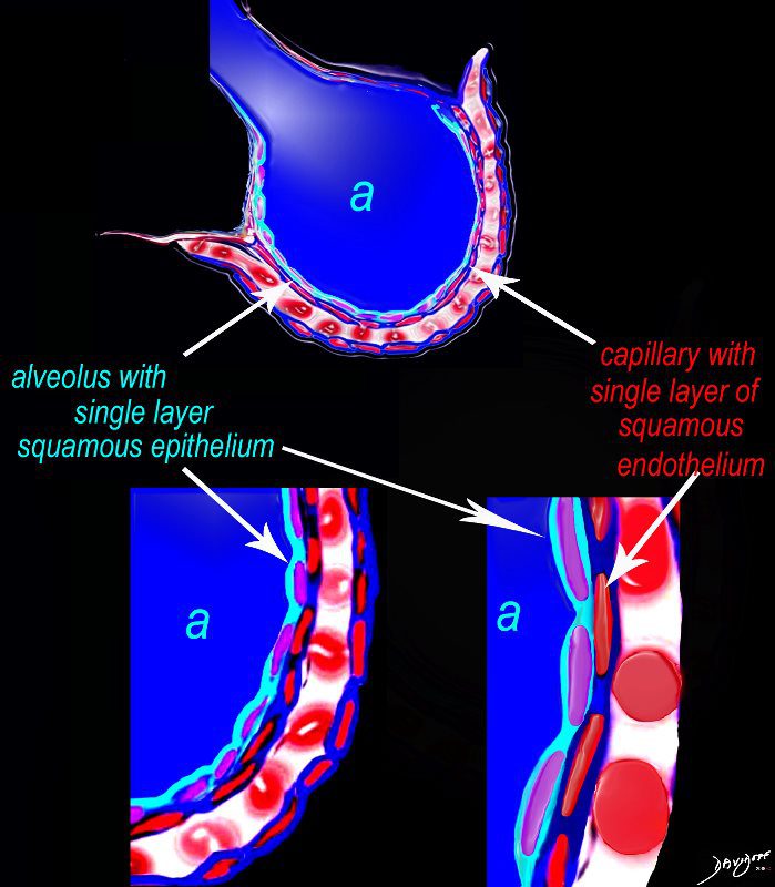
The diagram shows an alveolus (a) above, lined by a single layer of squamous cells, surrounded by a capillary with red cells which is also lined by a single layer of squamous endothelial cells . The images below show progressive magnification of the alveolar wall demonstrating the two thin layer of the alveolar membrane .
Ashley Davidoff MD TheCommonVein.net lungs-0028-low res
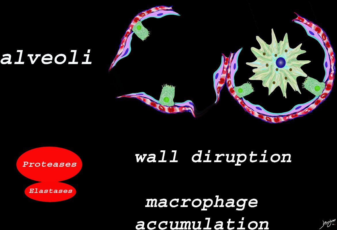
The effect of the proteases and and elastases cause destruction of the alveoli and loss of elasticity, and therefore overall function. The destruction leads to bullous disease
The accumulation of smokers macrophage, and in the case of Langerhans cell histiocytosis leads to space occupation of the alveoli also reducing function
Ashley Davidoff
TheCommonVein.net
lungs-00687
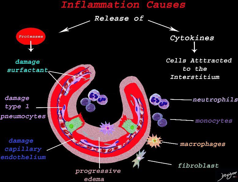
The cells of the immune system release cytokines, chemotactic agents and proteases. Immune cells , macrophages and fibroblasts are attracted to the interstitium. Some of proinflammatory agents are toxic to the cell lining causing damage to the surfactant, type 1 pneumocytes and the capillary endothelium. There is progressive edema.
Ashley Davidoff
TheCommonVein.net lungs-0692
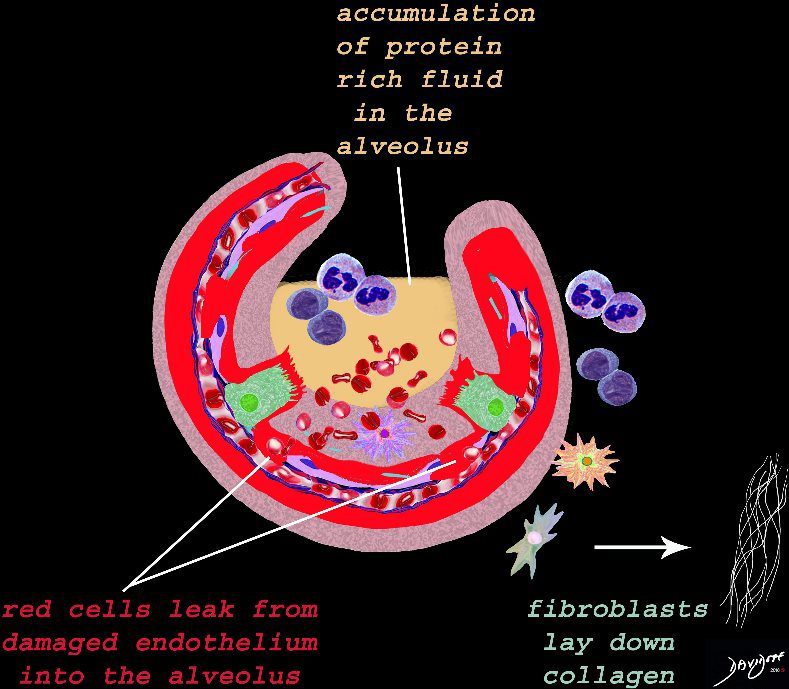
The damage to the endothelium of the capillary results in bleeding into the alveoli. The severe tissue damage and fluid exudation results in protein rich intra-alveolar fluid . The fibroblasts start to lay down collagen as part of the early repair process
Ashley Davidoff
TheCommonVein.net lungs-0693