
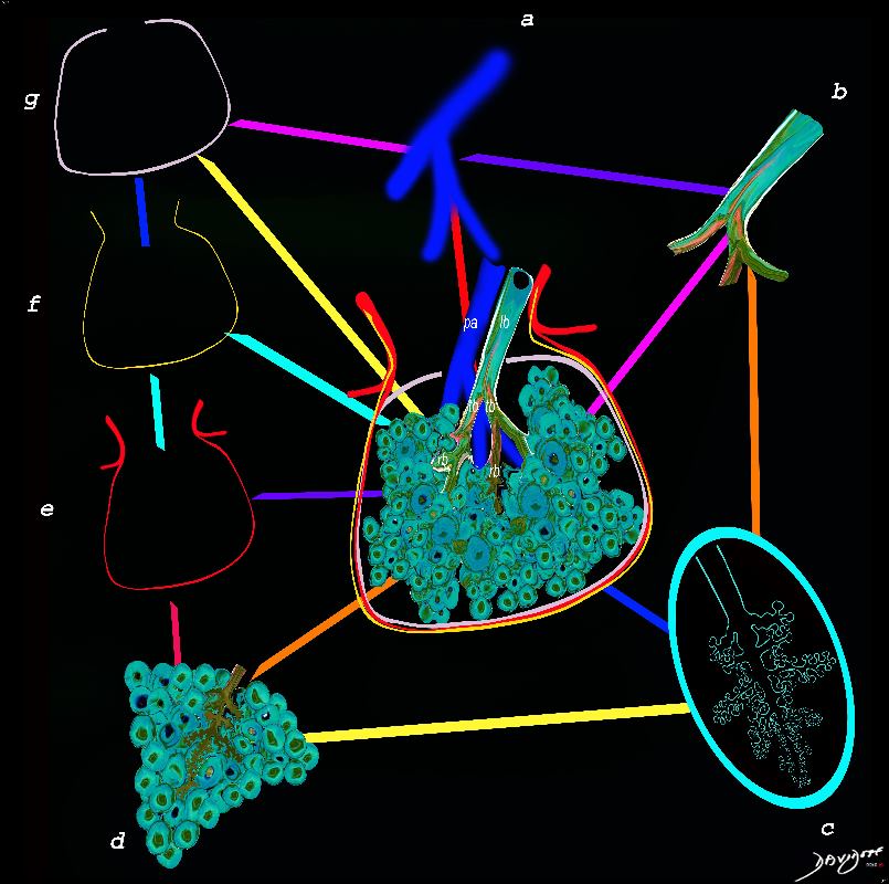
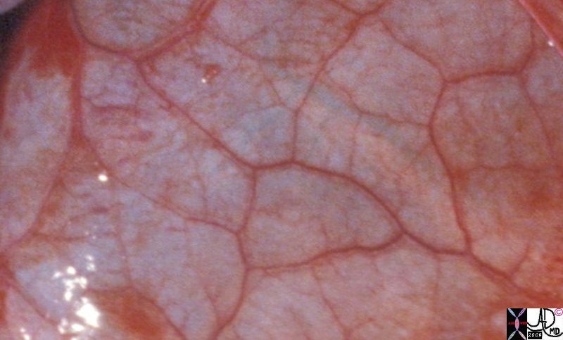
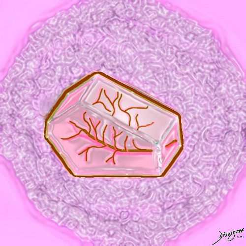
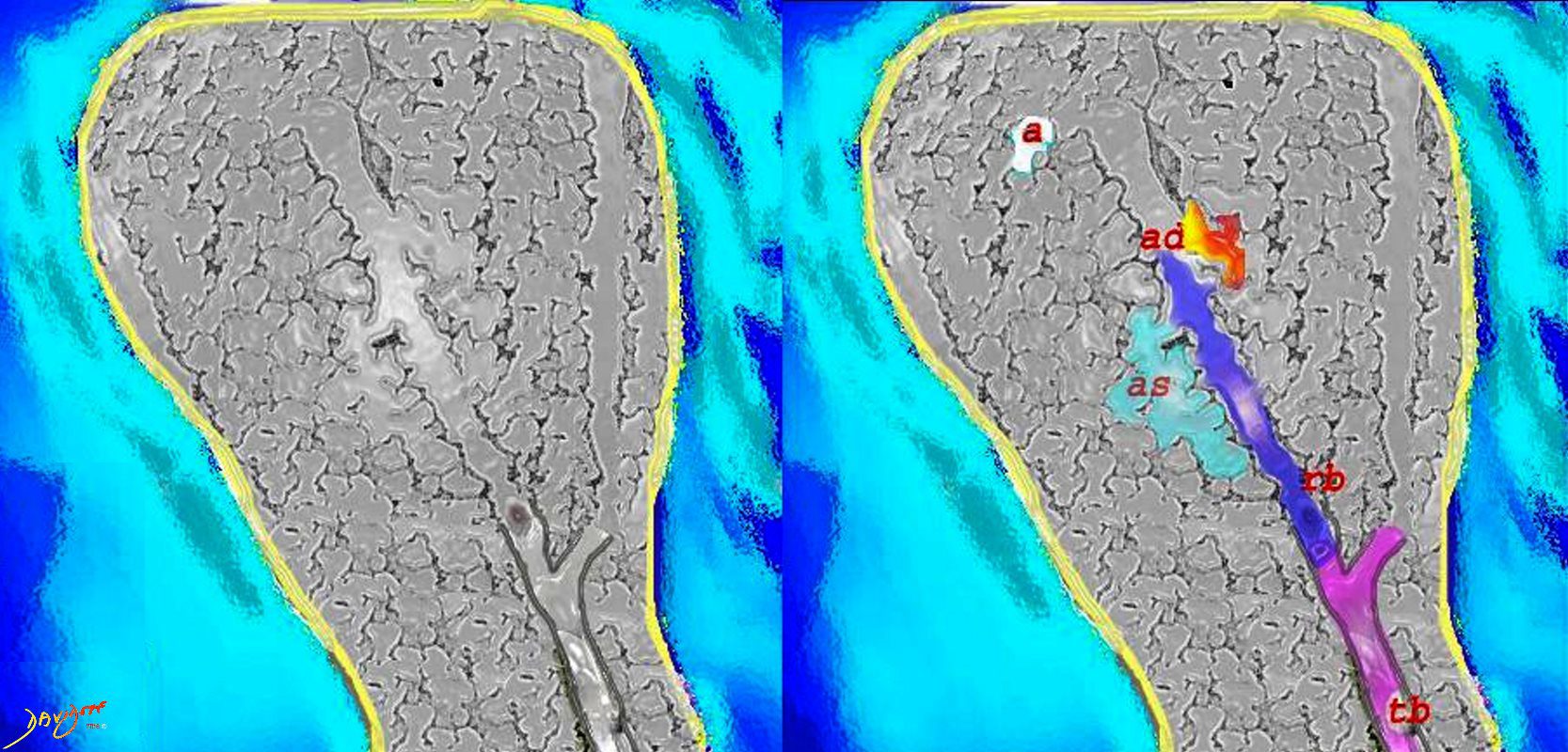
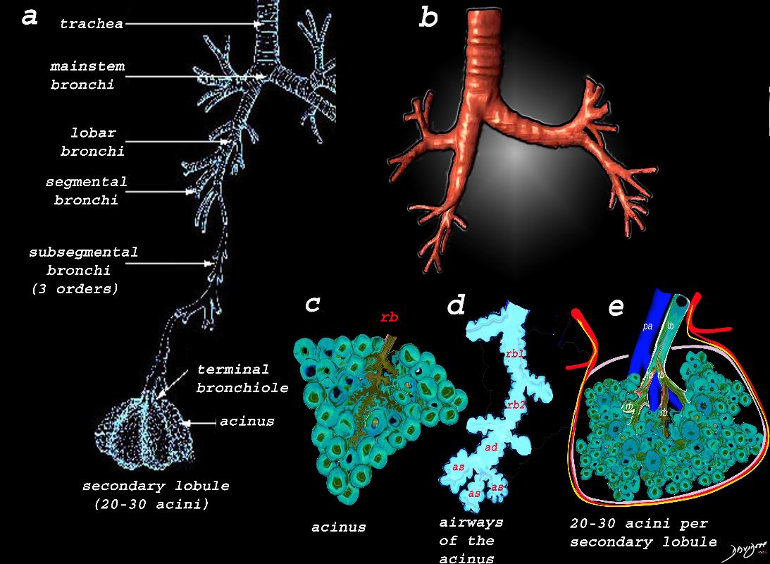
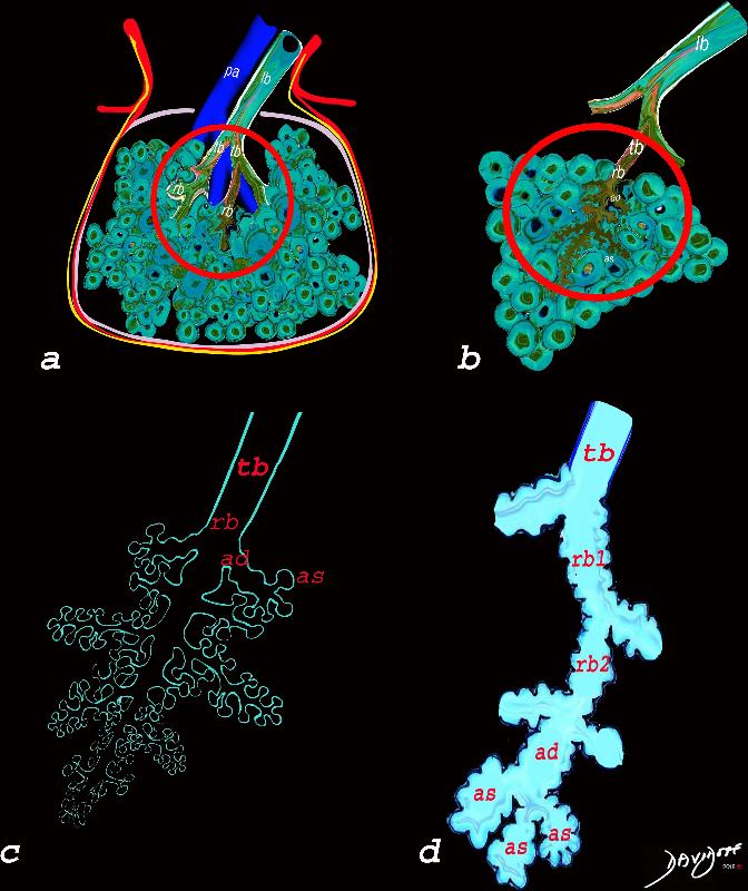
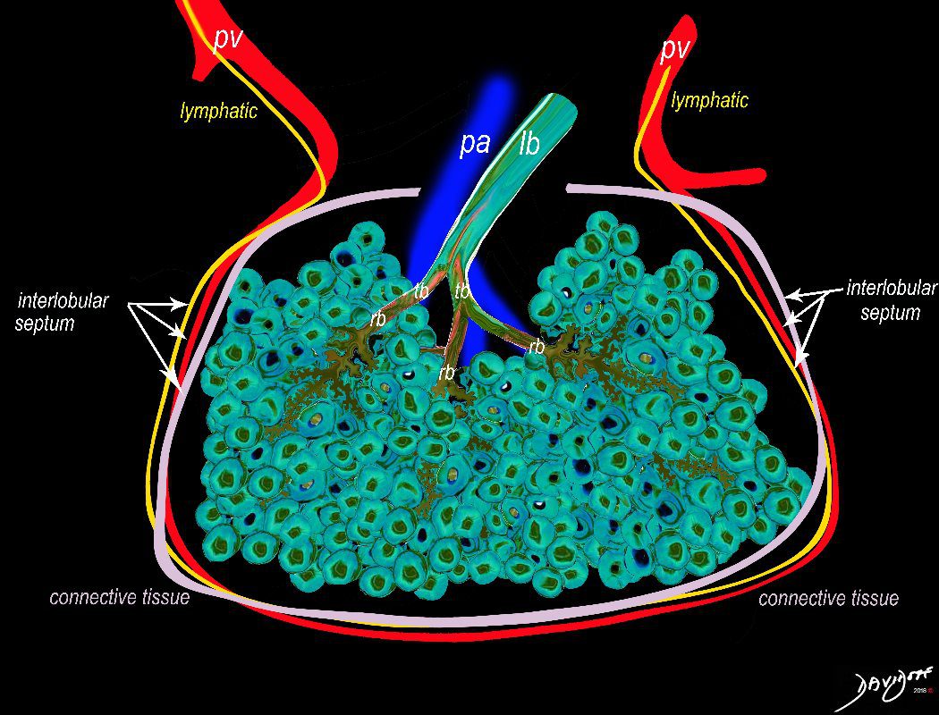
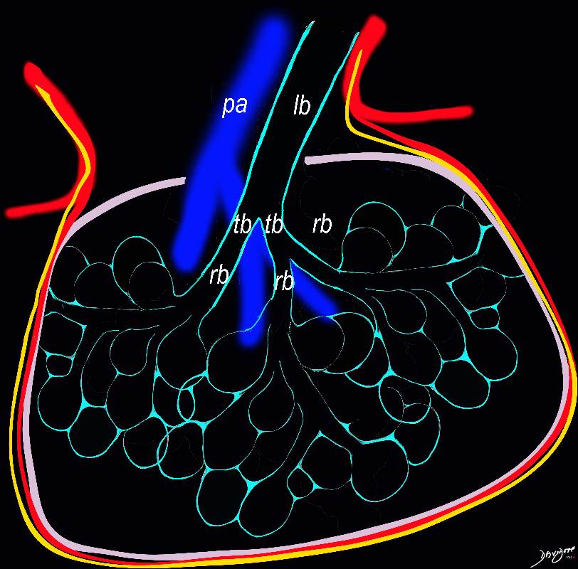
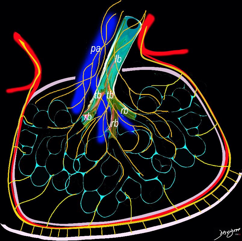
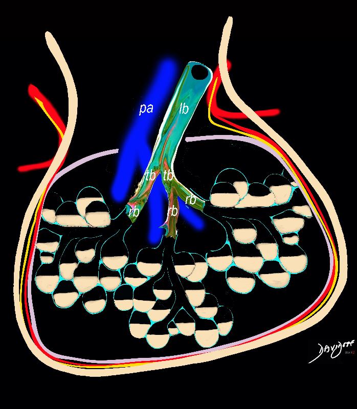
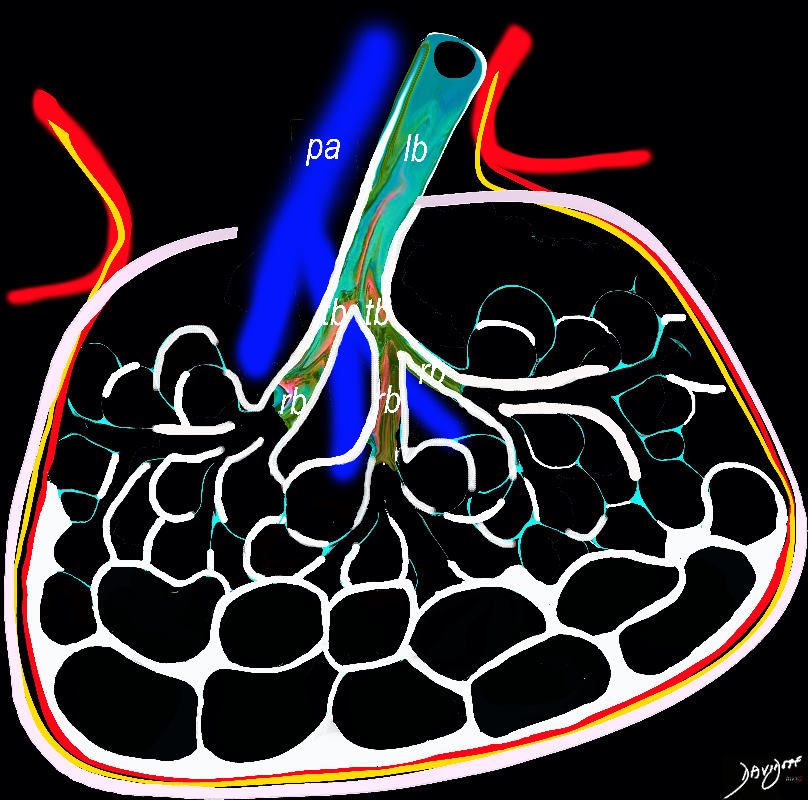
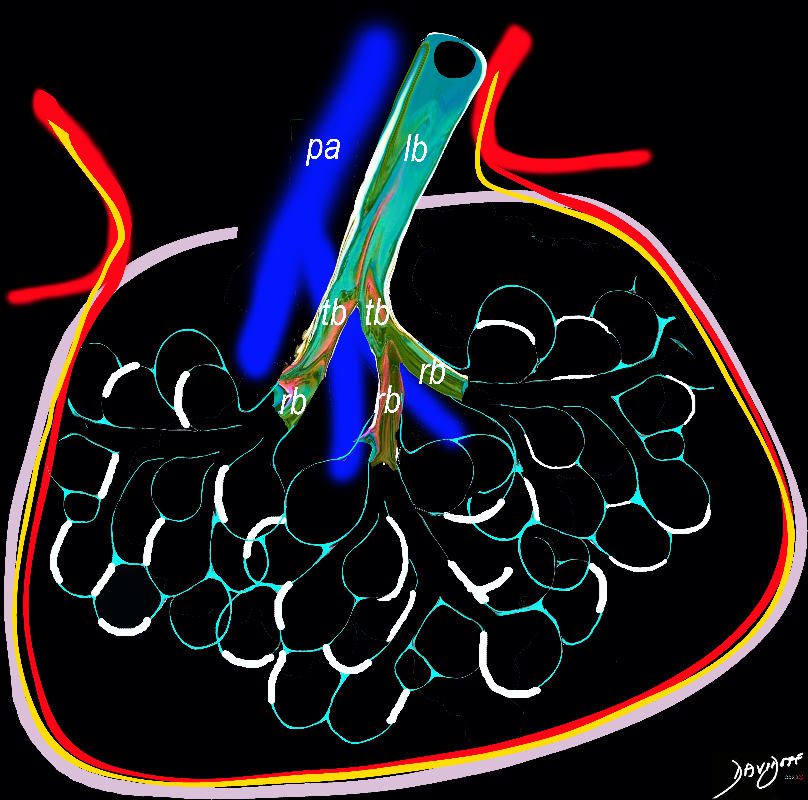
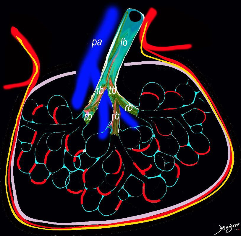
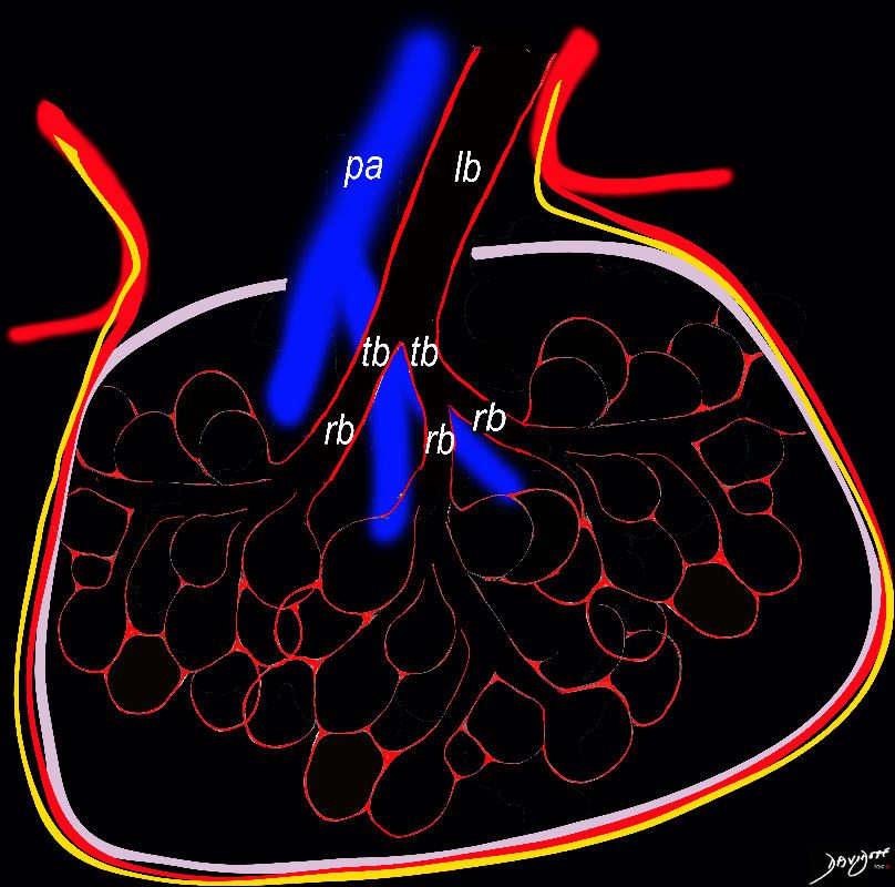
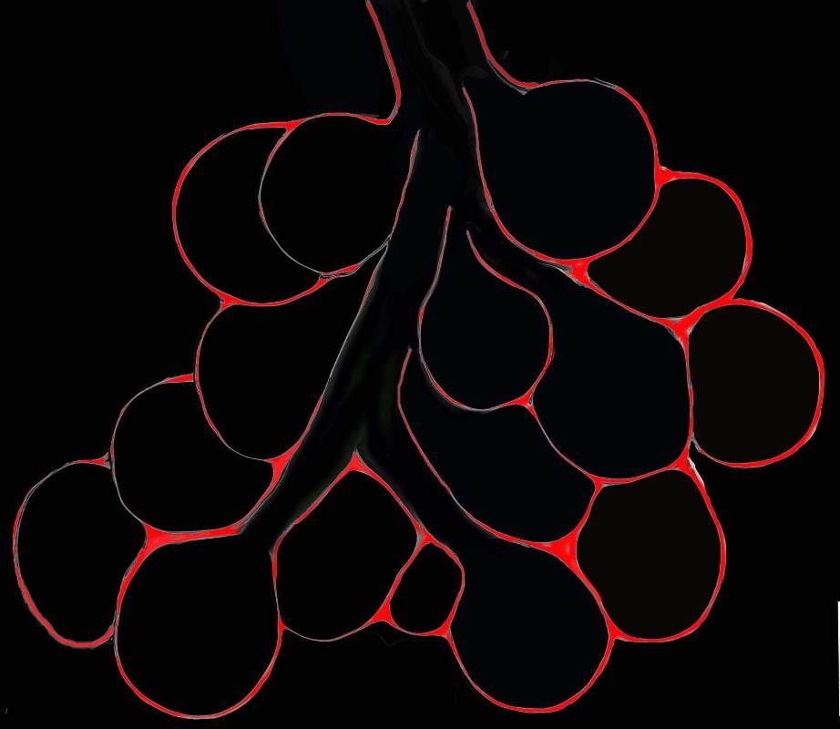
The secondary lobule is subtended by the lobular arteriole (a) and the lobular bronchiole (b) which which in turn branches into the respiratory bronchioles, alveolar ducts, and nd alveolar sacs (c) The acinus (d) consists of a respiratory bronchiole and its associated alveolar ducts, sacs, and alveoli and represents the functional unit of the lung.
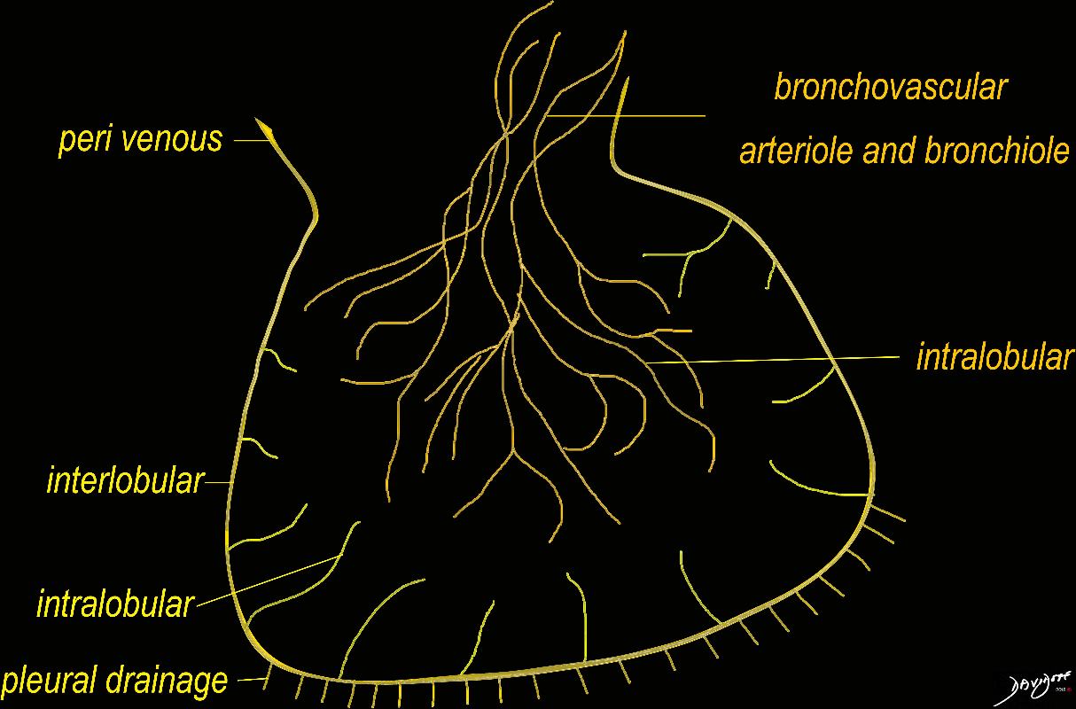
The secondary lobule is drained by the pulmonary venule (e) which runs in the interlobular septum also containing the lymphatics (f). The whole unit is housed and surrounded by a connective tissue framework (g) . The latter 3 structures form the interlobular septum.
Ashley Davidoff MD TheCommonVein.net lungs-0751
32649b
32649c06.8s
Ashley Davidoff MD TheCommonVein.net 31866collage
Davidoff MD TheCommonVein.net 32557bb03.8s
bundle Ashley Davidoff MD. The Common Vein.net lungs-0003
Ashley Davidoff MD TheCommonVein.net 42448b03
Ashley Davidoff MD. The Common Vein.net 42449b02
Ashley Davidoff MD TheCommonVein.net lungs-0008
Courtesy Ashley Davidoff MD
lungs-0028-low res
Ashley Davidoff MD. The Common Vein.net 42447b03b01
Ashley Davidoff MD. The Common Vein.net 42447b05b02
Ashley Davidoff MD TheCommonVein.net
lungs-0739
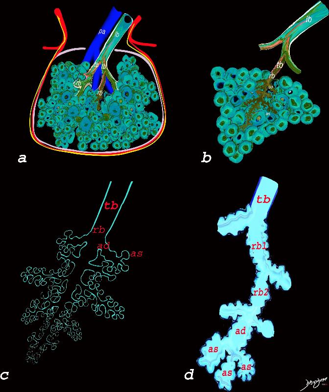
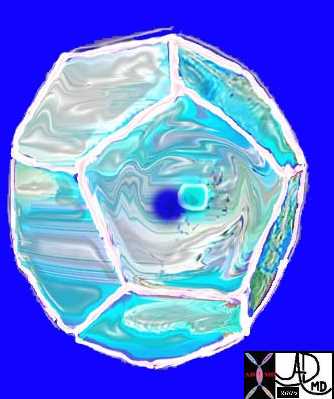
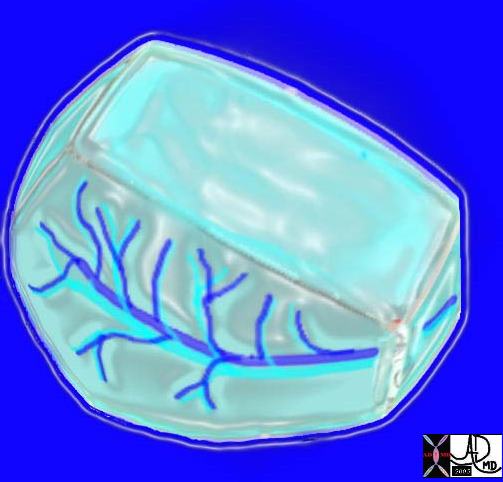
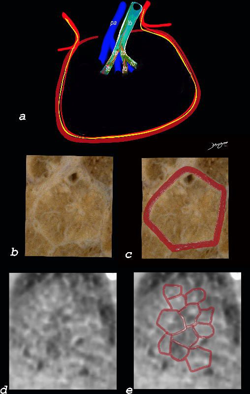
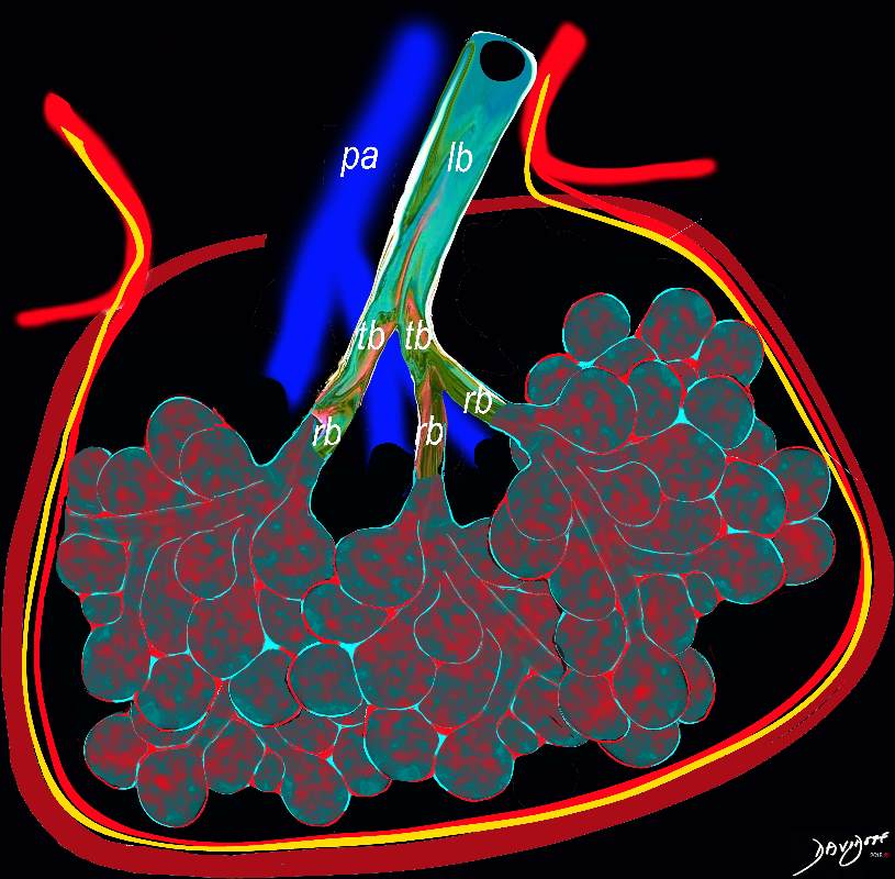
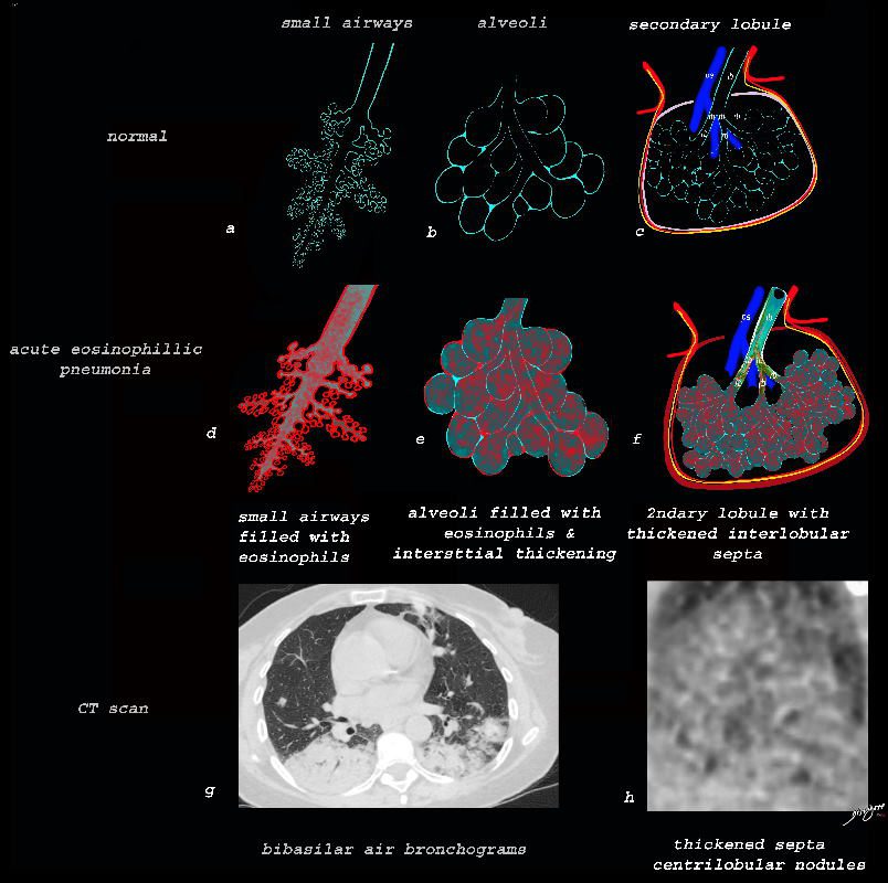
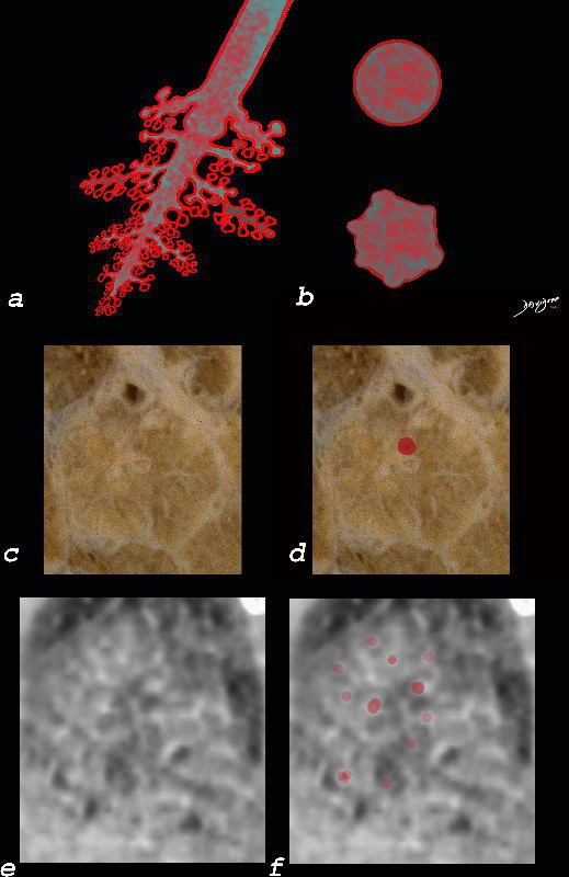
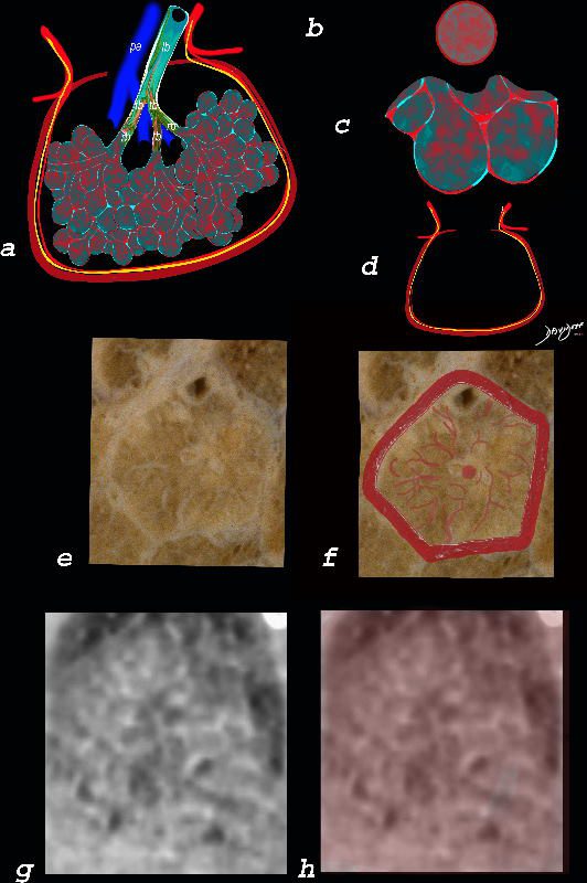
The diagram allows us to understand the the components and the position of the small airways starting in (a) which is a secondary lobule that is fed by a lobular bronchiole(lb) which enters into the secondary lobule and divides into terminal bronchioles (tb) which is the distal part of the conducting airways, and at a diameter of Ashley Davidoff MD TheCommonVein.net lungs-0744
Ashley Davidoff MD TheCommonVein.net lungs-0749
Courtesy Ashley Davidoff MD The CommonVein.net lungs-0036
Courtesy Ashley Davidoff MD The CommonVein.net lungs-0735
Ashley Davidoff MD TheCommonVein.net lungs-0767
Ashley Davidoff MD TheCommonVein.net lungs-0768
Diseases
NSIP
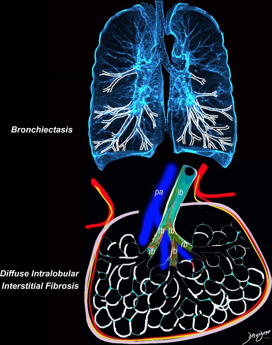
Broncho vascular and inter- alveolar interstitial fibrosis dominantly in the lower lobes but affecting the middle and upper lobes to lesser extent resulting in bronchiectasis and reticulations. The overall increase in density results in ground glass changes
Ashley Davidoff MD TheCommonvein.net lungs-0738 NSIP
Alveolar Proteinosis
Ashley Davidoff TheCommonVein.net lungs-0738b
Honeycomb
In patients with interstitial lung disease, the inflammatory process and interstitial fibrotic disease progresses and the walls between the alveoli are destroyed causing large subpleural, variably sized, subpleural, thick walled, stacked, cystic spaces . The appearance is reminiscent of a honeycomb and indicates end stage fibrosis
Ashley Davidoff MD thecommonvein.net lungs-0738bh
Interstitial Interalveolar Fibrosis
Ashley Davidoff TheCommonVein.net
lungs-0738b
Alveolitis
Ashley Davidoff TheCommonVein.net lungs-0736a01
Alveolitis and Inflammation of the Interlobular Septa
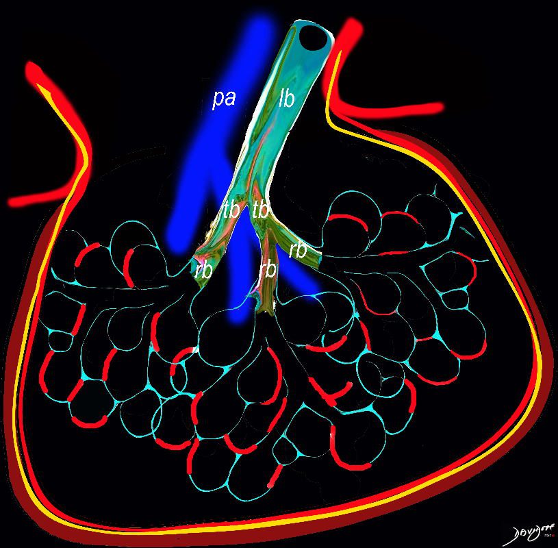
Diagram shows inflammation (red ) in the walls of the alveoli with thickening of the interlobular septa (maroon) . The increased density in the interalveolar septa and interlobular septa results in a ground glass opacity with and crazy paving appearance on CT scan
Ashley Davidoff TheCommonVein.net
lungs-0736a
Alveolitis
Ashley Davidoff TheCommonVein.net
lungs-0736
Alveolitis
Ashley Davidoff TheCommonVein.net
lungs-0736b
Ashley Davidoff MD The CommonVein.net lungs-0761
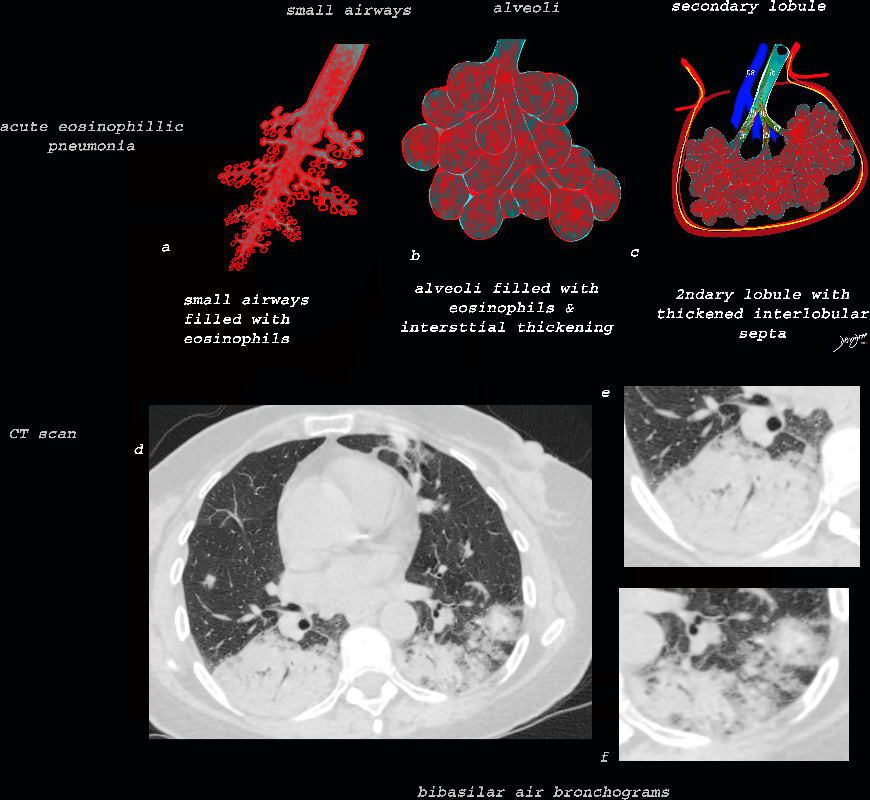
Eosinophilic Pneumonia
Ashley Davidoff TheCommonVein.net lungs-0758
Ashley Davidoff TheCommonVein.net lungs-0757b
Ashley Davidoff MD The CommonVein.net lungs-0760b
Ashley Davidoff MD The CommonVein.net lungs-0762
Ashley Davidoff MD The CommonVein.net lungs-0763