
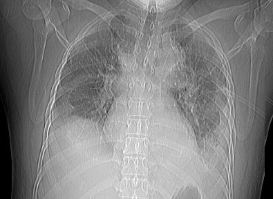
46 year-old female with bilateral pleural effusions, left greater than right. The scout film prior to the CT scan shows a left retrocardiac consolidation and a suggestion of a left sided complex pleural effusion. The liver appears enlarged.
Ashley Davidoff MD TheCommonvein.net
238Lu
Ashley Davidoff MD TheCommonvein.net
238Lu
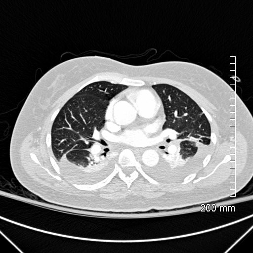
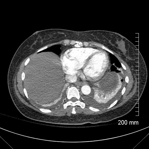
46-year-old female with bilateral pleural effusions, left greater than right. Axial CT shows bilateral pleural effusions. The undulating shape of the posterior surface of the posterior aspects of the lungs bilaterally suggests differing pressures on the lung parenchyma by the effusion and indicating loculation. There is sparing of the superior segments of the lower lobes bilaterally which both showing aeration
Ashley Davidoff MD TheCommonVein.net
238Lu
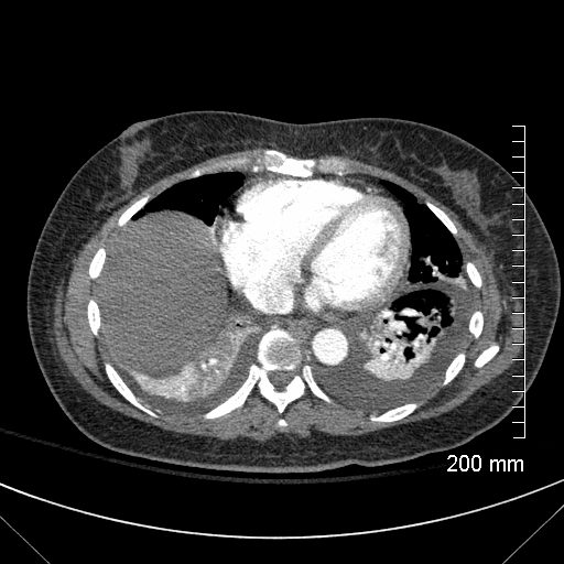
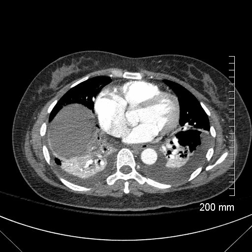
46-year-old female with bilateral pleural effusions, left greater than right. Axial CT shows bilateral pleural effusions. The flattening of the posterior surface of the right lung, undulations of the posterior surface of the left lung, and the sparing of the anterior segment of the left lower lobe suggests differing pressures on the lung parenchyma by the effusions and indicating bilateral loculation.
Ashley Davidoff MD TheCommonvein.net
238Lu
46 year-old female with bilateral pleural effusions, left greater than right. The scout film prior to the CT scan shows a left retrocardiac consolidation and a suggestion of a left sided complex pleural effusion. The liver appears enlarged.
Ashley Davidoff MD TheCommonvein.net
238Lu
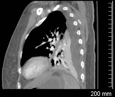
Compressive Atelectasis and Complex Pleural Effusions
Sagittal CT through the left lung shows undulations of the posterior surface of the left lung, and the suggesting differing pressures on the lung parenchyma by the effusions and indicating complexity and loculation.
Ashley Davidoff MD TheCommonVein.net
238Lu
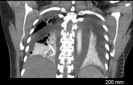
Coronal CT through the lungs show bilateral pleural effusions with compressive atelectasis
Ashley Davidoff MD TheCommonvein.net
238Lu
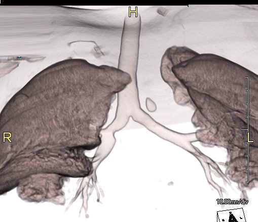
3D CT reconstructions show “naked” segmental airways to the lowers consistent with atelectasis. There is sparing of the superior segmental airways bilaterally.
Ashley Davidoff MD TheCommonVein.net
238Lu
Ashley Davidoff MD TheCommonVein.net
238Lu
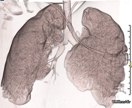
3D CT reconstructions show “naked” segmental airways to the lowers consistent with atelectasis. There is sparing of the superior segmental airways bilaterally.
Ashley Davidoff MD TheCommonVein.net
238Lu