
- 51 y.o. female with PMH of asthma/COPD, PAH on
- DIP (dxed by lung biopsy in 1999),
- Chest CT
- Stable reticulations and
- ground glass opacities at the periphery of the
lung bases, - component of honeycombing versus bronchiolectasis.
- air trapping in the lower lung field suggesting small
airway disease - Background severe centrilobular emphysema
Postsurgical changes from right upper lobe wedge resection with severe emphysematous changes.
CXR Desquamative Interstitial Pneumonia DIP and Emphysema
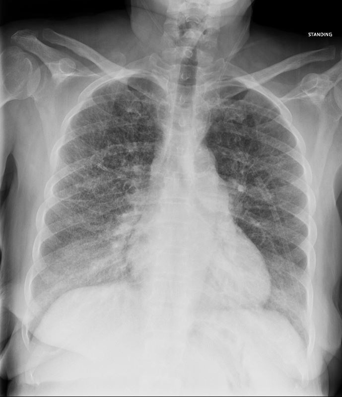
51-year-old female smoker with a history of COPD asthma and pulmonary hypertension presents with progressive dyspnea. Frontal chest Xray shows diffuse bilateral interstitial disease characterised by coarsening of the lung markings and an enlarged main pulmonary artery (MPA). Path confirmed diagnosis of DIP.
Ashley Davidoff MD TheCommonVein.net 252Lu 135957
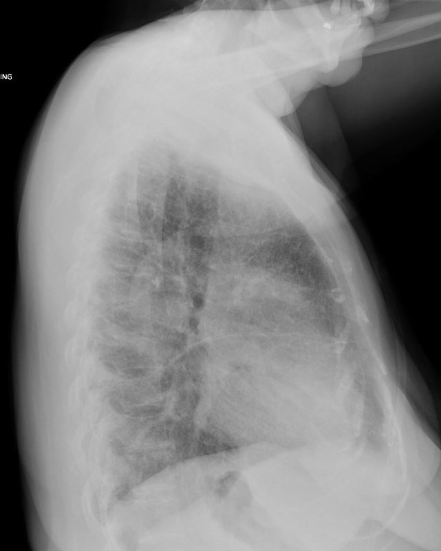
51-year-old female smoker with a history of COPD asthma and pulmonary hypertension presents with progressive dyspnea. Lateral chest Xray shows flattening of the diaphragms indicating hyperinflation and diffuse bilateral interstitial disease characterised by coarsening of the lung markings and an enlarged main pulmonary artery (MPA) The transverse fissure is thickened. Path confirmed diagnosis of DIP
Ashley Davidoff MD TheCommonVein.net 252Lu 135958
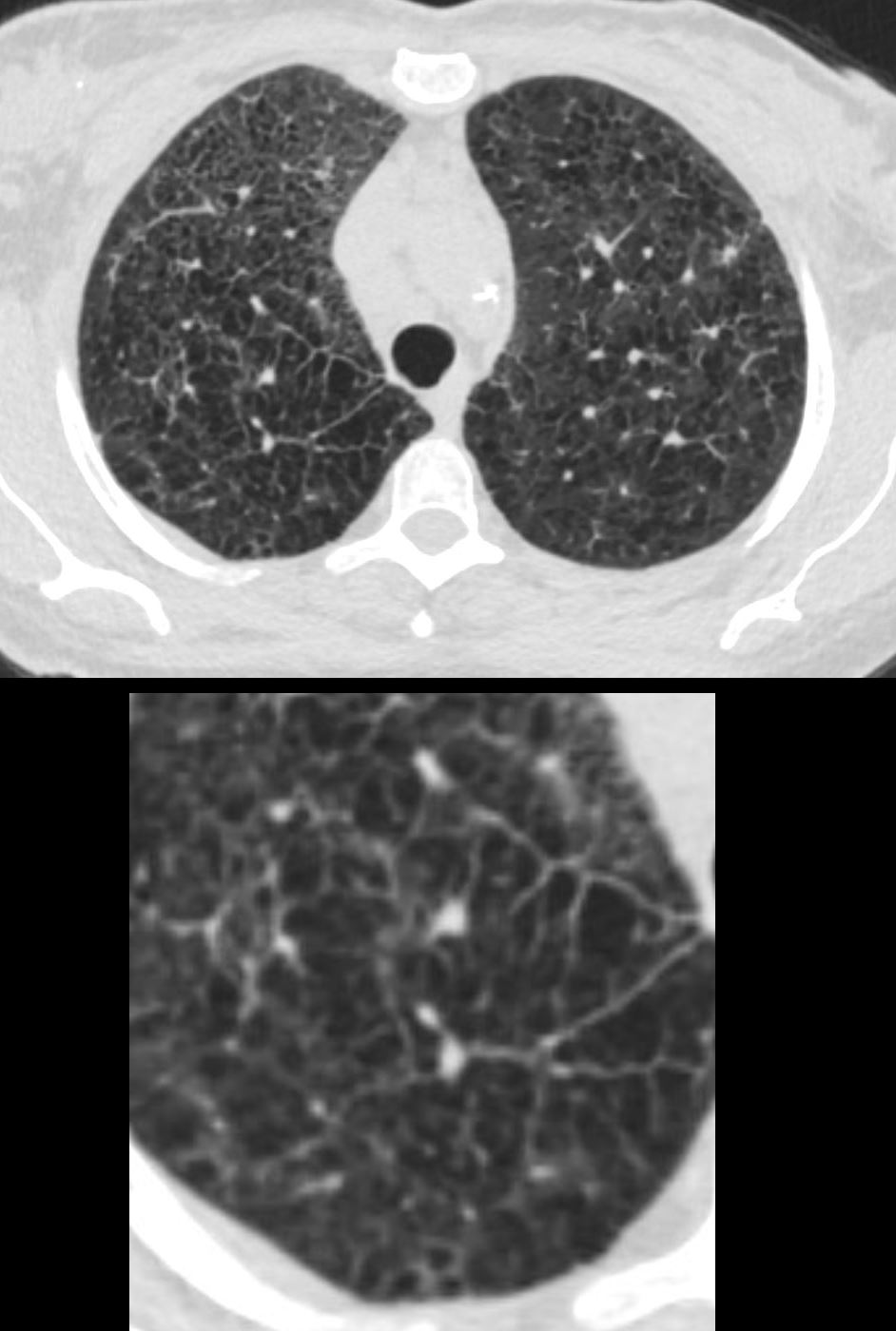
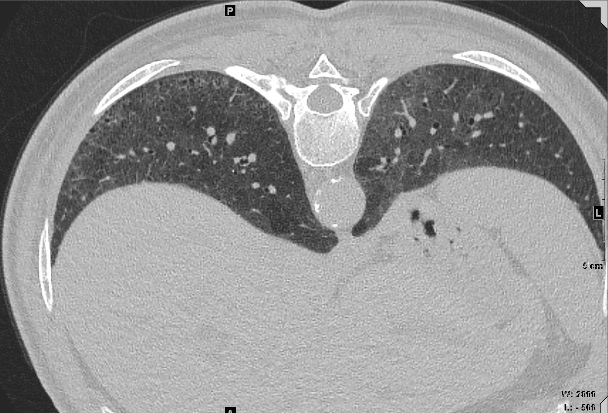
Emphysema and Bronchial Wall Thickening Upper Lung Fields
51-year-old female smoker with a history of COPD asthma and pulmonary hypertension presents with progressive dyspnea. Axial CT through the upper lung fields shows extensive changes of centrilobular emphysema associated with peribronchial thickening noted in the right upper lobe Path confirmed diagnosis of DIP
Ashley Davidoff MD TheCommonVein.net 252Lu 135961c
Enlarged Group of Secondary Lobules
51-year-old female smoker with a history of COPD asthma and pulmonary hypertension presents with progressive dyspnea. Axial CT through the upper lung fields shows extensive changes of centrilobular emphysema and an expanded group of secondary lobules noted in the right upper lobe Path confirmed a diagnosis of DIP
Ashley Davidoff MD TheCommonVein.net 252Lu 135963c
Fissural Irregularity and Thickened Septa
51-year-old female smoker with a history of COPD asthma and pulmonary hypertension presents with progressive dyspnea. Axial CT through the upper lung fields at the level of the carina shows extensive changes of centrilobular emphysema and ground glasses changes in the anterior segments – right more prominent than the left. In addition there is irregularity of the right major fissure (lower panel) seemingly as a result of the enlarged secondary lobule, and stress on the fissure by the interlobular septa. Path confirmed a diagnosis of DIP
Ashley Davidoff MD TheCommonVein.net 252Lu 135965c
Upper Lung Fields Transforming from
Emphysematous Changes to Ground Glass
51-year-old female smoker with a history of COPD asthma and pulmonary hypertension presents with progressive dyspnea. Axial CT through the upper lung fields at the level of the carina shows progression from extensive centrilobular changes to ground glass changes in the left anterior segment, and diffuse ground glass changes in the lower lobes. In addition, there is irregularity of the right major fissure seemingly as a result of the enlarged secondary lobule, and stress on the fissure by the interlobular septa. Path confirmed a diagnosis of DIP
Ashley Davidoff MD TheCommonVein.net 252Lu 135966
Emphysema Upper Lung Fields Transforming from Emphysematous Changes to Ground Glass
Thickened Irregular Fissure
51-year-old female smoker with a history of COPD asthma and pulmonary hypertension presents with progressive dyspnea. Axial CT through the upper lung fields at the level of the carina shows progression from extensive centrilobular changes to ground glass changes in the left anterior segment, and diffuse ground glass changes in the lower lobes. In addition, there is irregularity of the right major fissure (lower panel) seemingly as a result of the enlarged secondary lobule, and stress on the fissure by the interlobular septa. Path confirmed a diagnosis of DIP
Ashley Davidoff MD TheCommonVein.net 252Lu 135966c
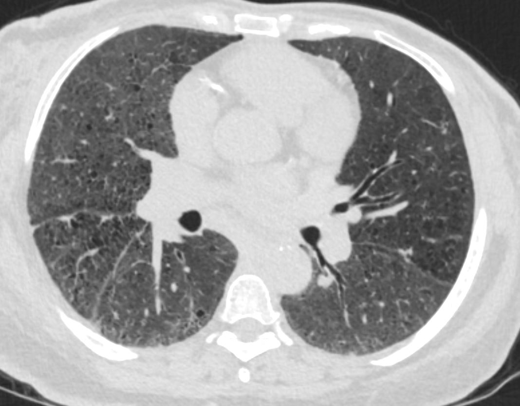
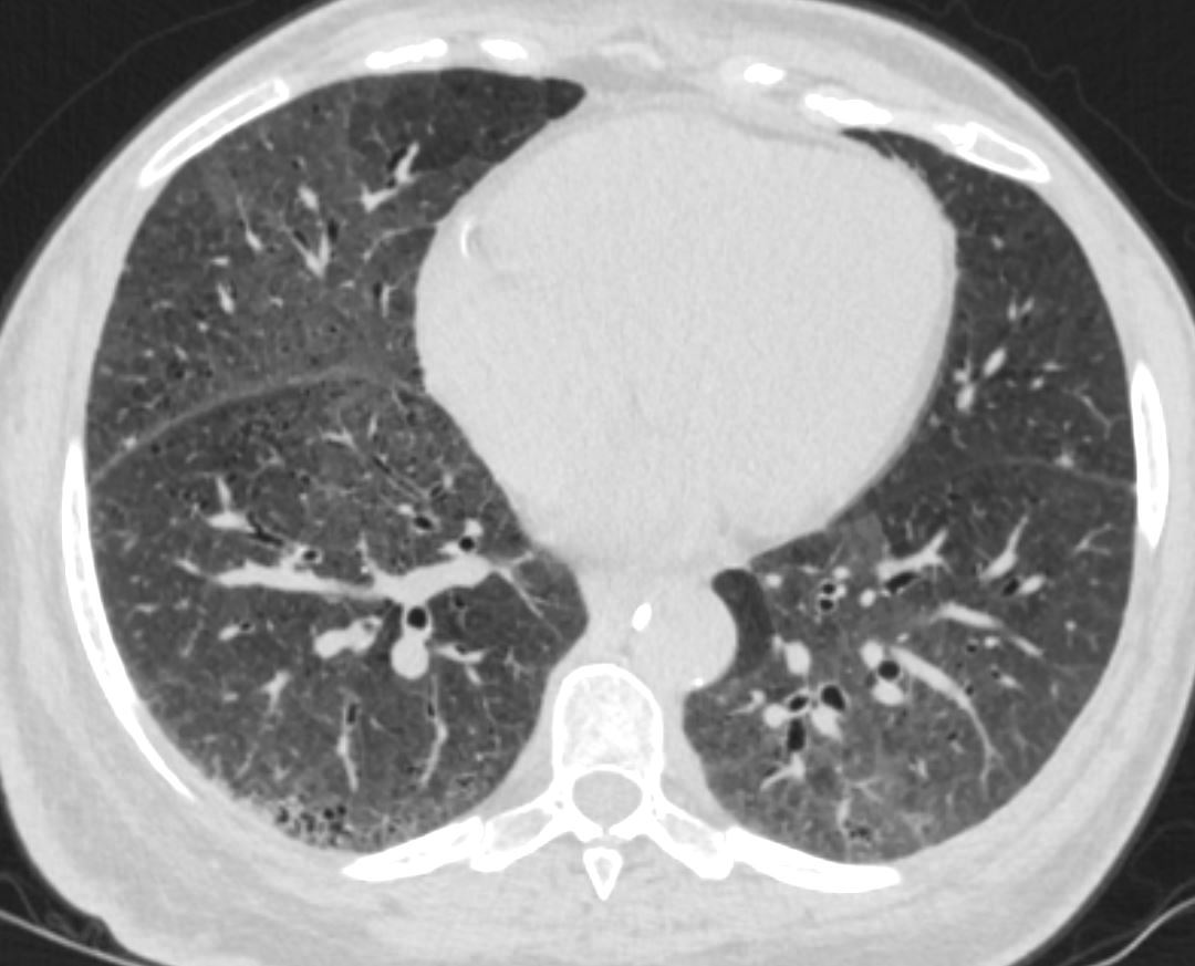
Patchy Ground Glass Changes
51-year-old female smoker with a history of COPD asthma and pulmonary hypertension presents with progressive dyspnea. Axial CT through the lower lung fields shows patchy ground glass changes in the middle lobe inferior ligula and lower lobes and some regions of mosaicism. There is mild subsegmental airway thickening. Early honeycomb changes are suggested in the right lower lobe. Pathology confirmed a diagnosis of DIP
Ashley Davidoff MD TheCommonVein.net 252Lu 135968
Patchy Ground Glass Changes
51-year-old female smoker with a history of COPD asthma and pulmonary hypertension presents with progressive dyspnea. Axial CT through the lower lung fields shows patchy ground glass changes in the middle lobe inferior ligula and lower lobes and some regions of mosaicism. There is mild subsegmental airway thickening. Early honeycomb changes are suggested in the right lower lobe. Pathology confirmed a diagnosis of DIP
Ashley Davidoff MD TheCommonVein.net 252Lu 135969
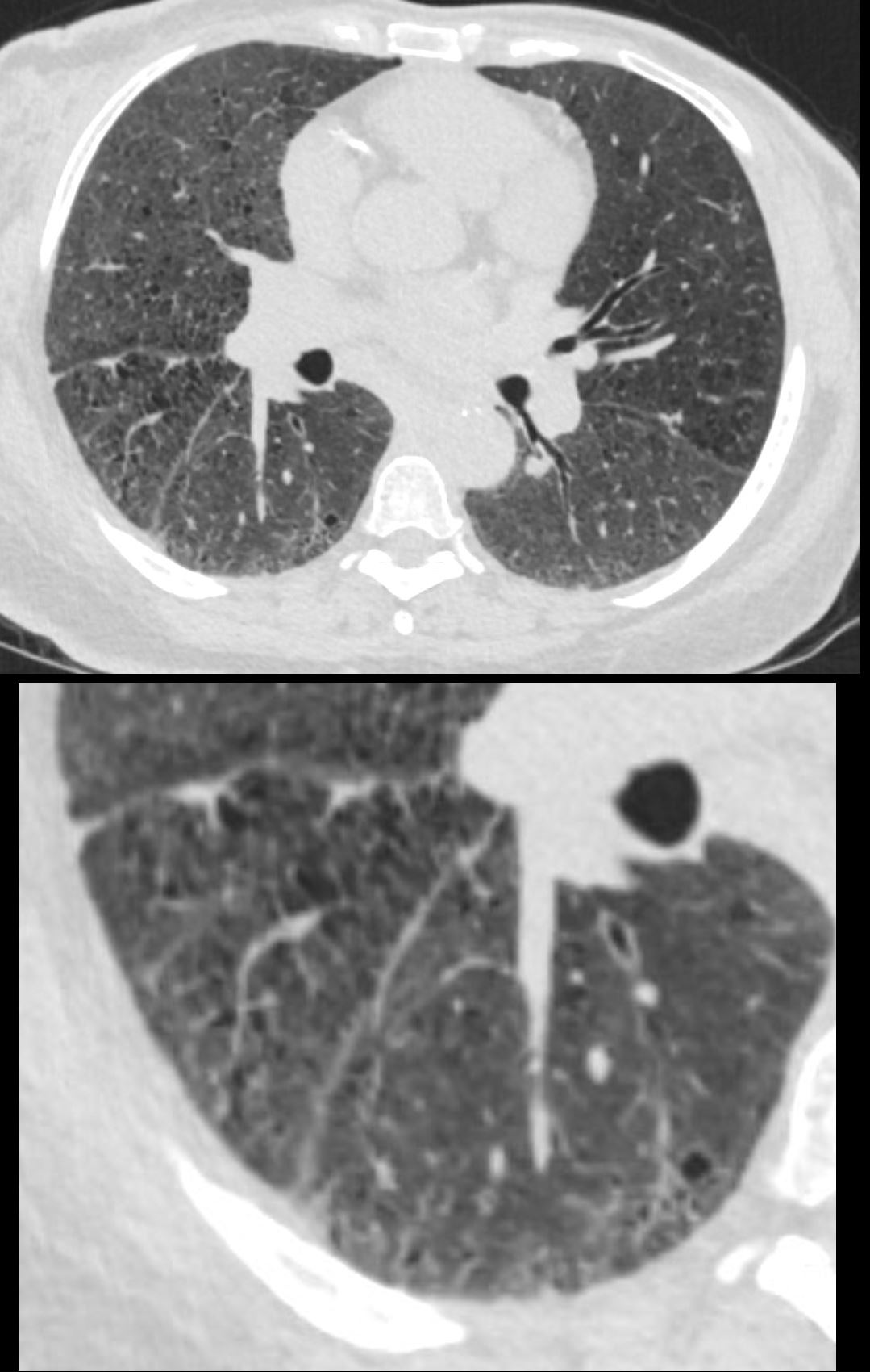
Patchy Ground Glass Changes
51-year-old female smoker with a history of COPD asthma and pulmonary hypertension presents with progressive dyspnea. Axial CT through the lower lung fields shows patchy ground glass changes in the middle lobe inferior ligula and lower lobes and some regions of mosaicism. Focal regions of interlobular septal thickening are noted left lower lobe (lower panel). Pathology confirmed a diagnosis of DIP
Ashley Davidoff MD TheCommonVein.net 252Lu 135969c
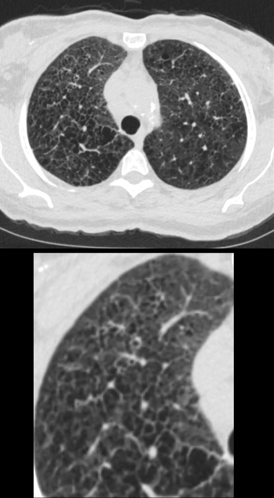
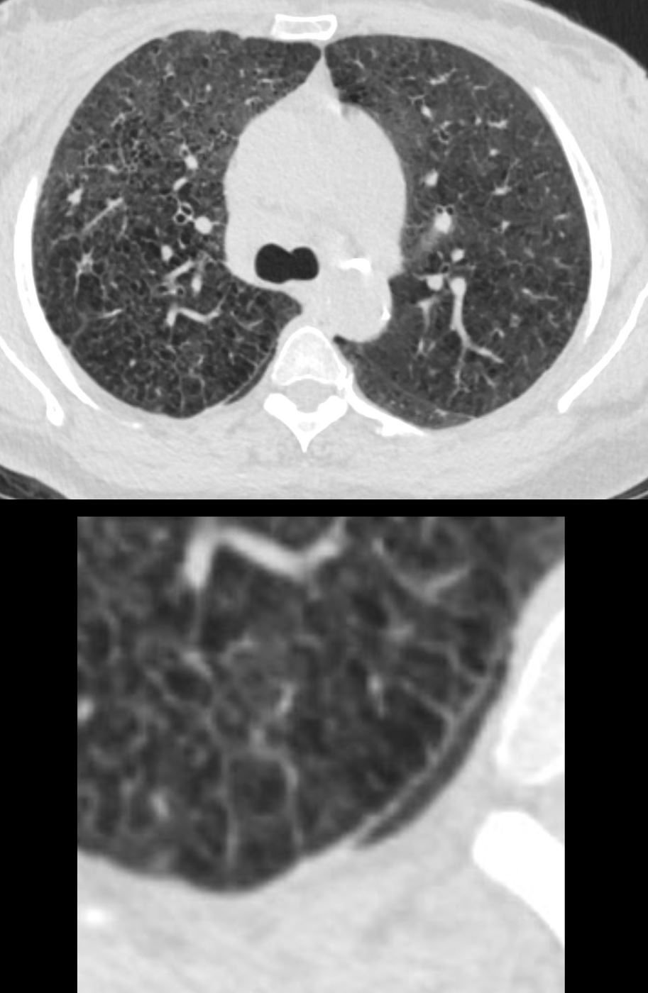
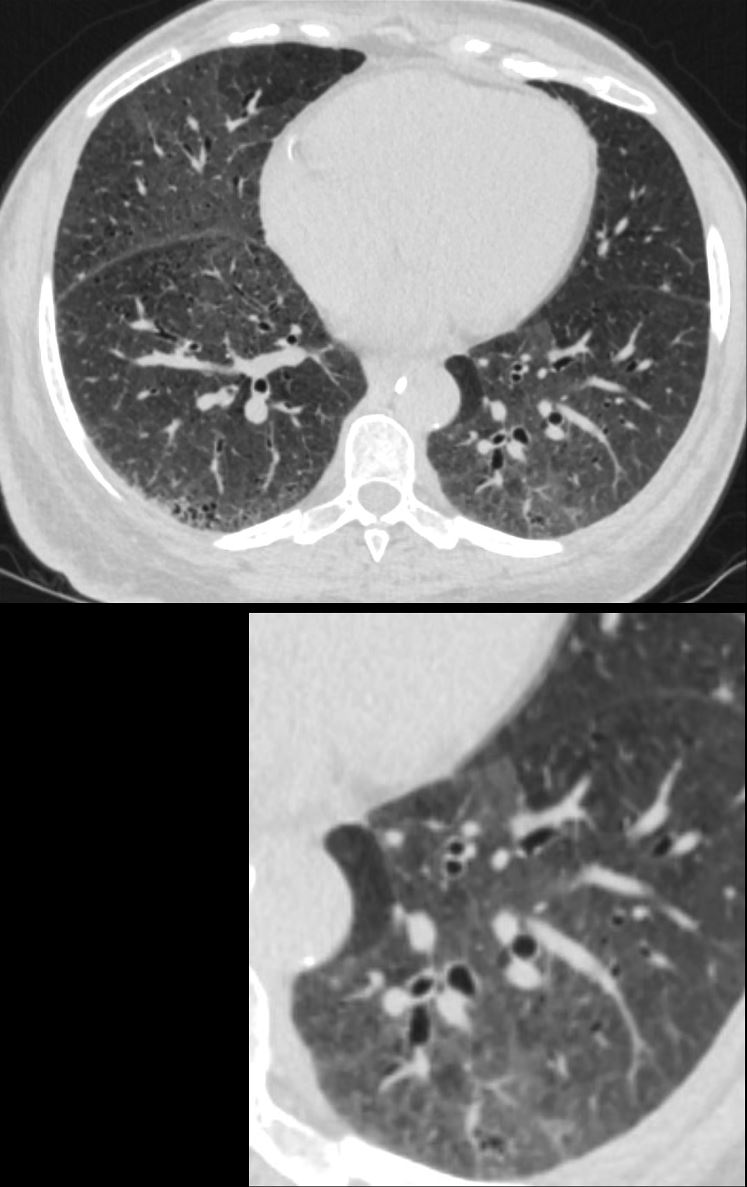
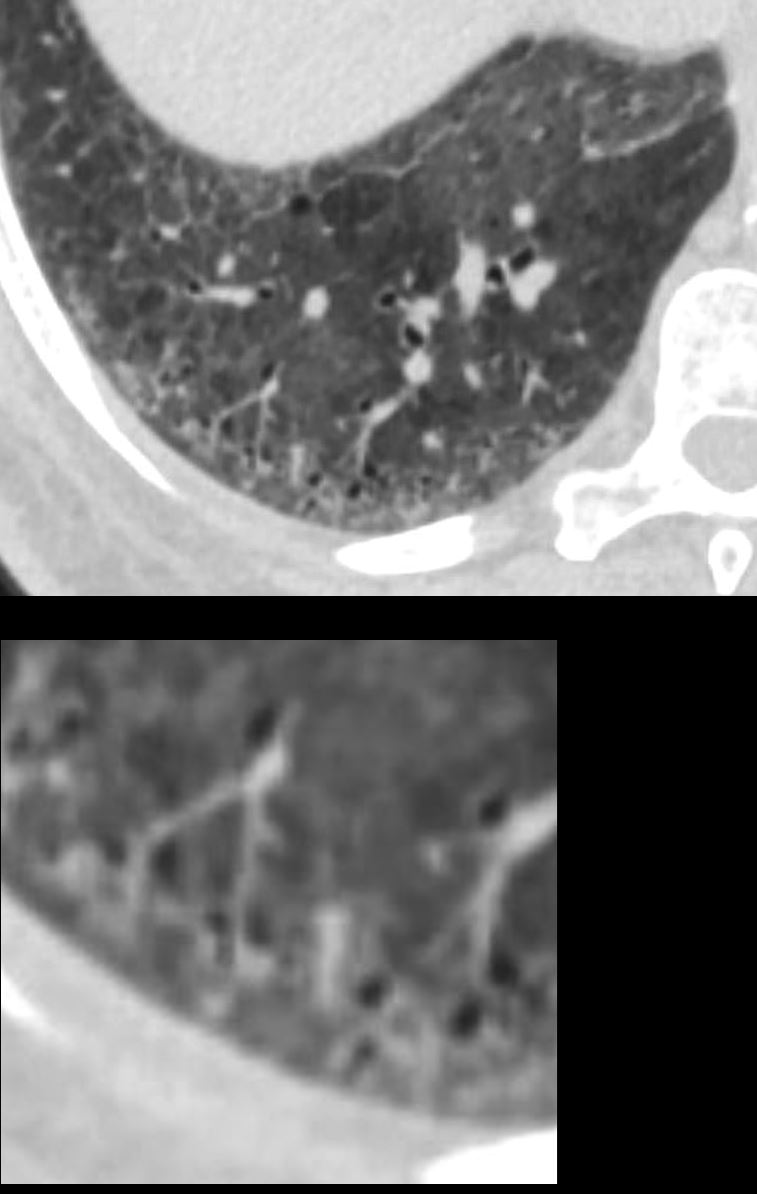
Patchy Ground Glass Changes and Bronchiolectasis
51-year-old female smoker with a history of COPD asthma and pulmonary hypertension presents with progressive dyspnea. Axial CT through the lower lung fields shows patchy ground glass changes in the middle lobe inferior ligula and lower lobes and some regions of mosaicism. Focal regions of interlobular septal thickening are noted left lower lobe and evidence of thick walled bronchiolectasis in the right lower lobe (lower panel). Pathology confirmed a diagnosis of DIP
Ashley Davidoff MD TheCommonVein.net 252Lu 135972c
Patchy Ground Glass Changes, Bronchiolectasis, and Mosaicism
51-year-old female smoker with a history of COPD asthma and pulmonary hypertension presents with progressive dyspnea. Axial CT through the lower lung fields shows patchy ground glass changes in the middle lobe inferior ligula and lower lobes and some regions of mosaicism. There is evidence of thick-walled bronchiolectasis in the right lower lobe Pathology confirmed a diagnosis of DIP
Ashley Davidoff MD TheCommonVein.net 252Lu 135976
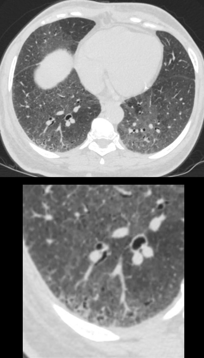
,Bronchiolectasis Mosaicism and the Secondary Lobul
51-year-old female smoker with a history of COPD asthma and pulmonary hypertension presents with progressive dyspnea. Axial CT through the right posterior recess shows patchy ground glass changes with some regions of mosaicism. The bronchovascular bundle subtending 2 secondary lobules is highlighted in the lower panel. The centrilobular arteriole and ectatic bronchiole are magnified
Pathology confirmed a diagnosis of DIP
Ashley Davidoff MD TheCommonVein.net 252Lu 135981c
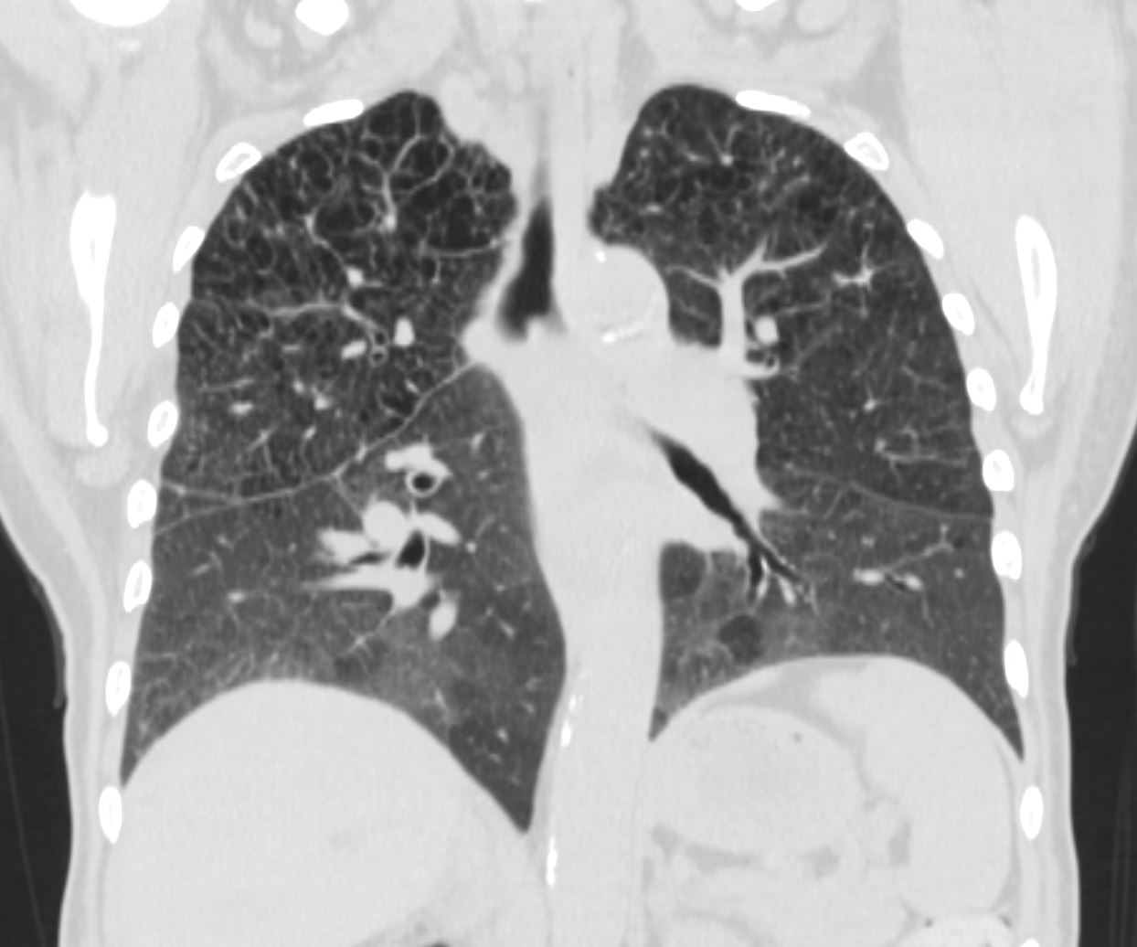
Emphysema Upper Lung Fields
Patchy Ground Glass Changes Lower Lung Fields
51-year-old female smoker with a history of COPD asthma and pulmonary hypertension presents with progressive dyspnea. Coronal CT through the mid lung shows upper lobe centrilobular emphysematous disease and patchy ground glass changes in the lower lobes
Pathology confirmed a diagnosis of DIP
Ashley Davidoff MD TheCommonVein.net 252Lu 135990
Emphysema Upper Lung Fields
Patchy Ground Glass Changes Lower Lung Fields
51-year-old female smoker with a history of COPD asthma and pulmonary hypertension presents with progressive dyspnea. Coronal CT through the posterior lungs shows upper lobe centrilobular emphysematous disease and patchy ground glass changes in the lower lobes with mosaicism.
Pathology confirmed a diagnosis of DIP
Ashley Davidoff MD TheCommonVein.net 252Lu 135993
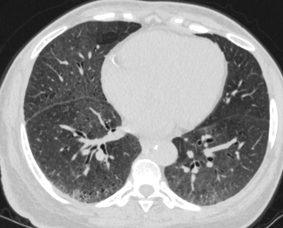
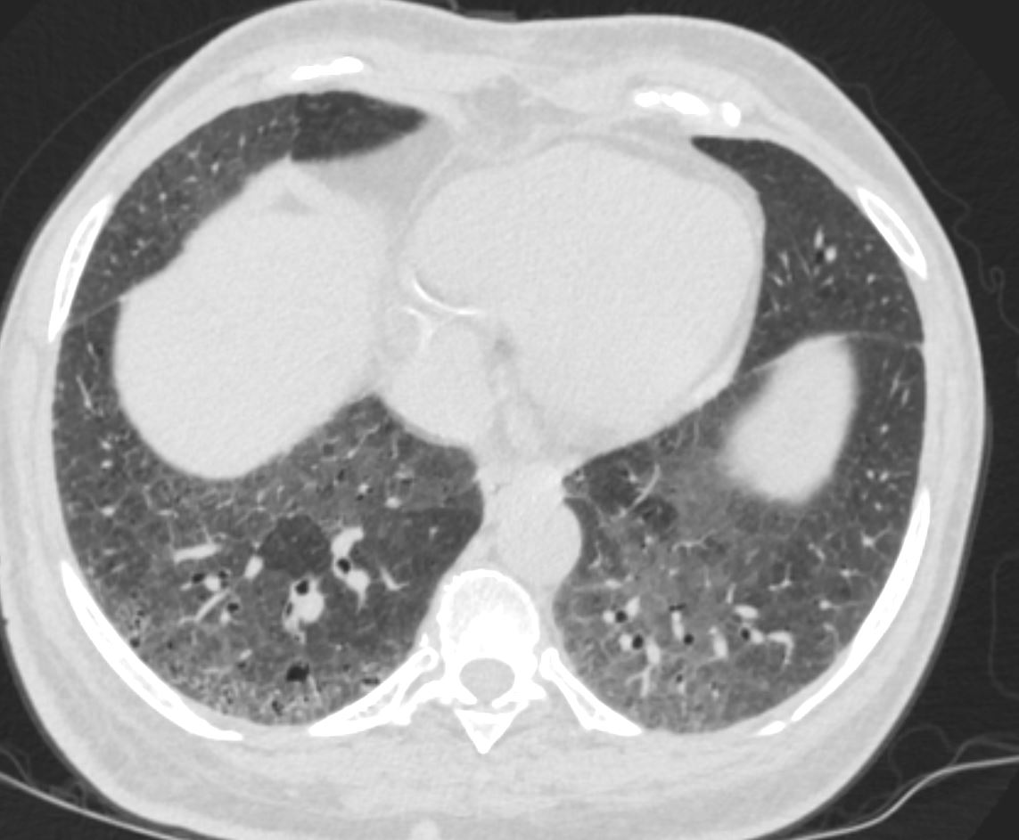
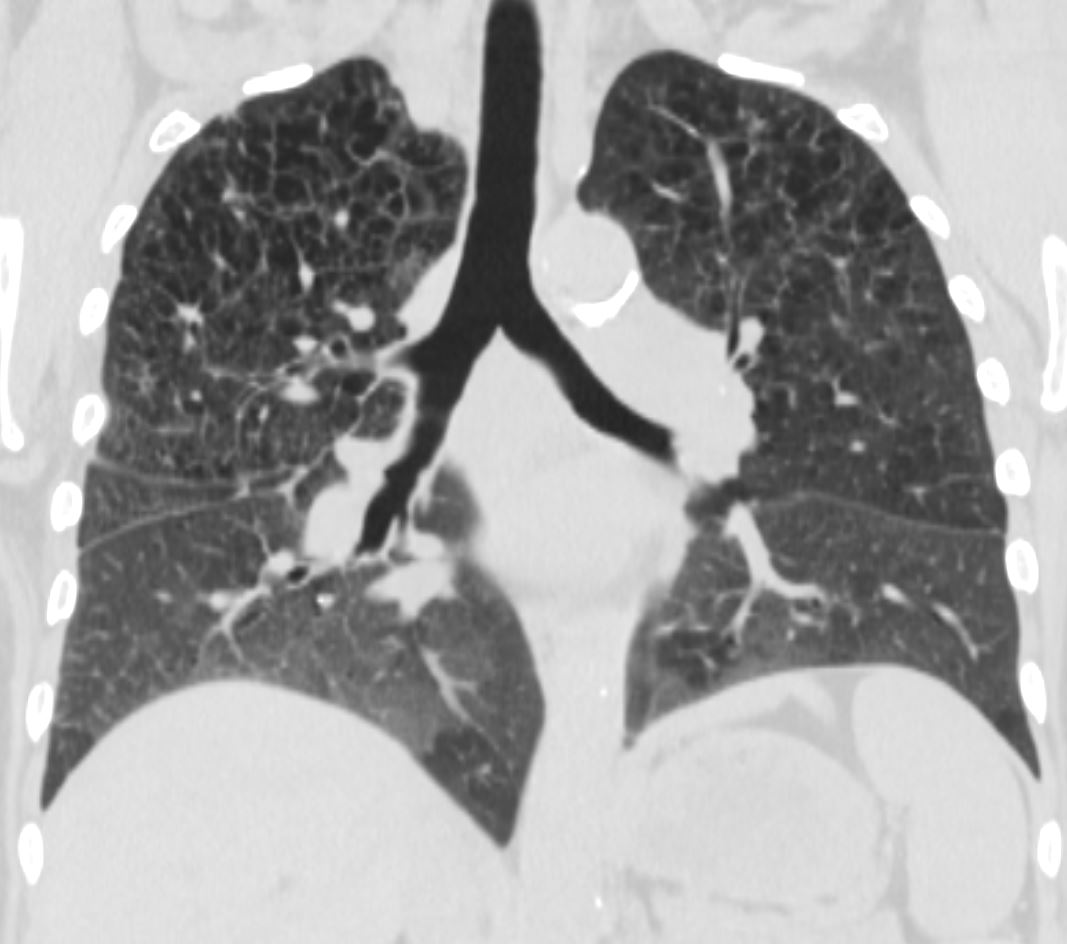
Diffuse Ground Glass Changes Lower Lung Fields and
Interlobular Septal Thickening (Crazy Paving)
51-year-old female smoker with a history of COPD asthma and pulmonary hypertension presents with progressive dyspnea. Coronal CT through the posterior lungs shows diffuse ground glass changes in the lower lobes with interlobular septal thickening
Pathology confirmed a diagnosis of DIP
Ashley Davidoff MD TheCommonVein.net 252Lu 135997
Emphysema Right Upper Lung Field-
Patchy Ground Glass Changes Lower Lung Field
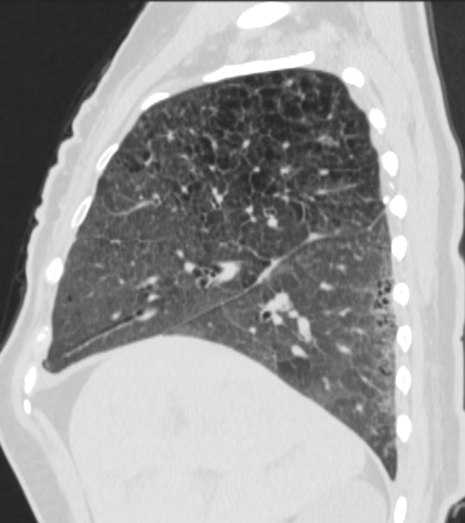
51-year-old female smoker with a history of COPD asthma and pulmonary hypertension presents with progressive dyspnea. Sagittal CT through the lateral right mid lung shows upper lobe posterior and superior segmental centrilobular emphysematous disease and ground glass changes in the anterior segment of the upper lobe and right lower lobe
Pathology confirmed a diagnosis of DIP
Ashley Davidoff MD TheCommonVein.net 252Lu 135998
Emphysema Right Upper Lung Field-
Patchy Ground Glass Changes Lower Lung Field
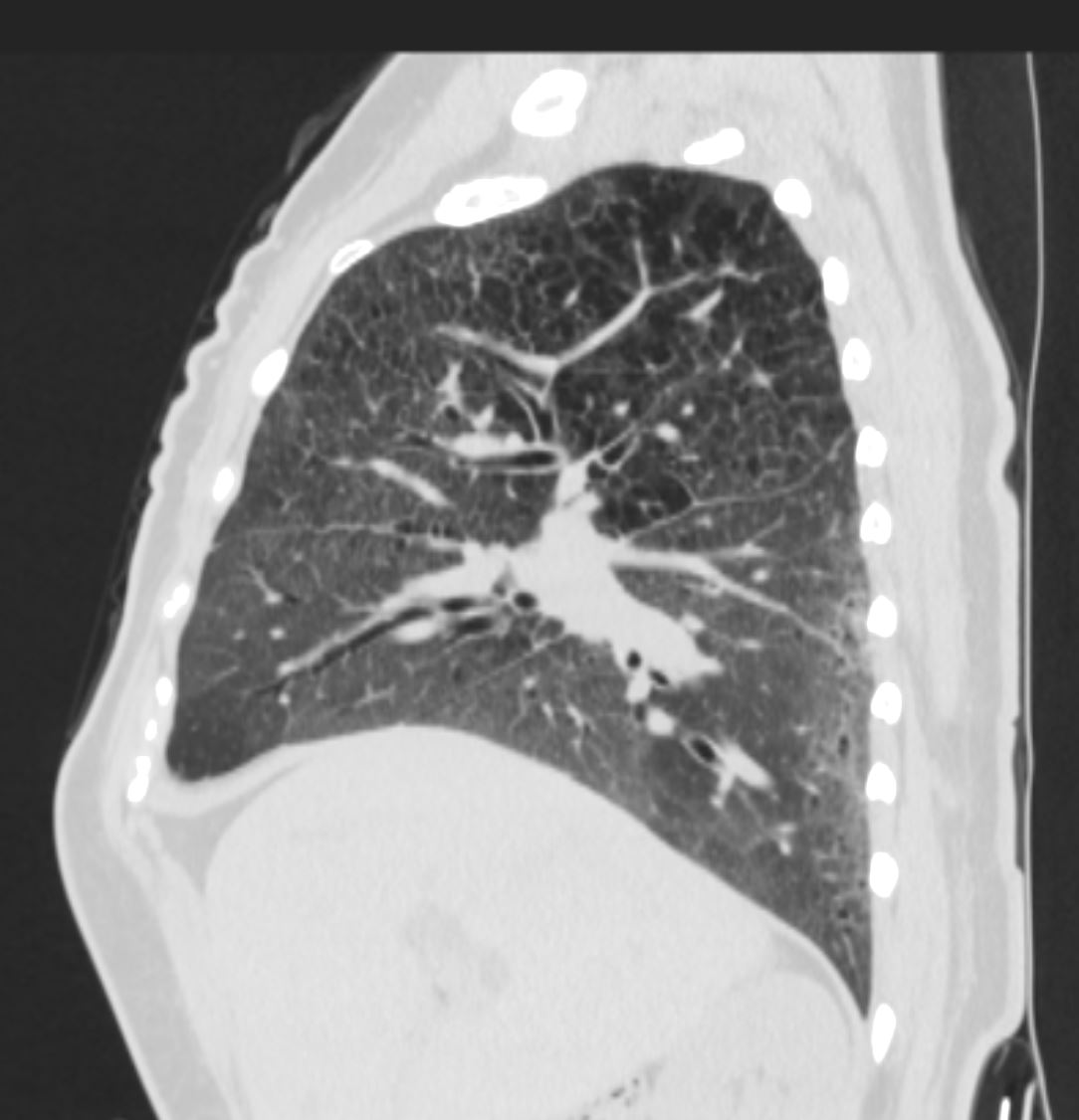
51-year-old female smoker with a history of COPD asthma and pulmonary hypertension presents with progressive dyspnea. Sagittal CT through the medial right mid lung shows upper lobe centrilobular emphysematous disease and patchy ground glass changes in the middle and right lower lobe There is thickening and irregularity of the minor fissure
Pathology confirmed a diagnosis of DIP
Ashley Davidoff MD TheCommonVein.net 252Lu 135999
Emphysema Left Upper Lung Field-
Patchy Ground Glass Changes Lower Lung Field
Thickened Septa
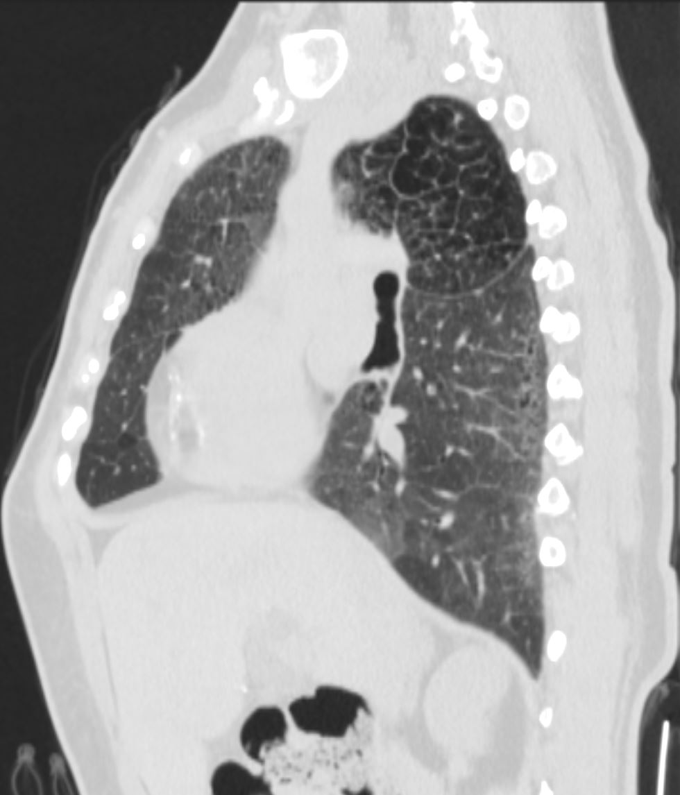
51-year-old female smoker with a history of COPD asthma and pulmonary hypertension presents with progressive dyspnea. Sagittal CT through the medial left lung shows upper lobe centrilobular emphysematous disease with enlarged secondary lobules, and patchy ground glass changes in the lingula and left lower lobe There is smooth thickening of the interlobular septa posteriorly
Pathology confirmed a diagnosis of DIP
Ashley Davidoff MD TheCommonVein.net 252Lu 136000
Emphysema Left Upper Lung Field- Ground Glass Changes Lower Lung Field with Crazy Paving Pattern
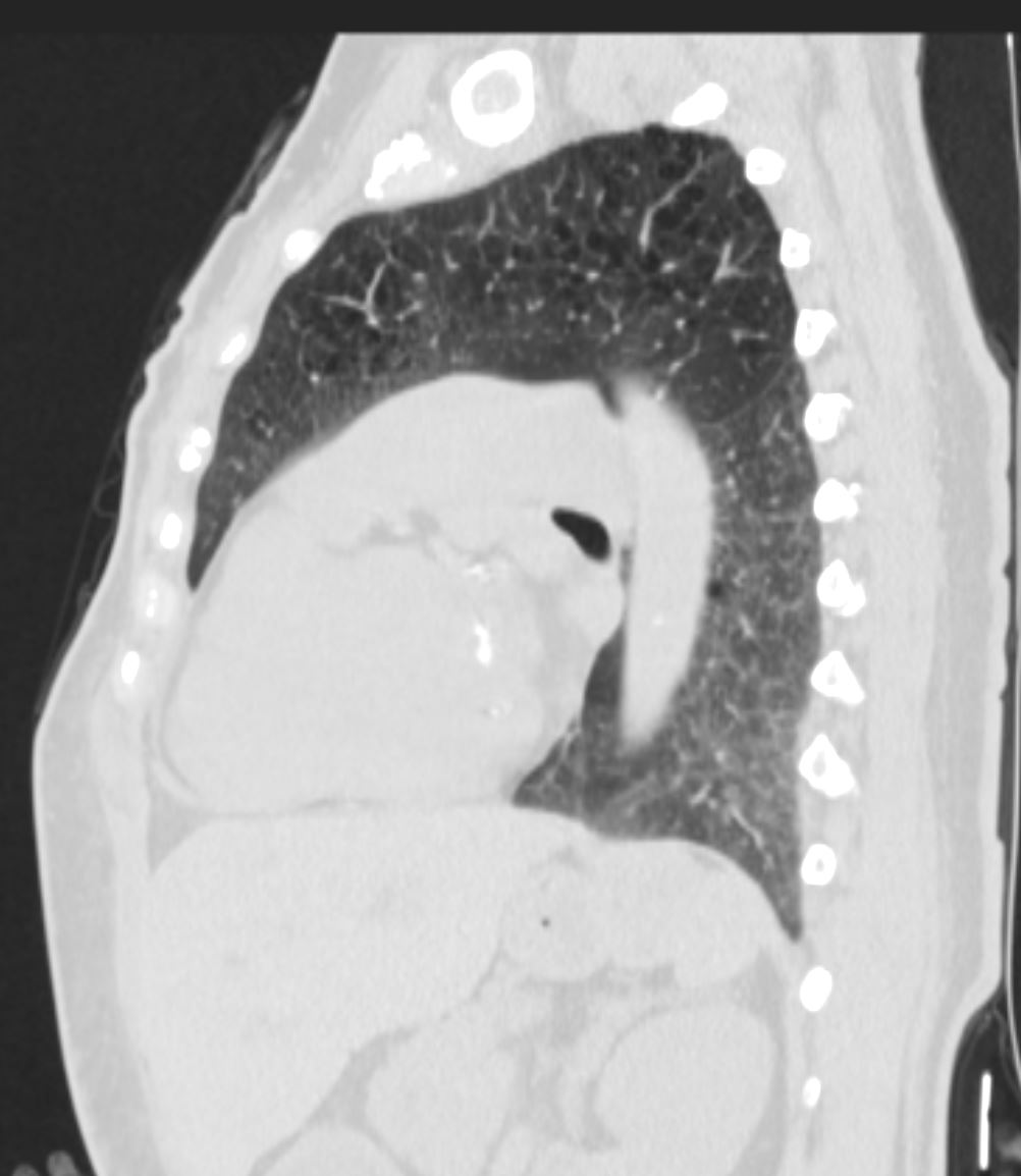
51-year-old female smoker with a history of COPD asthma and pulmonary hypertension presents with progressive dyspnea. Sagittal CT through the left lung shows upper lobe centrilobular emphysematous disease, and ground glass changes in the left lower lobe. There is smooth thickening of the interlobular septa posteriorly and a crazy paving pattern
Pathology confirmed a diagnosis of DIP
Ashley Davidoff MD TheCommonVein.net 252Lu 136001
Patchy Ground Glass Changes and Mosaicism and Air Trapping – Inspiration Expiration Views
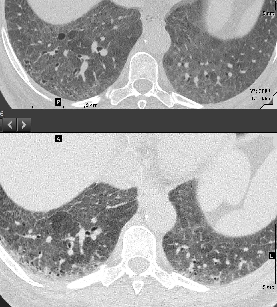
51-year-old female smoker with a history of COPD asthma and pulmonary hypertension presents with progressive dyspnea. Axial CT through the right posterior recesses at end inspiration (upper panel) and end expiration (lower panel) confirms the presence of air trapping indicating the presence of mild small airway disease
Pathology confirmed a diagnosis of DIP
Ashley Davidoff MD TheCommonVein.net 252Lu 135987
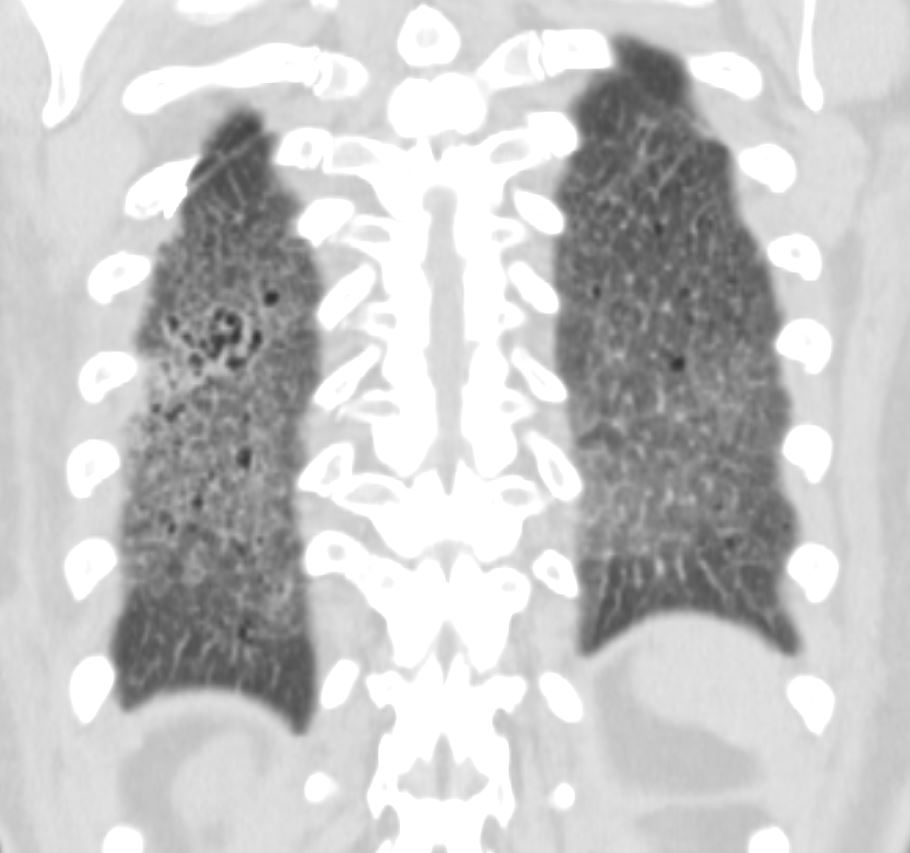
Persistence of Interstitial Changes on Prone Imaging
51-year-old female smoker with a history of COPD asthma and pulmonary hypertension presents with progressive dyspnea. Axial CT in prone position through the right posterior recesses confirms the presence of persistent interstitial lung disease
Pathology confirmed a diagnosis of DIP
Ashley Davidoff MD TheCommonVein.net 252Lu 135988
Enlarged Main Pulmonary Artery and Pulmonary Hypertension
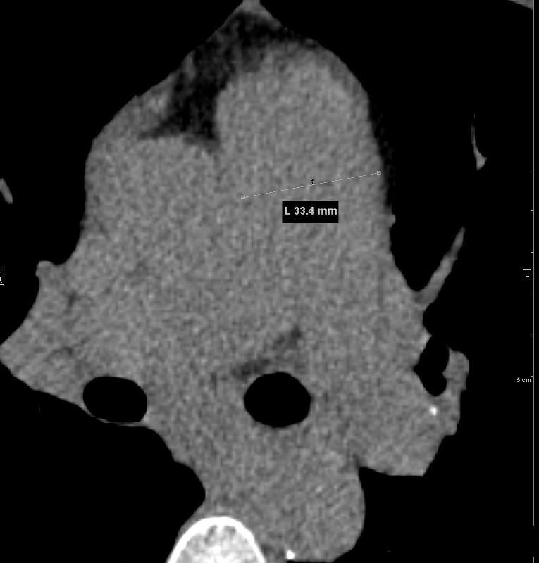
The main pulmonary artery is enlarged and measures 3.3cms indicating pulmonary hypertension
Ashley Davidoff MD TheCommonVein.net 252Lu 135986