
See also Cardiac Module of Faces of Dextrocardia
Parts
Size
Shape
Position
Character
Time Associated Findings
Infection
Inflammation
Malignancy
Mechanical
Severe Pectus Excavatum and Dextrocardia
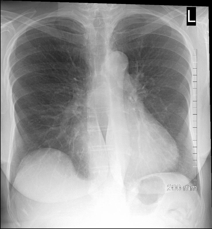
68-year-old female presents with a severe pectus excavatum. CXR in the frontal view shows horizontal orientation of the ribs and distortion of the right heart border
Ashley Davidoff MD TheCommonVein.net 270Lu 121391
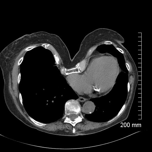
68-year-old female presents with severe pectus excavatum. CT in the axial plain shows significant depression of the sternum, and dextrocardia
Ashley Davidoff MD TheCommonVein.net 270Lu 121393
Atelectasis
Trauma
Metabolic
Circulatory-
Hemorrhage
Immune Infiltrative Idiopathic
Iatrogenic
White Out S/P Left Pneumonectomy
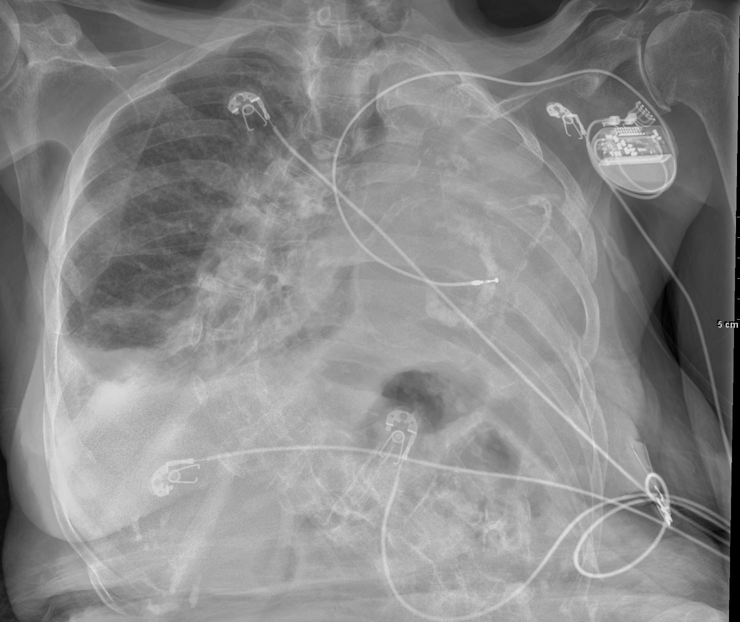
Frontal CXR of a 98-year-old woman showing a left sided white out secondary to a pneumonectomy. The soft tissue structures of the mediastinum have all shifted into the left hemithorax accounting for the white out. The calcified mitral annulus, calcified coronary arteries and right ventricle (RV pacemaker lead) confirm the diagnosis of acquired dextrocardia. There is hyperinflation of the right lung which crosses the midline associated with a small effusion . A significant dextro-thoracic scoliosis with a compensatory levoscoliosis of the lumbar spine is present
Ashley Davidoff MD TheCommonVein.net 269Lu 136234
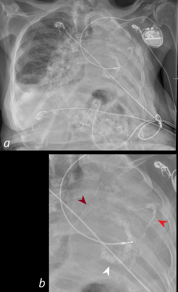
Frontal CXR of a 98-year-old woman showing a left sided white out secondary to a pneumonectomy. The soft tissue structures of the mediastinum have all shifted into the left hemithorax accounting for the white out. The calcified mitral annulus (b, white arrowhead), calcified right coronary artery – RCA (b, maroon arrowhead) and left anterior descending (LAD) – (b, bright red arrowhead) and right ventricle (RV pacemaker lead) confirm the diagnosis of acquired dextrocardia. There is hyperinflation of the right lung which crosses the midline associated with a small right effusion. A significant dextro-thoracic scoliosis with a compensatory levoscoliosis of the lumbar spine is present
Ashley Davidoff MD TheCommonVein.net 269Lu136234cL
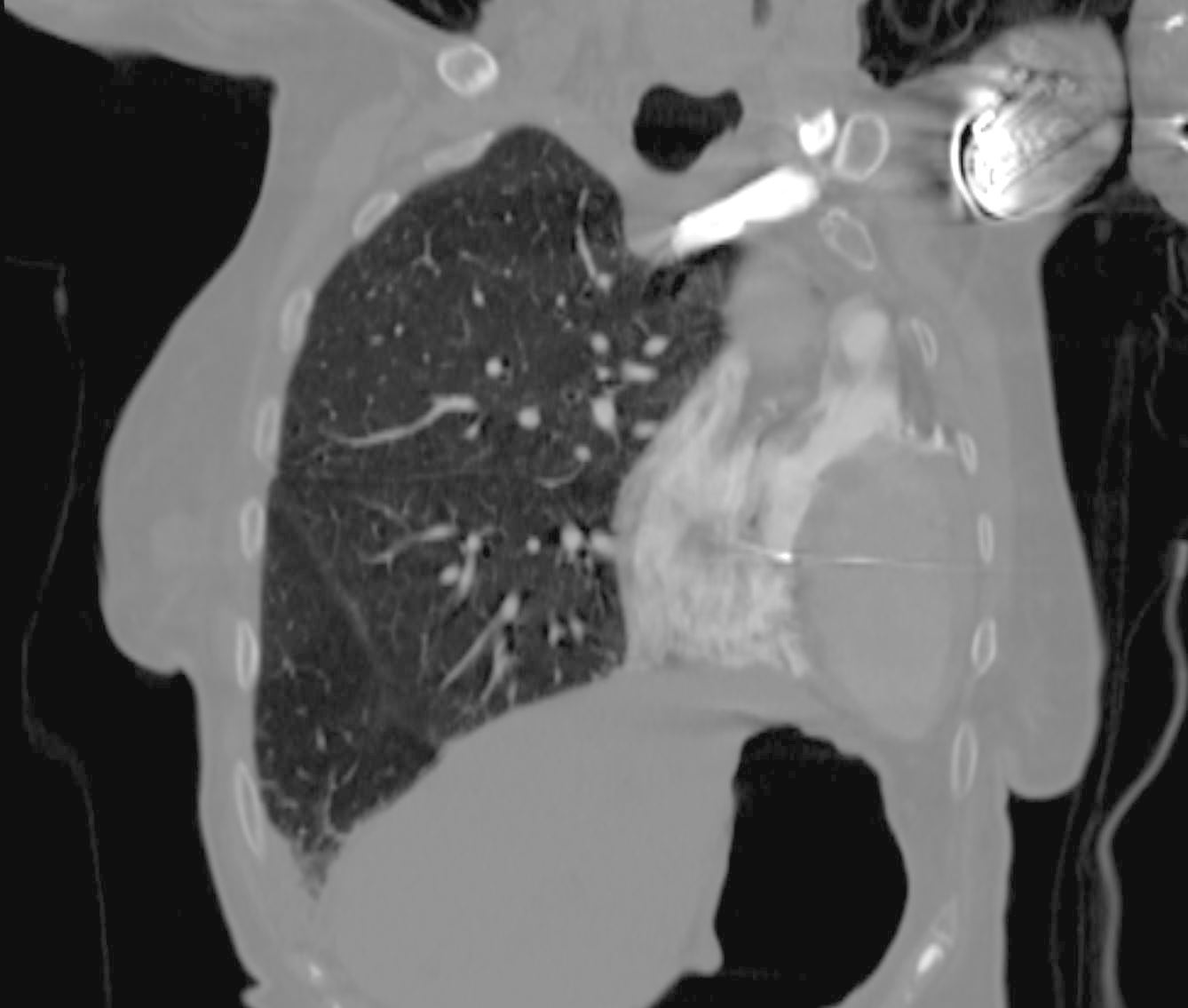
Coronal CT of a 98-year-old woman showing a left sided white out on CXR secondary to a pneumonectomy shows a leftward shift of cardiac structures including the contrast filled right ventricle (RV) and the oval shaped left ventricle (LV) which occupy the left hemithorax. The tip of the pacing lead is noted in the RV septum. There is hyperinflation of the right lung which crosses the midline associated with a small right effusion.
Ashley Davidoff MD TheCommonVein.net 269Lu 136235b