
Bronchial Disease
Mild Disease
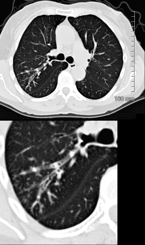
ABPA 3 years prior
CT Scan RUL
72 year old female with asthma presented 3 years prior with acute dyspnea.
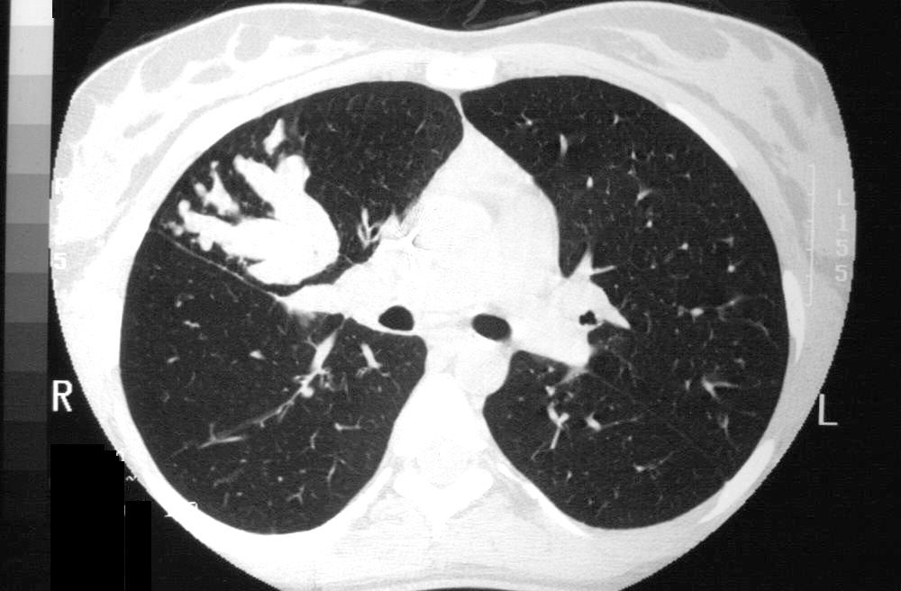
CT in the axial plane shows thickening of the segmental and subsegmental airways of the posterior segment of the right upper lobe with mucoid impaction
Ashley Davidoff MD TheCommonVein.net 294Lu 135116b.0001c
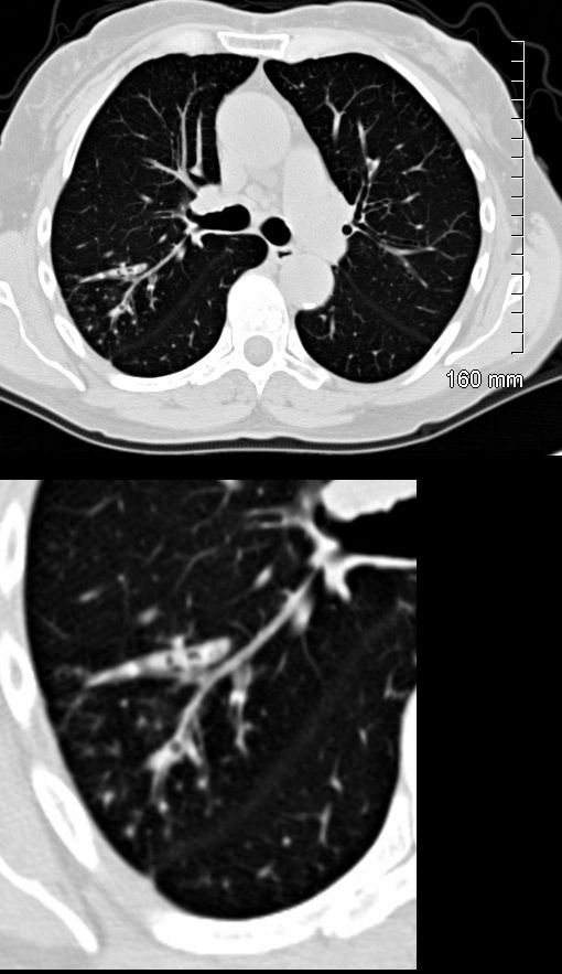
ABPA 3 years prior
CT Scan RUL
72 year old female with asthma presented 3 years prior with dyspnea.
CT in the axial plane shows thickening of the segmental and subsegmental airways of the posterior segment of the right upper lobe with mucoid impaction
Ashley Davidoff MD TheCommonVein.net 294Lu 135116bc.0001
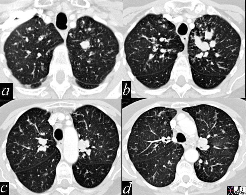
Finger in Glove Bronchiectasis and Tree in Bud
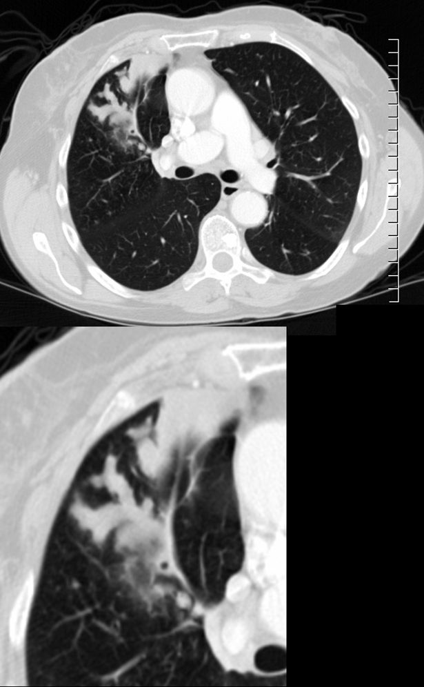
CT scan with contrast shows finger in glove appearance of the anterior segmental airways of the right upper lobe ) with a focal region of subsegmental atelectasis medially. The finger in glove sign results thick, mucus plugs within the bronchi due to the exaggerated inflammatory and immune response caused by Aspergillus fumigatus leading to airway inflammation, mucus production, bronchial wall thickening, and bronchiectasis.
Ashley Davidoff MD The CommonVein.net 294Lu 117966c
CT scan through the chest shows medium sized bronchi, bronchioles and small airways impacted with fluid. This collage is presented to reveal tree in bud changes resulting from impaction in the smaller terminal bronchioles and respiratory units. The tree-in-bud pattern also results in small centrilobular nodules connected to multiple branching linear structures of similar caliber from a single stalk. Originally it was felt to result from endobronchial spread of Mycobacterium tuberculosis, but is is now recognized in diverse entities including peripheral airway diseases caused by infection (bacterial, fungal, viral, or parasitic), congenital disorders, idiopathic disorders (obliterative bronchiolitis, pan bronchiolitis), aspiration or inhalation of foreign substances, immunologic disorders, connective tissue disorders and peripheral pulmonary vascular diseases such as neoplastic pulmonary emboli.
In this case there are also dilated medium sized airways, impacted with soft tissue characteristic of the finger in glove sign and most likely due to allergic bronchopulmonary aspergillosis (ABPA)
Ashley Davidoff MD Ashley Davidoff MD TheCommonVein.net
221Lu 47113c01
47114c01 bronchi lungs fx dilated enlarged impacted with sft tissue finger in glove dx allergic bronchopulomonary aspergillosis ABPA aspergillus dx infection inflammation CTscan Davidoff MD 221Lu 47114c01
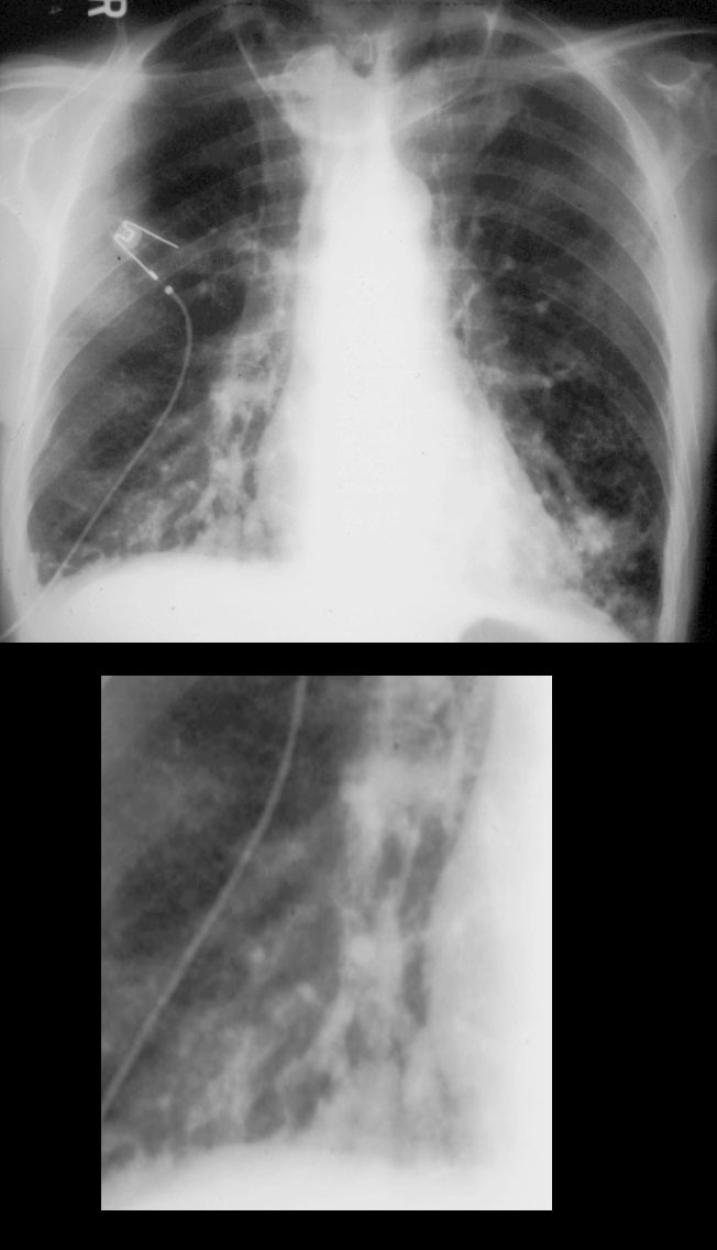
60 year old male with history of asthma, allergic bronchopulmonary aspergillosis (ABPA)
CXR suggests hyperinflation, with tubular ectasia and soft tissue prominence of the bronchovascular bundle in the right lobe (magnified in lower image)
Ashley Davidoff TheCommonVein.net 221Lu 14644c
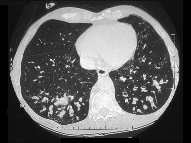
60 year old male with history of asthma, allergic bronchopulmonary aspergillosis (ABPA)
CT scan shows bibasilar bronchiectasis and soft tissue/fluid impaction of the bronchovascular bundles
Ashley Davidoff TheCommonVein.net 221Lu 14645
60 year old male with history of asthma, allergic bronchopulmonary aspergillosis (ABPA)
CT scan shows upper lobe bronchiectasis and soft tissue/fluid impaction in the left upper lobe reminiscent of the finger in glove appearance of ABPA
Ashley Davidoff TheCommonVein.net 221Lu 47112
19 year old female with cystic fibrosis and bronchiectasis
CT scan through the upper lung fields shows mucin filled subsegmental bronchi of the right upper lobe with morphology reminiscent of the “finger in glove” sign
Courtesy Priscilla Slanetz MD MPH TheCommonVein.net

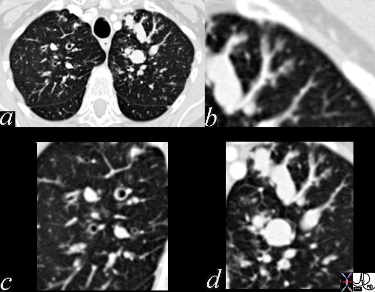
CT – with Noted Tubular Structures in the Upper Lobes
77 year old female with history of asthma, allergic bronchopulmonary aspergillosis (ABPA) and COPD
CXR shows prominent bronchovascular bundles in the upper lung fields (green arrowheads a, and b) . CT shows fluid filled bronchiectatic airways (green arrowheads in image d, which is a magnified image of c) reminiscent of the finger in glove appearance of ABPA)
Ashley Davidoff TheCommonVein.net 227Lu 135161cL03
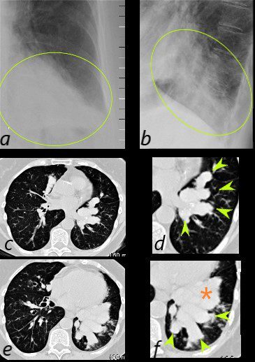
77 year old female with history of asthma, allergic bronchopulmonary aspergillosis (ABPA) and COPD
CXR shows LLL infiltrate in the PA (green oval in a) and lateral views (green oval in d and f) which reflect mucus filled bronchiectatic airways magnified image s of the CT scan of the LLL ) reminiscent of the finger in glove appearance of ABPA. There is a LL infiltrate possibly atelectatic (orange asterisk)
Ashley Davidoff TheCommonVein.net 227Lu 135161cL03
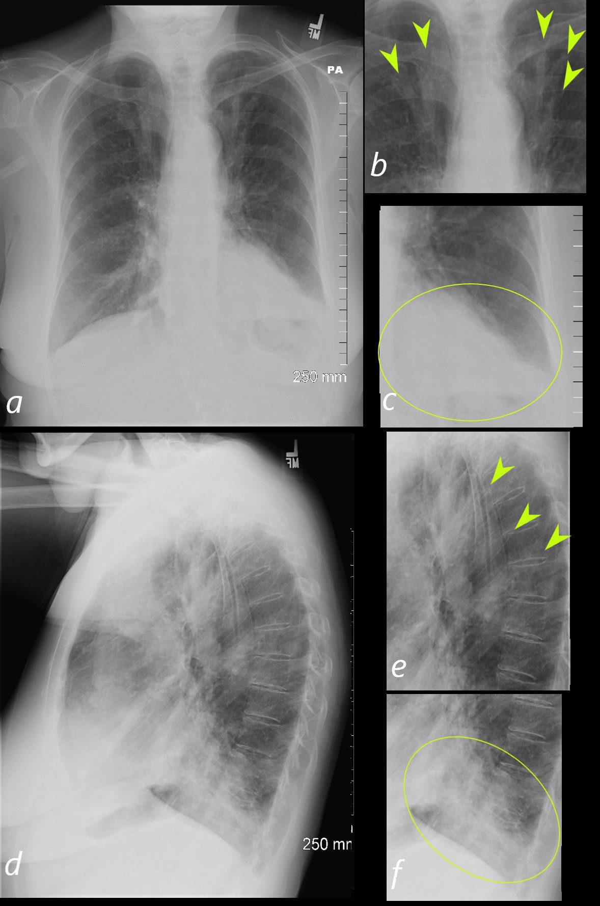
77 year old female with history of asthma, allergic bronchopulmonary aspergillosis (ABPA) and COPD
CXR in the PA projection(a) shows prominent tubular structures in the in the upper lung fields (green arrowheads in b) more prominent than the expected blood vessels seen in the hila. There is an infiltrate in the LLL with silhouetting of the left hemidiaphragm (green oval in c)
On the lateral examination (d) there is a suggestion of hyperinflation, and the tubular structures noted in b, are also appreciated, but are less obvious (green arrowheads in e). The consolidation in the left lower lobe is better appreciated (outlined by the green oval in f)
Ashley Davidoff TheCommonVein.net 227Lu 135161cL
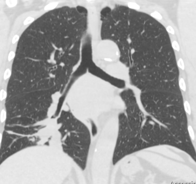
56-year-old male presents with chronic cough dyspnea and weight loss. CT scan in coronal projection shows an appearance reminiscent of finger in glove in the right lower lobe. There s segmental and subsegmental thickening of the airways in the upper lobes, and paraseptal emphysema. Micronodules in the upper lobes suggest smoker’s bronchiolitis. The subcarinal esophageal mass was diagnosed as a leiomyoma, Pathology of the right lower process was a squamous cell carcinoma
Ashley Davidoff MD TheCommonVein.net 267Lu 136219
Parenchymal Disease
Atelectasis
Right Upper Lobe Atelectasis
Allergic Bronchopulmonary Aspergilloses (ABPA) and Atelectasis and Luftsichel Sign
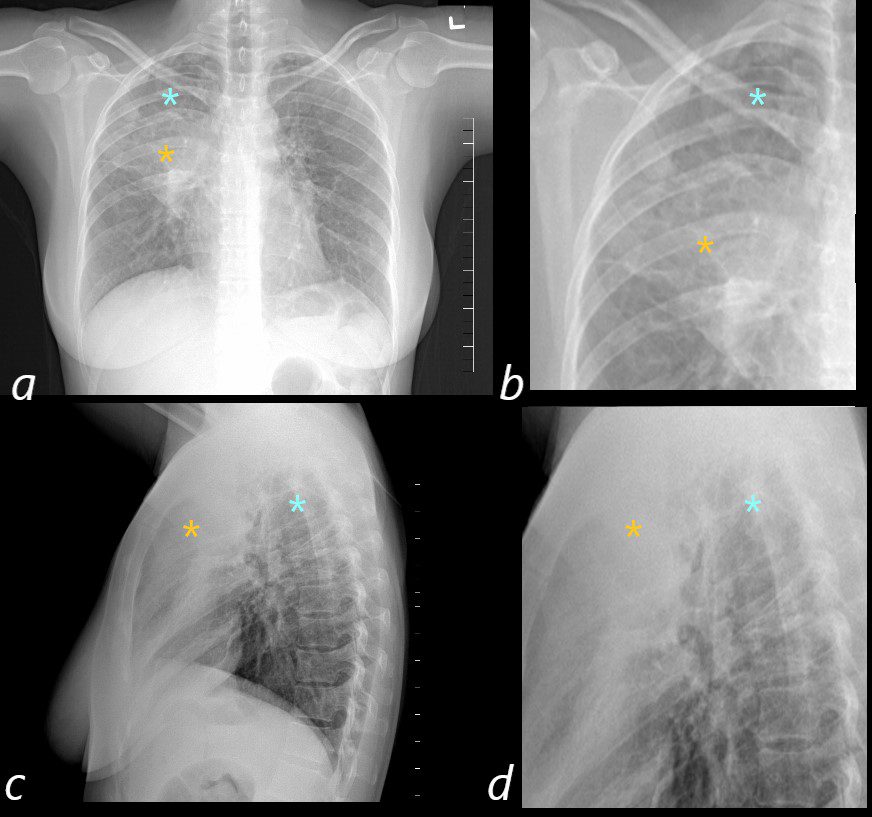
50 year old female with a history of asthma represents with productive cough. CXR in the PA projection (a magnified in b) shows an ill defined density in the right upper lobe of the lung (orange asterisk) and a relatively lucent right apex (blue asterisk Luftsichel sign)) with only minimal elevation of the right hemidiaphragm and minimal rightward mediastinal deviation . The lateral CXR shows a poorly defined density of the atelectatic RUL (orange asterisks c and D) filling in the retrosternal air space, with the hyperinflated right lower lobe reaching the right apex (orange asterisks c and d) . The significantly hyperinflated right lower lobe likely reduces the overall volume loss and hence the subtle compensatory changes of the elevated right hemidiaphragm and the mediastinal shift.
Ashley Davidoff MD TheCommonVein.net
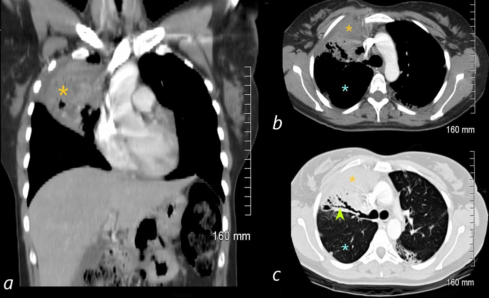
ABPA
The coronal image is relatively anterior and hence presents as a dense consolidation of atelectasis (orange asterisk, a) In the axial images the hyperinflated RLL is seen posteriorly (teal asterisks in b and c) The region of varicose bronchiectasis is noted posteriorly (lime green arrow, c) When the net density of these 3 findings (consolidation, hyperinflated RLL and bronchiectasis in the LUL) are superimposed on CXR they present with a difficult interpretation since it is the overall net density that gets reflected. The CT scan helps us understand the findings
Ashley Davidoff MD TheCommonVein.net
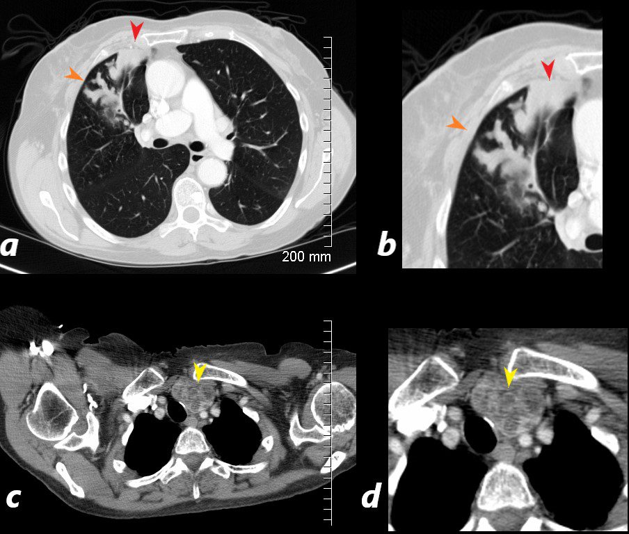
CT scan with contrast shows finger in glove appearance of the anterior segmental airways of the right upper lobe (orange arrowheads a, b) with a focal region of subsegmental atelectasis (red arrow head a,b).
The lower panel shows a cluster of low density lymph nodes in the anterior mediastinum in the retro-clavicular region (yellow arrowheads c,d) .
Ashley Davidoff MD The CommonVein.net

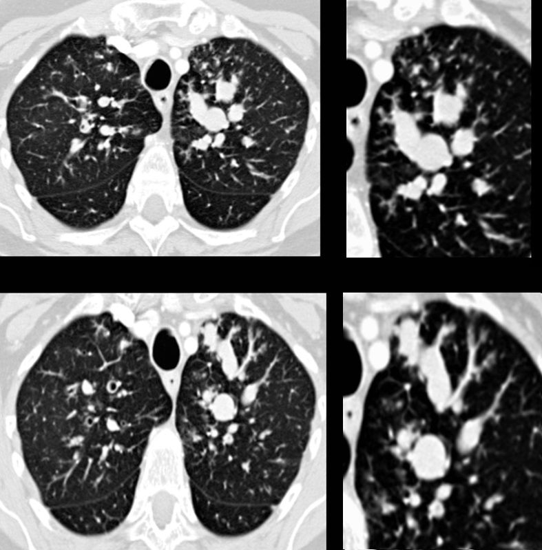
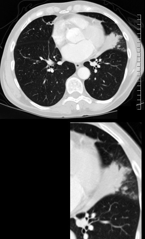
CT Scan RUL
72 year old female with asthma presented 2 years prior with acute dyspnea.
CT in the axial plane shows subsegmental atelectasis of an anterior subsegment of the RUL with impaction and bronchiectasis of visualized anterior segmental airways and a posterior subsegmental airway
Ashley Davidoff MD TheCommonVein.net 294Lu 135116c.0017
CT scan with contrast shows finger in glove appearance of the anterior segmental airways of the right upper lobe ) with a focal region of subsegmental atelectasis medially. The finger in glove sign results thick, mucus plugs within the bronchi due to the exaggerated inflammatory and immune response caused by Aspergillus fumigatus leading to airway inflammation, mucus production, bronchial wall thickening, and bronchiectasis.
Ashley Davidoff MD The CommonVein.net 294Lu 117966c
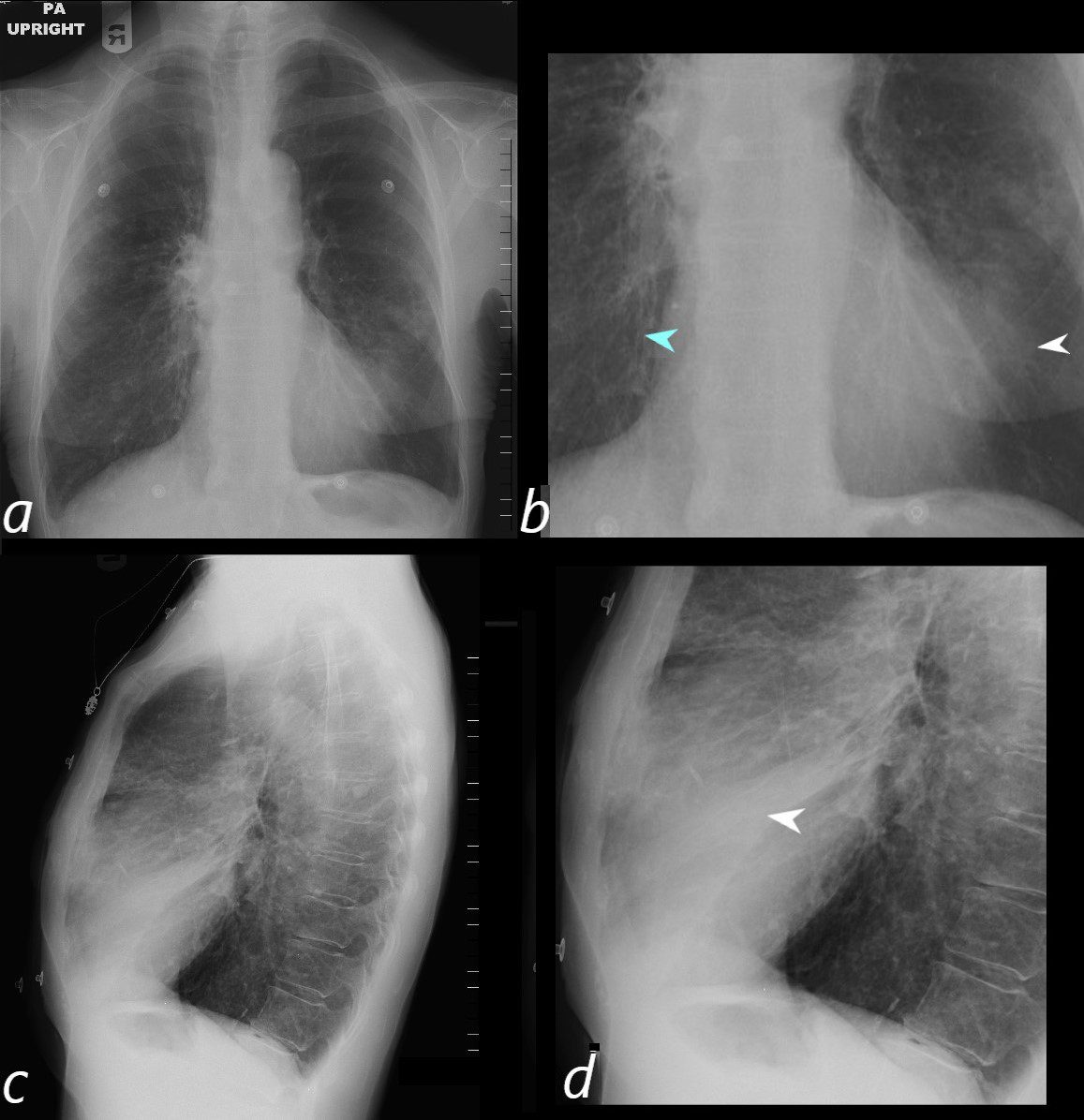
72 year old female with asthma presents with a cough and dyspnea
CXR in frontal and lateral projection show hyperinflated lung fields characterized by flattening of the hemidiaphragms, pectus carinatum and a barrel chest. There is partial silhouetting of the left heart border resulting from segmental consolidation of the medial segment of the ligula (b, white arrowhead) and confirmed on the lateral exam (d, white arrowhead). There is thickening of the airways subtending the right lower lobe (b, teal arrowhead).
Ashley Davidoff MD TheCommonVein.net 294Lu 117965cL
ABPA Current CT Scan
72 year old female with asthma with cough and chronic dyspnea.
CT in the axial plane shows subsegmental atelectasis of the lingula likely caused by mucoid impaction
Ashley Davidoff MD TheCommonVein.net 294Lu 117969c
Right Middle Lobe
Segmental Atelectasis
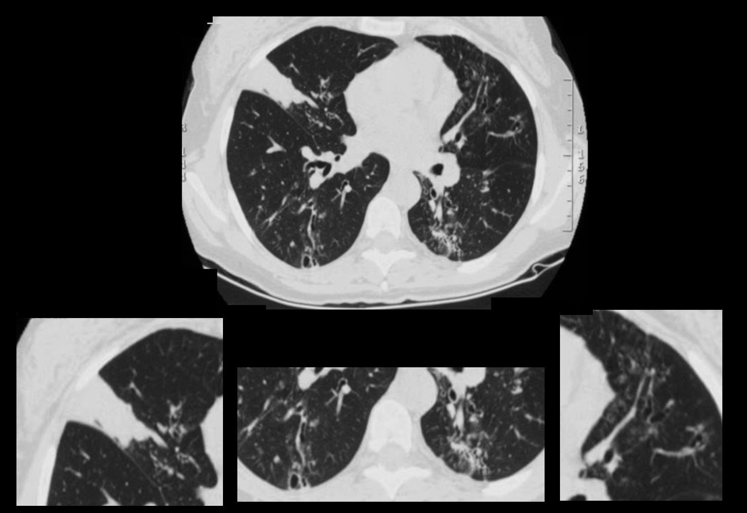
CT Allergic Bronchopulmonary Aspergillosis (ABPA)
48 year old female with a history of asthma presents with productive cough. CT scan 18 months prior confirms atelectasis in the middle lobe (upper panel and right lower panel) . There is diffuse mild multicentric foci of bronchial wall thickening in the segmental and subsegmental airways of the middle lobe, lingula and the lower lobes bilaterally (upper panel magnified in lower 3 panels).
Ashley Davidoff MD TheCommonVein.net
Pneumonia
Lymphadenopathy
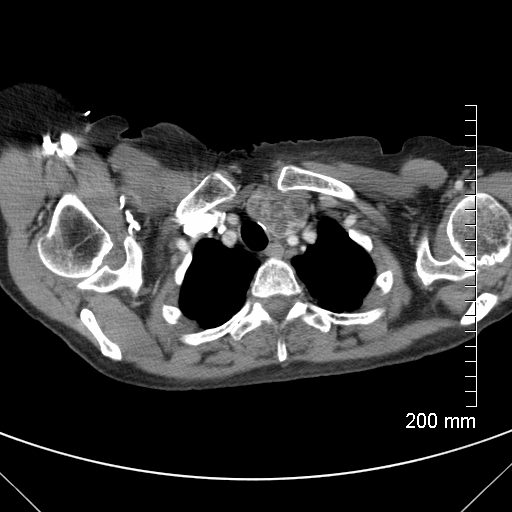
CT scan with contrast shows a cluster of low density lymph nodes in the anterior mediastinum in the retro-clavicular region
Ashley Davidoff MD The CommonVein.net 294Lu 117970
CT scan with contrast shows finger in glove appearance of the anterior segmental airways of the right upper lobe (orange arrowheads a, b) with a focal region of subsegmental atelectasis (red arrow head a,b).
The lower panel shows a cluster of low density lymph nodes in the anterior mediastinum in the retro-clavicular region (yellow arrowheads c,d) .
Ashley Davidoff MD The CommonVein.net 294LU 117972b