- Lipoid pneumonia is an uncommon disease
- caused by the presence of lipid in the alveoli.
- Two types
- exogenous (exogenous lipoid pneumonia) or
- inhaled nose drops with an oil base, or
- accidental inhalation of cosmetic oil.
- Amiodarone is an anti-arrythmic known to cause this condition.
- Fire breather’s pneumonia
- from the inhalation of hydrocarbon fuel
- Gastroesophageal reflux.
- endogenous/idiopathic (endogenous lipoid pneumonia)
- obstructed airway is, it is often the case that
- lipid-laden macrophages and giant cells fill the lumen
- distal to the obstruction,
- of the disconnected airspace.
- exogenous (exogenous lipoid pneumonia) or
- Two types
- Resulting in
- Clinicically
- insidious onset
- dyspnea and/or cough.
- insidious onset
- Imaging
- consolidations,
- ground-glass attenuation,
- airspace nodules and
- ‘crazy-paving’ pattern. However, the radiological
-

Lipoid Pneumonia
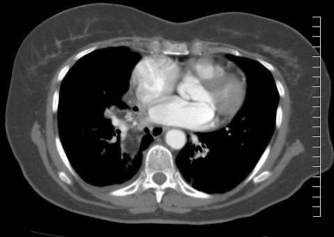
82 year old female who treated her colds with Vaseline aspiration and subsequently developed lipoid pneumonia
CT scan shows a right lower lobe infiltrate involving the superior segment
Ashley Davidoff MD TheCommonVein.netLipoid Pneumonia
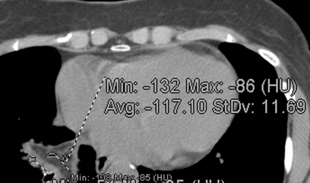
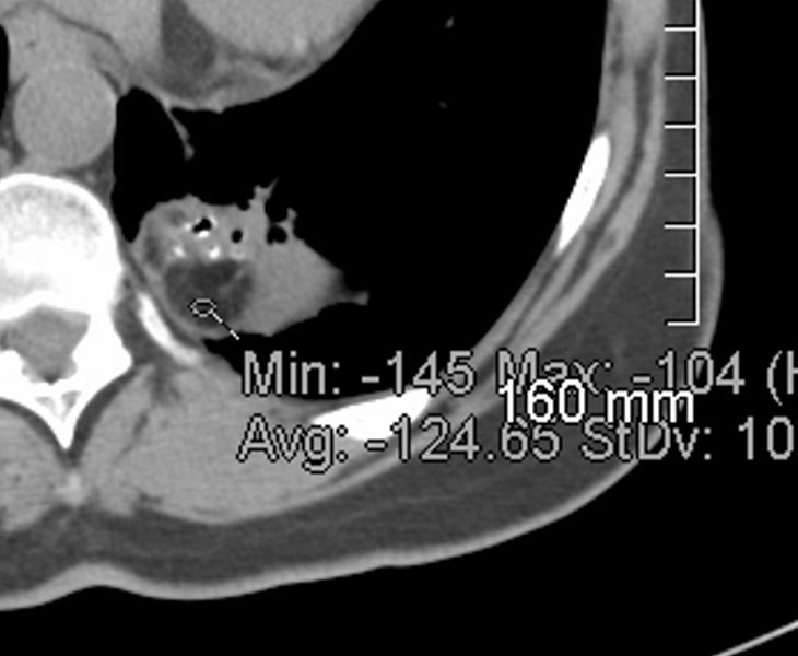
82 year old female with lipoid pneumonia from vaseline aspiration
Density measurements confirm an average of -117 Hounsfield units
Ashley Davidoff MD TheCommonVein.netLipoid Pneumonia
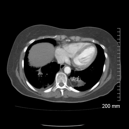
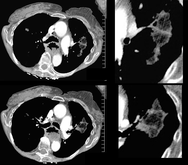
82 year old female who treated her colds with Vaseline aspiration and subsequently developed lipoid pneumonia
CT scan shows a left lower lobe fat containing infiltrate involving the superior segment
Ashley Davidoff MD TheCommonVein.netLipoid Pneumonia
82 year old female with lipoid pneumonia from vaseline aspiration
Density measurements confirm an average of -124 Hounsfield units
Ashley Davidoff MD TheCommonVein.net
- Pathologically,
- lipid-laden macrophages.
-
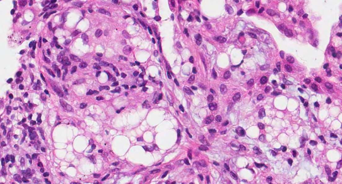
Lipid laden macrophages in a patient with lipoid pneumonia
Courtesy pathologyoutlines.com/
- Lab
- Lipid-laden macrophages in respiratory samples from
- sputum,
- bronchoalveolar lavage fluid or
- fine-needle aspiration cytology/biopsy from lung lesions.
- Lipid-laden macrophages in respiratory samples from
- Treatment protocols for this illness are
- poorly defined.
- Clinicically
A second Patient with achalasia and chronic aspiration
78 year old female with achalasia and likely chronic aspiration
There is a fatty infiltrate in the left upper lobe.
Note the dilated esophagus with air fluid levels
Ashley Davidoff MD TheCommonVein.net
134411c
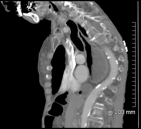
78 year old female with achalasia and likely chronic aspiration
Note the distended esaophagus with air fluid level
Ashley Davidoff MD TheCommonVein.net
-
-
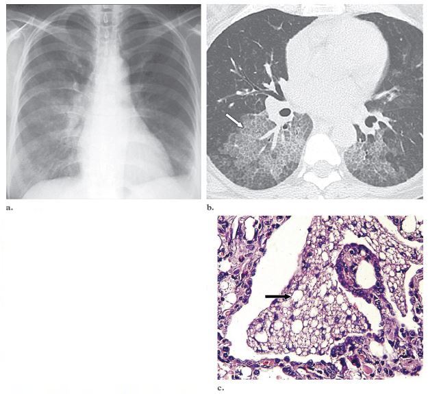
Lipoid pneumonia in a 64-year-old
woman with a 20-year history of scleroderma who presented with progressive dyspnea and a dry cough.
(a) Posteroanterior chest radiograph shows bilateral,
asymmetric, scattered areas of increased opacity in the
air space, which have a predominantly perihilar and
basal distribution. (b) High-resolution CT scan shows
geographic ground-glass attenuation in association with
interlobular thickening and intralobular lines (arrow).
The results of bronchoalveolar lavage and transbronchial biopsy were nondiagnostic. (c) Photomicrograph
(original magnification, 250; hematoxylin-eosin
stain) of a specimen from open lung biopsy shows numerous lipid-laden macrophages that fill and distend
the alveoli (arrow) and interstitium.
Rossi, S.E et al “Crazy-Paving” Pattern at Thin-Section CT of the Lungs: RadiologicPathologic Overview Radiographics Volume 23 – Number 6, 2003
-