
74 year old alcoholic male with bilateral aspiration
74 year old male alcoholic with bilateral basilar lobar atelectasis caused by bilateral aspiration
CT scan near the AP window shows reactive lymph nodes, gynecomastia, an infiltrate in the superior segment of the right lobe of the lung and bilateral pleural effusions
Ashley Davidoff MD TheCommonVein.net RnD image
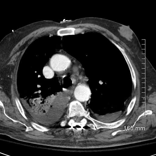
74 year old male alcoholic with bilateral basilar lobar atelectasis caused by bilateral aspiration
CT scan at the level of the carina shows right main bronchus filled with aspirated content associated with an infiltrate in the right lobe of the lung with both focal consolidations and ground glass infiltrates and bilateral pleural effusions
Ashley Davidoff MD TheCommonVein.net RnD image
Airless Lungs with Airways Filled with Aspirated Material
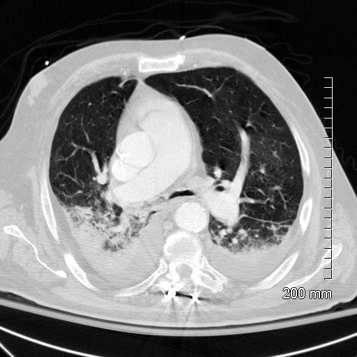
74 year old male alcoholic with bilateral basilar lobar atelectasis caused by bilateral aspiration
CT scan shows airless lower lobes with small bilateral effusions.
Ashley Davidoff MD TheCommonVein.net
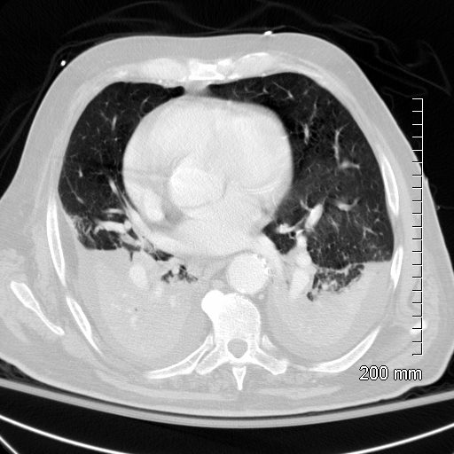
74 year old male alcoholic with bilateral basilar lobar atelectasis caused by bilateral aspiration
CT scan shows airless lower lobes with small bilateral effusions.
Ashley Davidoff MD TheCommonVein.net RnD image
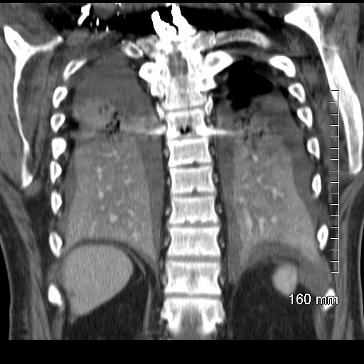
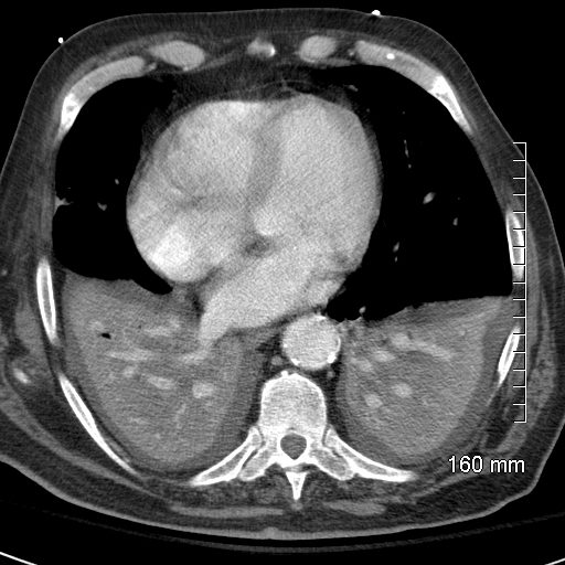
74 year old male alcoholic with bilateral basilar lobar atelectasis caused by bilateral aspiration
CT scan shows airless lower lobes with small bilateral effusions.
Ashley Davidoff MD TheCommonVein.net
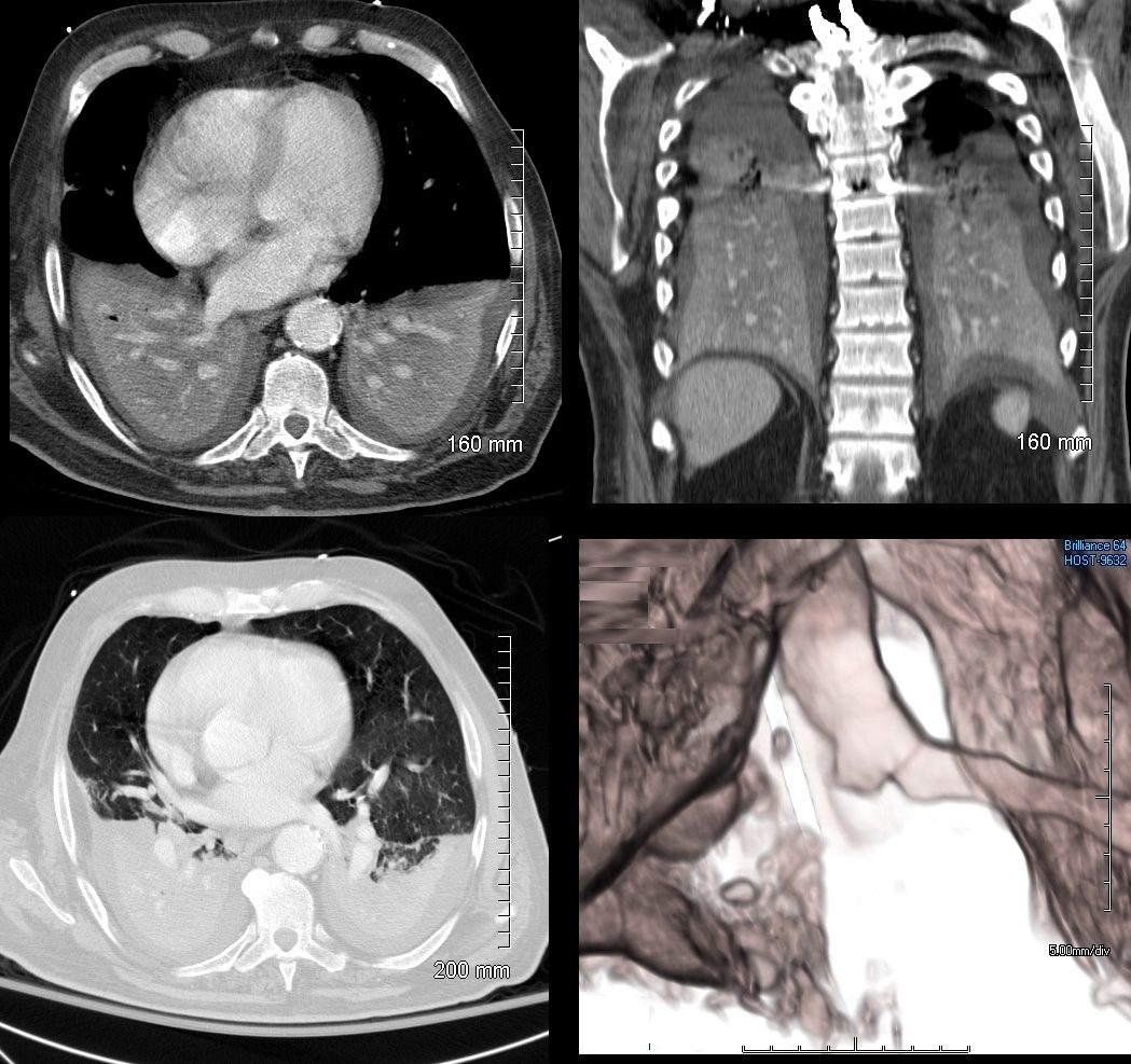
74 year old male alcoholic with bilateral basilar lobar atelectasis caused by bilateral aspiration
CT scan shows airless lower lobes with small bilateral effusions. 3D reconstruction shows total obstruction of the right mainstem bronchus, and patent proximal mainstem bronchus
Ashley Davidoff MD TheCommonVein.net
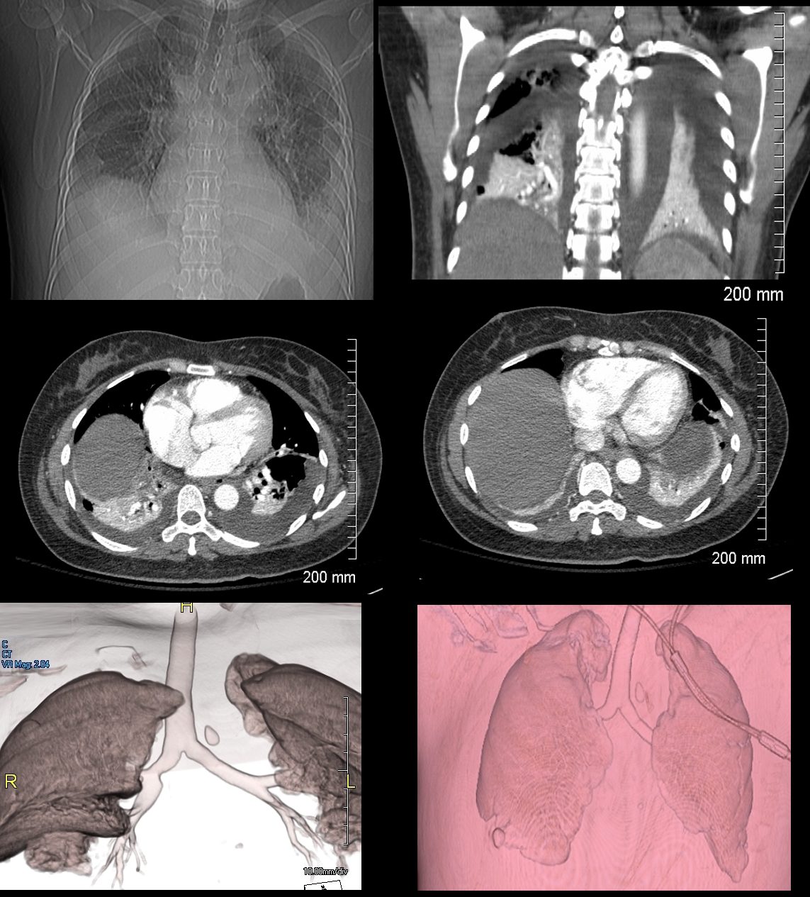
46-year-old female presents with a dyspnea and a cough. Imaging of the chest shows cardiomegaly with bilateral moderate sized pleural effusion with crescentic region of compressive atelectasis noted on the axial images at the bases and crowding of the bronchovascular bundles best evaluated on the coronal image. The 3D reconstructions show functionally “bare” lower lobe segmental airways.
Ashley Davidoff MD TheCommonVein.net