
Infection Inflammation Malignancy Mechanical/Atelectasis Trauma Metabolic Circulatory- Hemorrhage Immune Infiltrative Idiopathic Iatrogenic Idiopathic
Infection TB
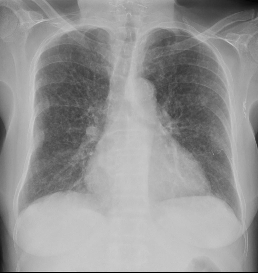
60-year-old immunocompromise female presents with a cough and weight loss CXR shows a diffuse miliary pattern. Final diagnosis was mycobacterium tuberculosis. Associated findings include healed right sided rib fractures and surgical clips in the left axilla
Ashley Davidoff MD TheCommonVein.net 265Lu 136197
CT Miliary Tuberculosis
Nodules – Bronchovascular Bundle, Centrilobular, Fissural and Pleural Distribution
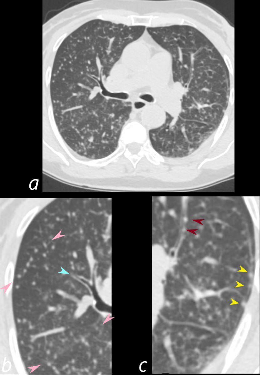
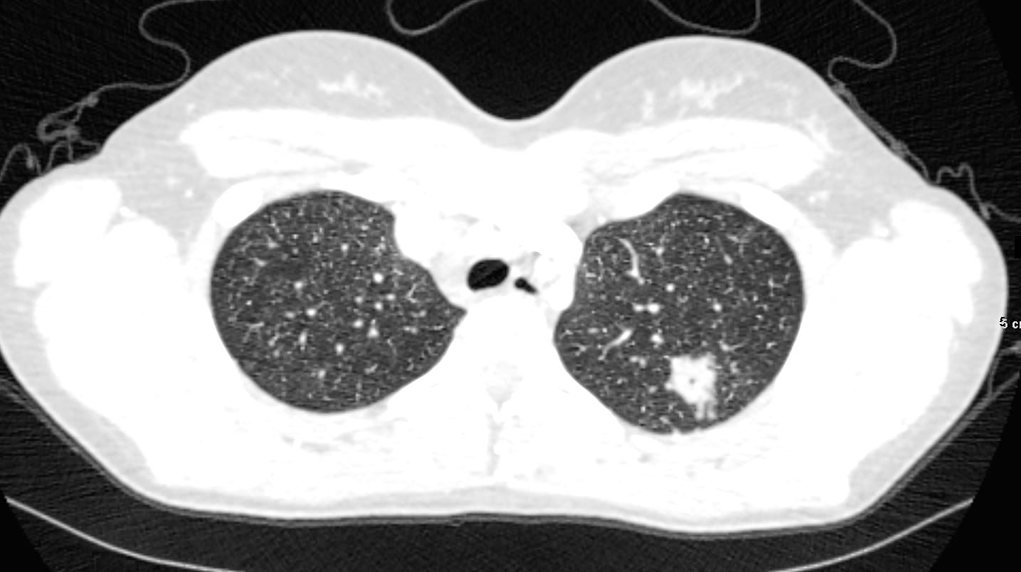
60-year-old immunocompromised female presents with a cough and weight loss. Axial CT shows miliary nodules throughout both lung fields. Some of these nodules are centrilobular or distributed along the bronchovascular bundles (c, maroon arrowheads) and others are fissural based (b, pink arrowheads) and along the pleura (yellow arrowheads suggesting at least a lymphatic distribution. There is bronchial wall thickening (b teal arrowhead). She responded well to treatment and final diagnosis was mycobacterium tuberculosis.
Ashley Davidoff MD TheCommonVein.net 265Lu 136202cL
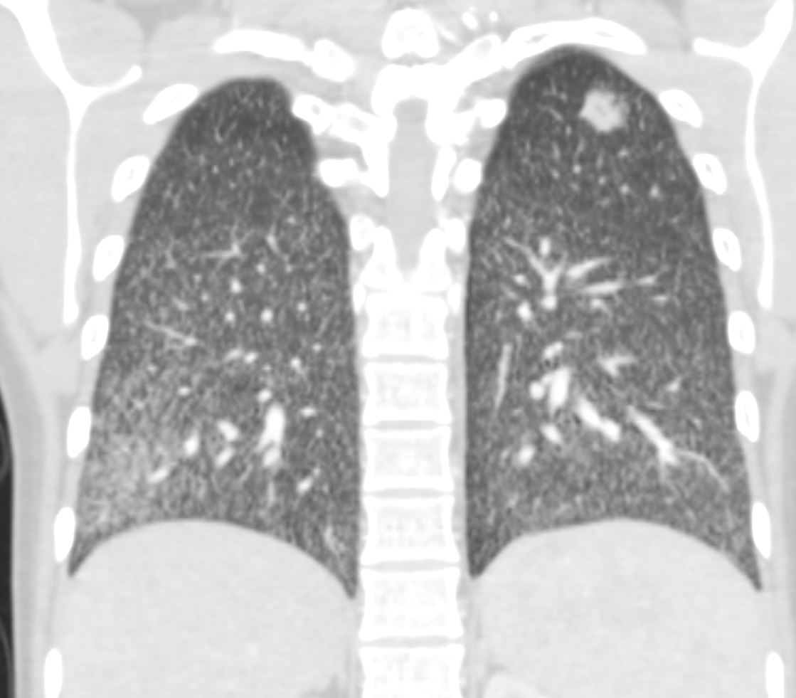
60-year-old immunocompromised female presents with a cough and weight loss. Coronal CT shows miliary nodules throughout both lung fields. The nodules appear to be distributed along the bronchovascular bundles and the lymphatics and are noted in centrilobular, fissural and pleural locations. She responded well to treatment and final diagnosis was mycobacterium tuberculosis.
Ashley Davidoff MD TheCommonVein.net 265Lu 136204c
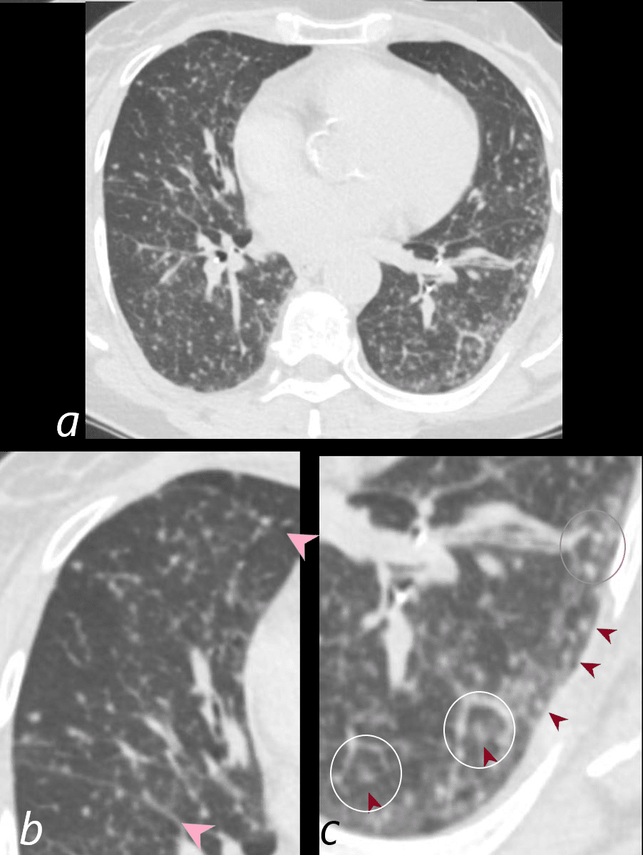
CT Miliary Tuberculosis
Centrilobular Nodules Suggesting Arterial Small Airway and or Lymphatic Involvement
Also Fissural Nodules and Pleural Nodules
60-year-old immunocompromised female presents with a cough and weight loss. Axial CT shows miliary nodules throughout both lung fields. Some of these nodules are centrilobular (c, maroon arrowheads) and others are fissural based (b, pink arrowheads). In some of the secondary lobules there are 2 centrilobular nodules indicating involvement of the airway and arteriole and or the lymphatics (c white rings). One lobule shows centrilobular and interlobular nodules (c gray ring anteriorly). She responded well to treatment and final diagnosis was mycobacterium tuberculosis.
Ashley Davidoff MD TheCommonVein.net 265Lu 136204cL
Infection Histoplasmosis
22-year-old female presents with flu like symptoms. CT shows left apical cavitating nodule and extensive diffuse bilateral micronodular miliary disease.
Ashley Davidoff MD TheCommonVein.net 131716
22-year-old female presents with flu like symptoms. 3 weeks later a chest CT shows a cavitating nodule in the left upper lobe, and extensive diffuse bilateral micronodular miliary disease.
Ashley Davidoff MD Ashley Davidoff MD TheCommonVein.net 131706