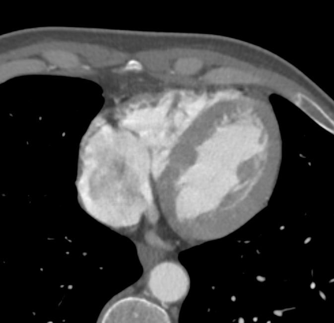
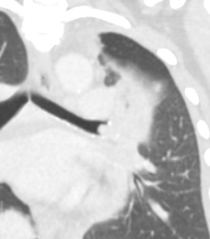
Left Hilar Carcinoid Tumor Tip of the Iceberg Sign
Axial CT scan at the level of the hila in a 56-year-old male shows a 4.2cms vascular mass in the distal left mainstem bronchus impinging on the lumen of the lingula airways. The mass demonstrates the tip of the iceberg sign with the larger portion of the tumor extending beneath the bronchial surface. This is a feature that is characteristic of carcinoid tumors.
Ashley Davidoff MD TheCommonVein.net 261Lu 118381c
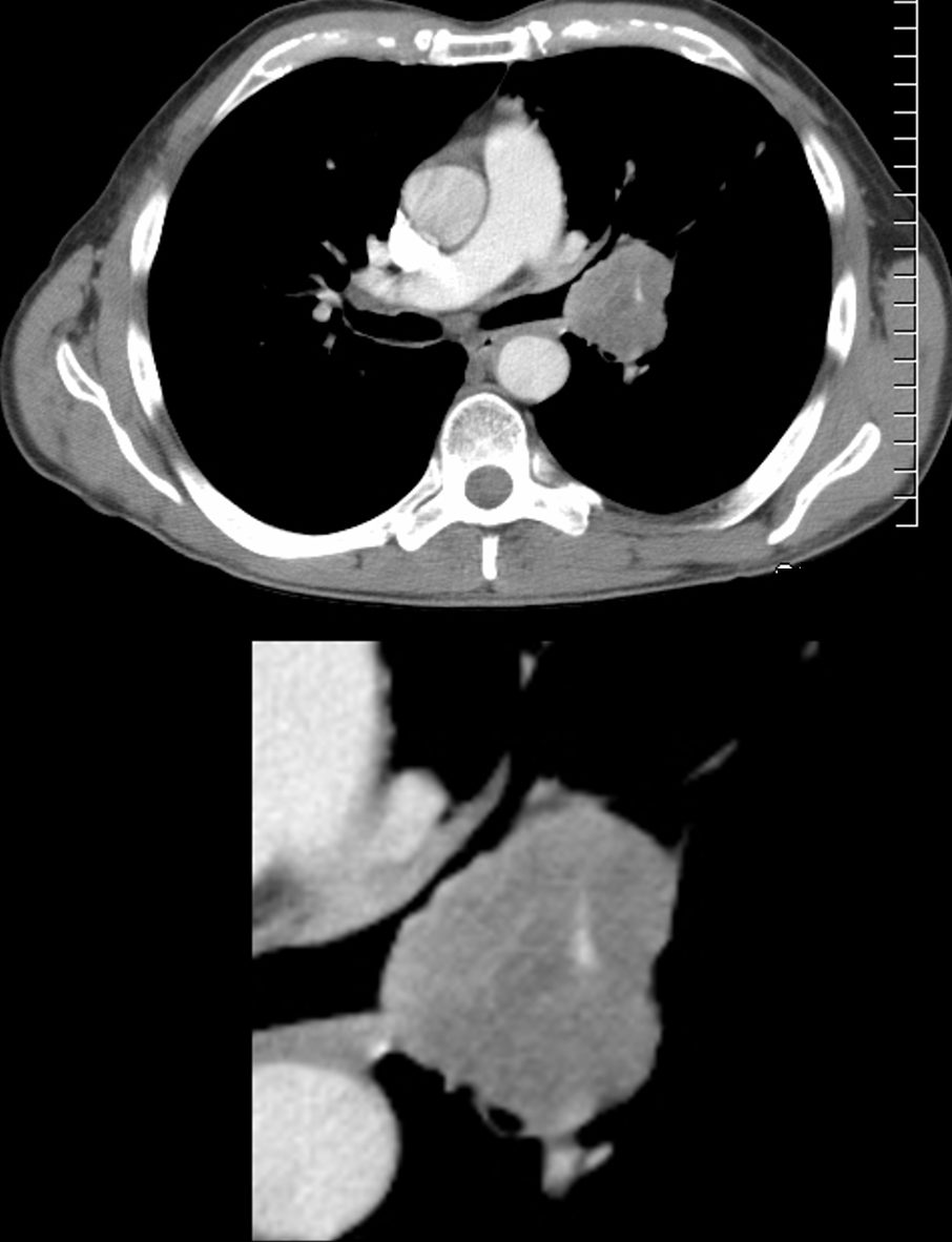
Carcinoid Tumor Causing Post Obstructive Atelectasis
65 year old female presents with a cough. CT shows a mass (green) in proximal portion of the right lower lobe bronchus with post obstructive atelectasis in the superior segment of the right lower lobe (yellow) pathology revealed carcinoid tumor
Ashley Davidoff MD TheCommonVein.net
75679c02
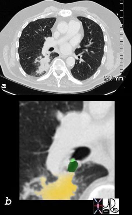
CT Obstructing Nodule in the Left Main Stem Bronchus
58-year-old female presents with a cough Coronal CT shows a nodule at the branching of the more horizontally oriented left mainstem bronchus with post obstructive atelectasis of the lingula and mild hyperinflation of the upper lobe segments.
Pathology revealed findings consistent with a carcinoid tumor of the left bronchus.
Ashley Davidoff MD TheCommonVein.net 257Lu 136118
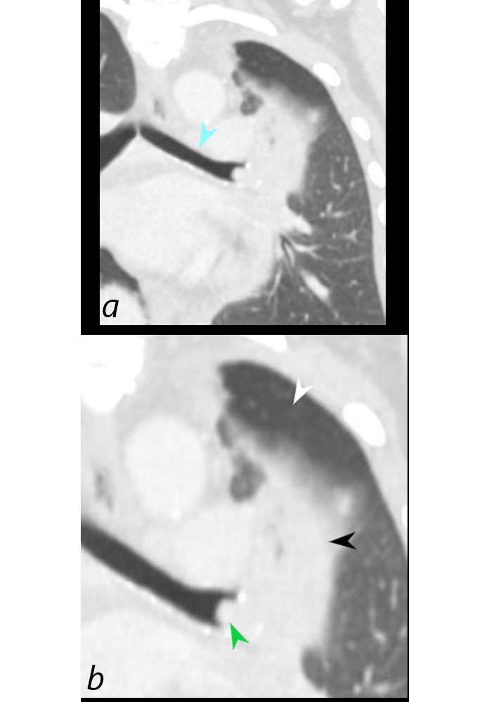
58-year-old female presents with a cough. Coronal CT shows a nodule (green arrowhead, b) at the branching of the more horizontally oriented left mainstem bronchus (teal arrow, a) with post obstructive atelectasis of the lingula (black arrowhead, b) and hyperinflation of the superior aspect of the lower lobe (white arrowhead) which occupies portion of the left apex. (Luftsichel sign)
Pathology revealed findings consistent with a carcinoid tumor
Ashley Davidoff MD TheCommonVein.net 257Lu 136118c
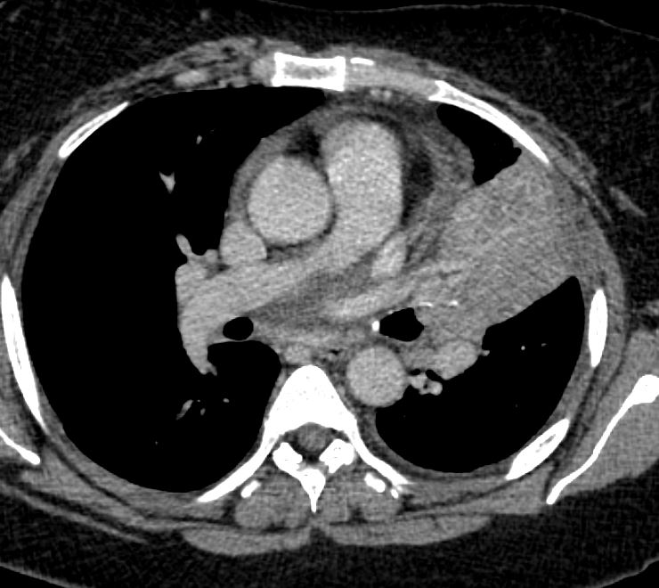
58-year-old female presents with a cough. CT in the axial plane on soft tissue windows shows an obstructing lesion in the left mainstem bronchus of the lung with post obstructive atelectasis of the lingula (black arrowhead) and a small portion of aerated left upper lobe anteriorly (white arrowhead).
Pathology revealed findings consistent with a carcinoid tumor of the left bronchus.
Ashley Davidoff MD TheCommonVein.net 257Lu 136114
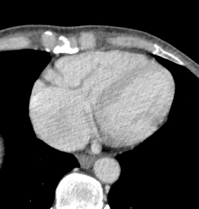
Carcinoid Tumors Manifestations in the Heart