
Bronchitis and Focal Bronchiectasis
69 year old female presents with a cough. CXR frontal view shows diffuse mild increase in lung markings in the right mid and lower lung fields. There is mild elevation of the right hemidiaphragm
Ashley Davidoff MD TheCommonVein.net
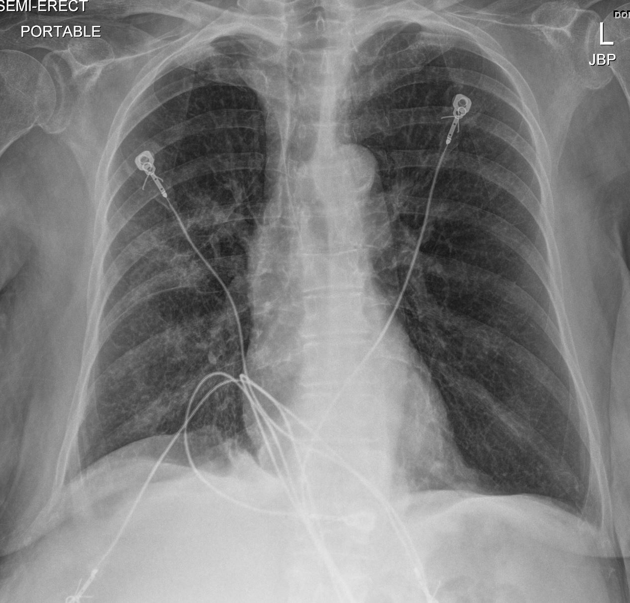
69 year old female presents with a cough. CXR frontal view shows diffuse mild increase in lung markings in the right mid and lower lung fields. There is mild elevation of the right hemidiaphragm
Ashley Davidoff MD TheCommonVein.net
Bronchitis and Focal Bronchiectasis
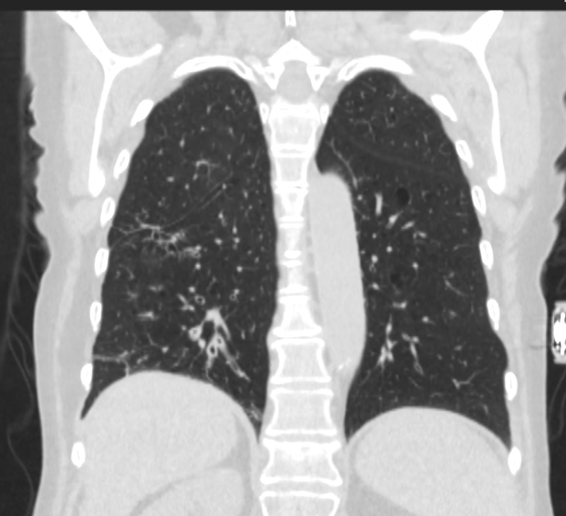
69 year old female presents with a cough. CT in the coronal plane shows thickening of the segmental and subsegmental airways (basilar segments), with foci of bronchiectasis (apical segment). There is mild elevation of the right hemidiaphragm
Ashley Davidoff MD TheCommonVein.net
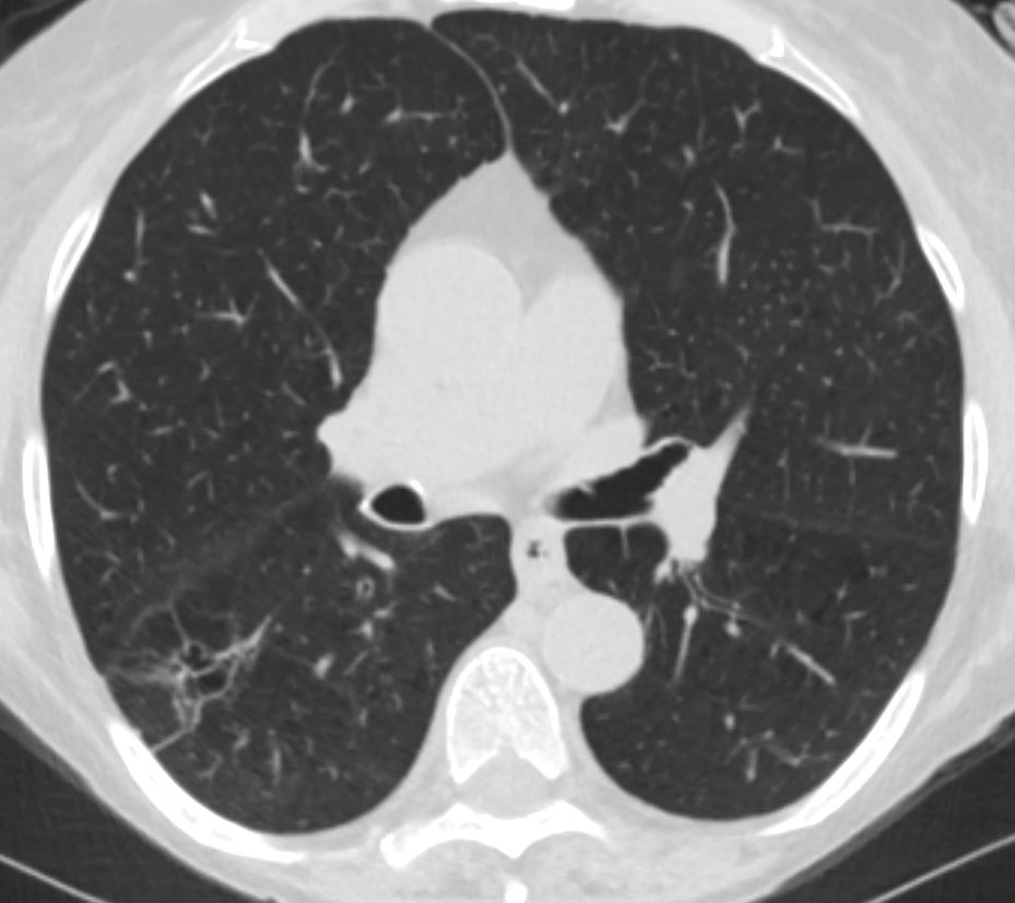
Bronchitis Right Lower Lobe
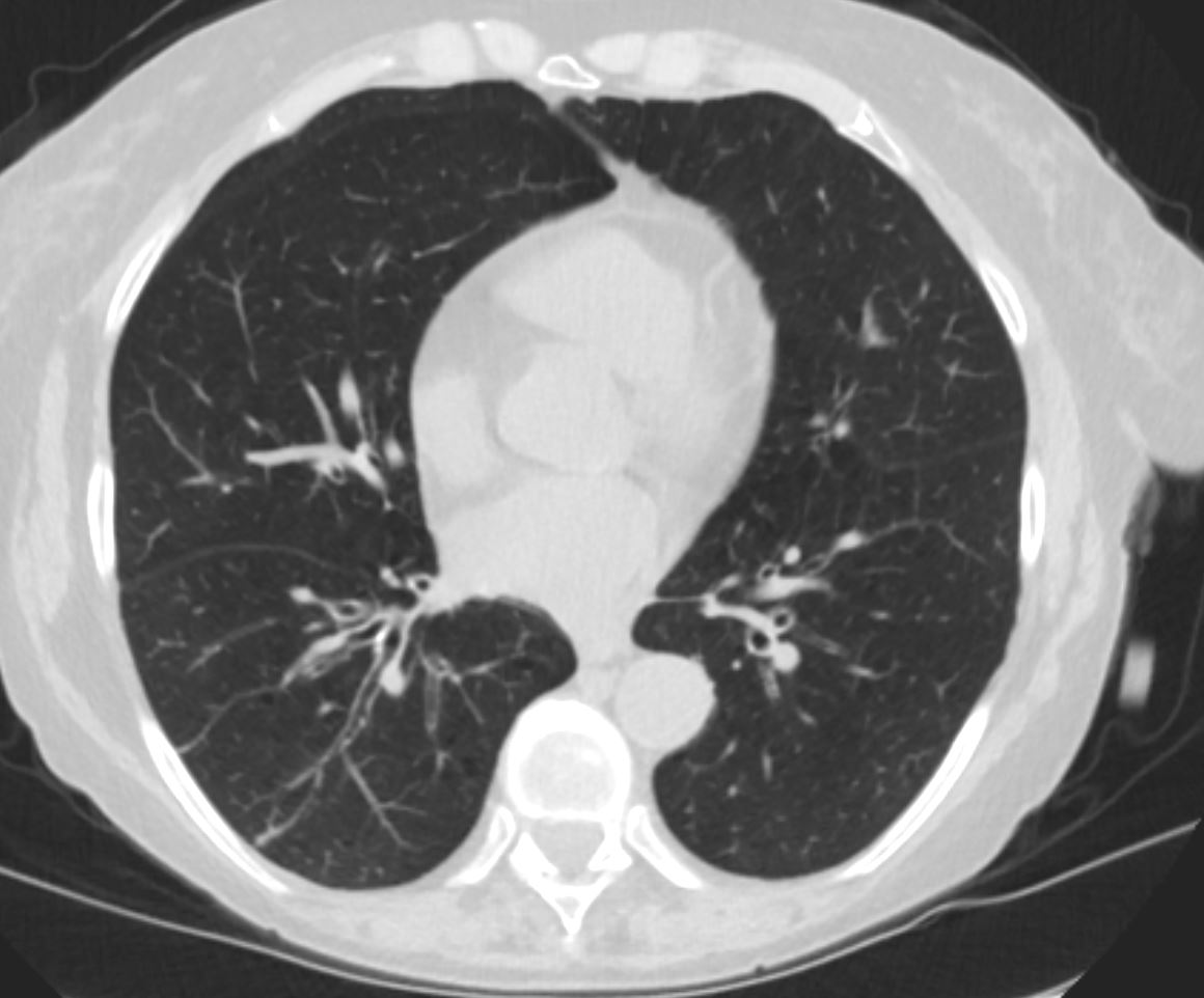
69 year old female presents with a cough. CT in the axial plane shows thickening of the segmental and subsegmental airways (basilar segments),in the right lower lobe compared to the normal appearing airways in the left lower lobe
Ashley Davidoff MD TheCommonVein.net
69 year old female presents with a cough. CT in the axial plane shows thickening of the subsegmental airways (basilar segments),in the right lower lobe compared to the normal appearing airways in the left lower lobe. Note also thickening of the interlobular septa that contribute to the reticular CXR findings
Ashley Davidoff MD TheCommonVein.net
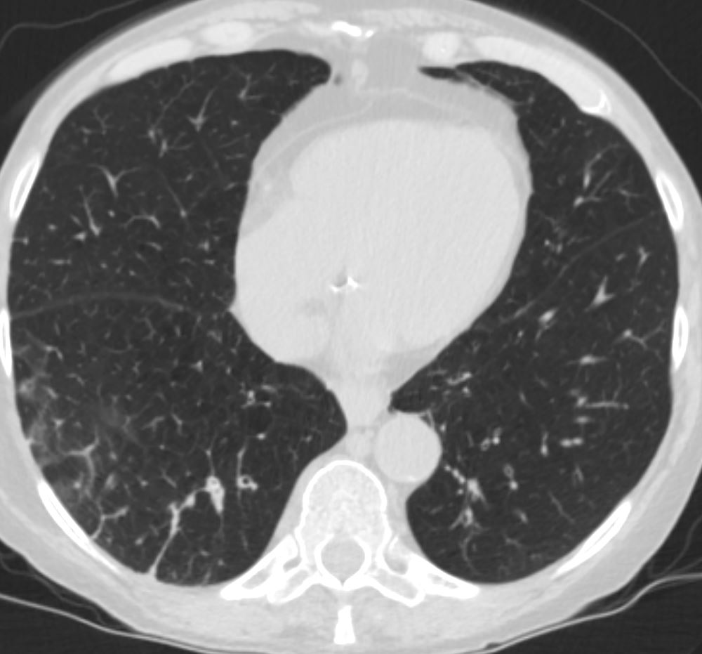
Bronchiectasis Right Lower Lobe
69 year old female presents with a cough. CT in the axial plane shows a focal region of bronchiectasis in the right lower lobe. Note the associated linear scarring that contributes to the reticular CXR findings
Ashley Davidoff MD TheCommonVein.net