
Ashley Davidoff
TheCommonVein.net
Ashley Davidoff MD

TheCommonVein.net 32679
Ashley Davidoff MD TheCommonVein.net lungs-0702
Ashley Davidoff MD TheCommonVein.net lungs-0701
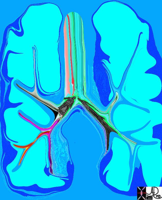
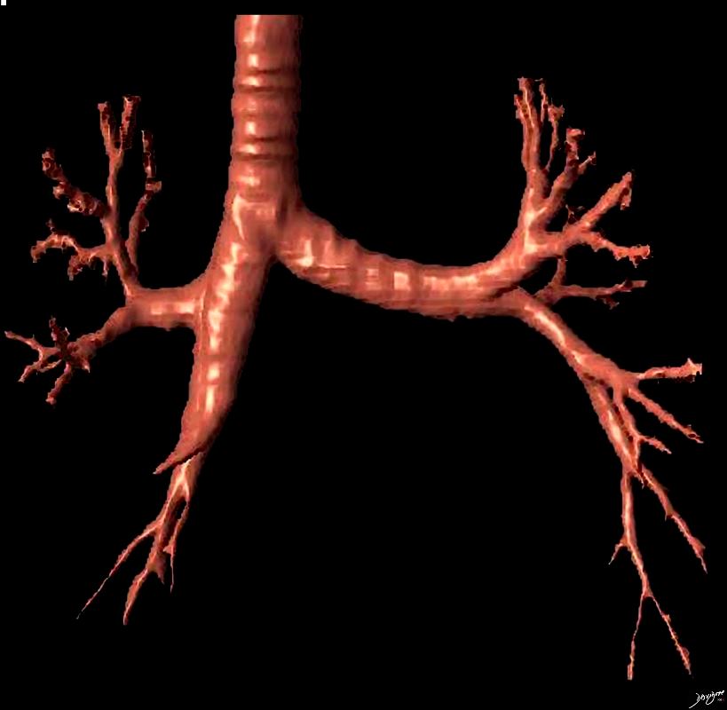
To remember the difference in the sizes of the mainstem bronchi think of 2 very different men in the airways. The right – short, stout and cute, and the left – tall thin and gracile
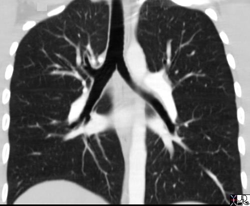
The carinal angle is about 85 degrees
Ashley Davidoff MD TheCommonVein.net 42474b14b

To remember the difference in the sizes of the mainstem bronchi think of 2 very different men in the airways. The right – short, stout and cute, and the left – tall thin and gracile
The carinal angle is about 85 degrees
Ashley Davidoff MD TheCommonVein.net 42474b10.8b
The Carinal Angle
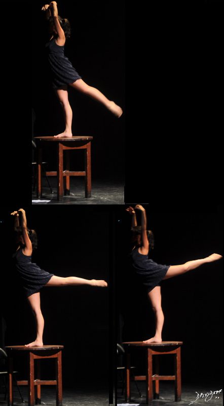
A dancer demonstrates a normal carinal angle (upper image) and as she continues to extend her left leg, (lower images) the angle becomes greater than 80 degrees and in terms of the carinal angle becomes abnormal.
Ashley Davidoff MD
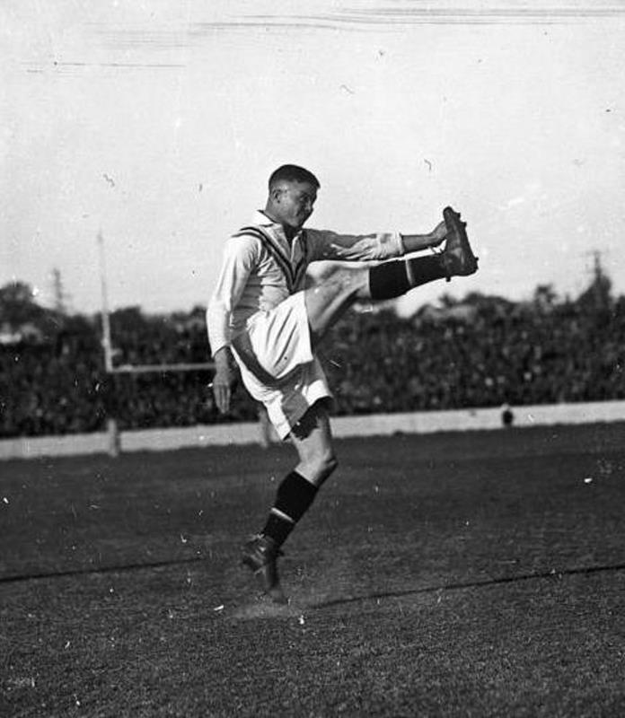
Jim Sullivan kicking for England, in a rugby league match against Australia, 1933
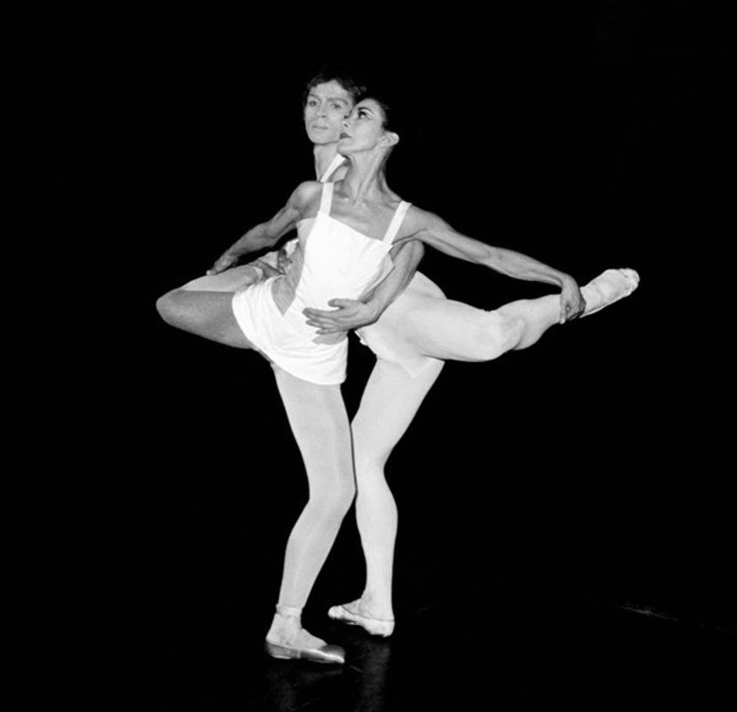
Margot Fonteyn dancing with Rudolf Nureyev during a rehearsal of Roland Petit’s Paradise Lost at Covent Garden in London in 1967 (PA Archive/PA)
Ashley Davidoff M.D.
TheCommonVein.net 32682
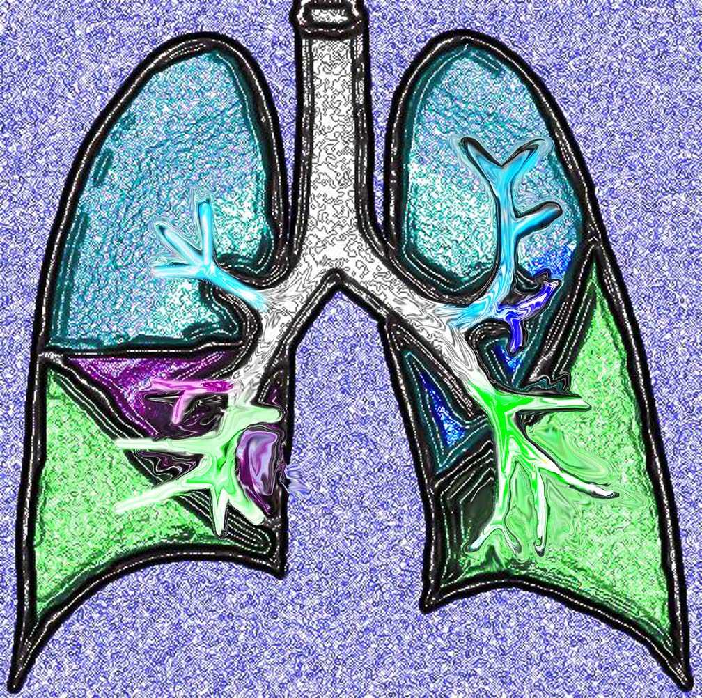
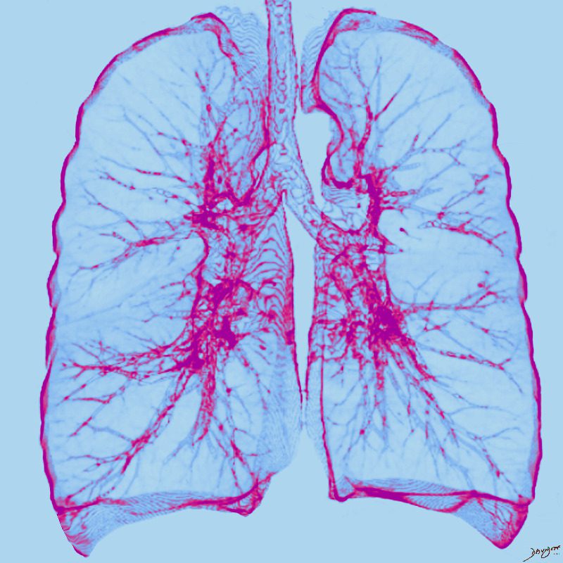
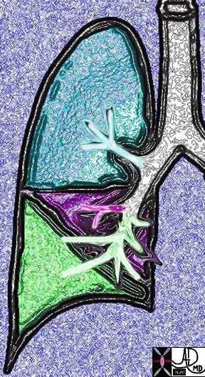
This diagram shows the basic division of the tracheobronchial tree into lobes. The right lung is divided into right upper (RUL) (teal) right middle, (RML pink) and right lower lobe (RLL green). The left lung is divided into left upper (LUL teal), which includes the lingula(dark blue), and left lower lobe (LLL= green). Note that the two mainstem bronchi are of unequal length and size. The right mainstem is short and fat while the left is long and thin. This irregular dichotomous branching pattern is characteristic of the branching pattern of all the conducting systems within the lungs.
Ashley Davidoff
TheCommonVein.net
32686b05
This diagram shows the segmental branches of the right bronchial system. The RUL has three branches, the apical, posterior and anterior segments. (teal overlay) The middle lobe has two segmental branches called lateral and medial segments. (pink) The right lower lobe has five: the superior, anterior basal, lateral basal, posterior basal and medial basal segments. Ashley Davidoff MD. TheCommonVein.net 32686b03
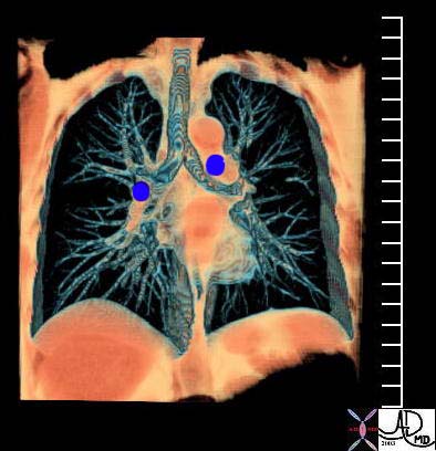
This coronal reformat shows the position of the main branch pulmonary arteries relative to the position of their respective bronchi. While the RPA runs under the right mainstem bronchus, the LPA runs above the left mainstem.
Courtesy Ashley Davidoff MD TheCommonVein.net 32620b01
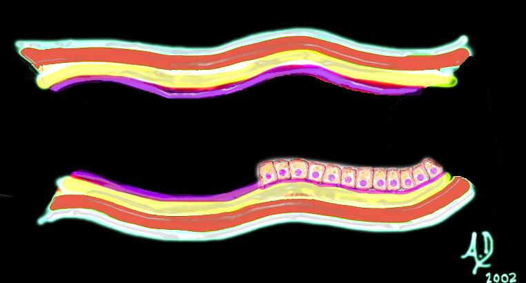
Histology

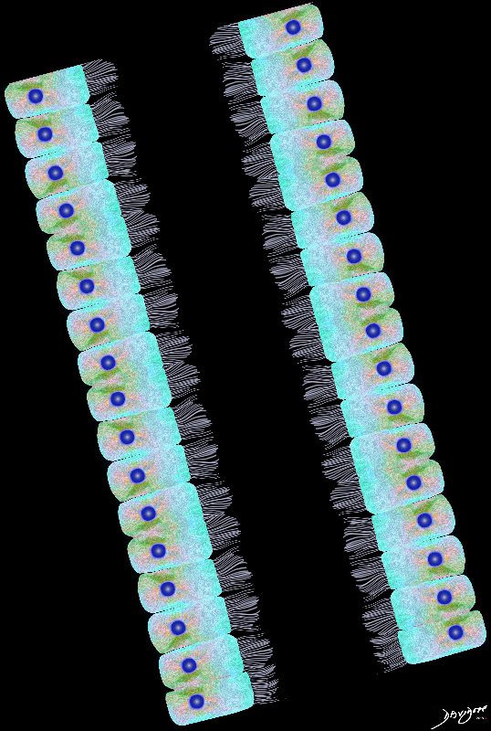
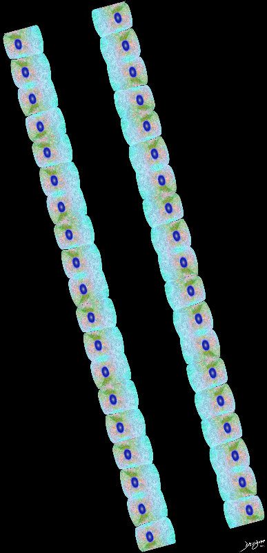
Airways are lined by a pseudostratified ciliated columnar epithelium interspersed with mucus secreting goblet cells
Ashley Davidoff
TheCommonVein.net lungs-00674b01-lo res
Ashley Davidoff
TheCommonVein.net
lungs-00675-lo-res
Ashley Davidoff
TheCommonVein.net
Ashley Davidoff MD
key words art mucosa submucosa muscularis adventitia serosa histology 32347 tube colon small bowel lung bronchus bronchi esophagus stomach large bowel bile duct ureter tube principles
Ashley Davidoff TheCommonVein.net
Physiology

Ashley Davidoff MD TheCommonVein.net lungs-0703

Ashley Davidoff MD TheCommonVein.net lungs-0704
Trees in the Lungs
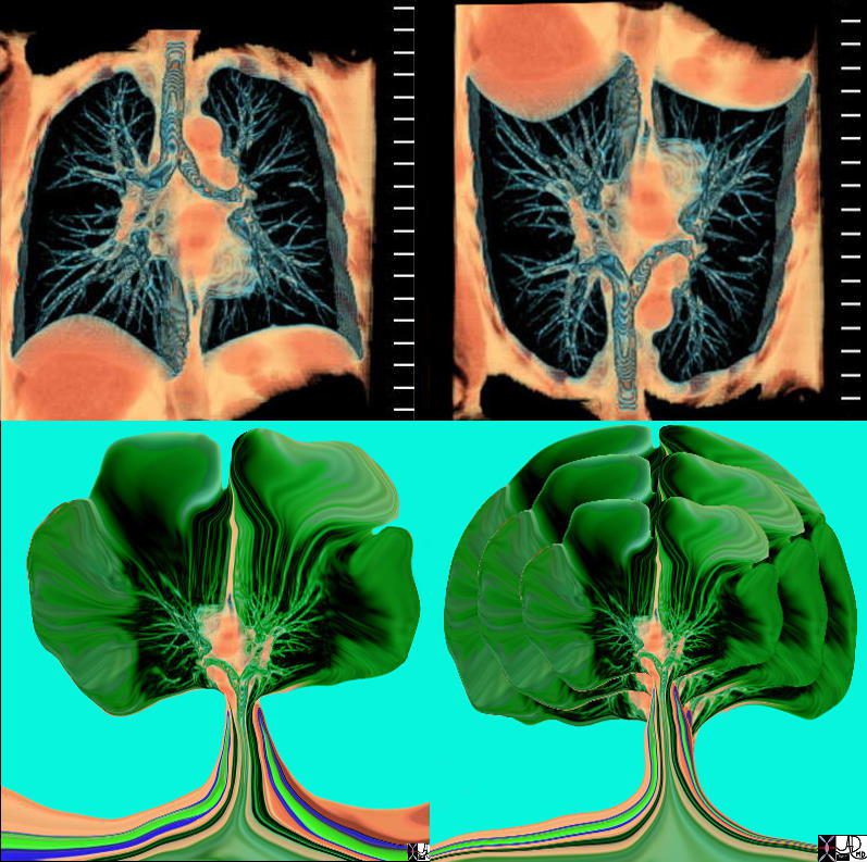
The tracheobronchial tree turned upside down shows it’s similarity to the branching pattern of a tree.
keywords lung bronchus tracheobronchial tree airway tree the common vein applied biology
Ashley Davidoff MD TheCommonVein.net
32620c02.800

Tracheobronchial Tree
Tree, flower, tracheobronchial tree, trachea bronchi lung
Ashley Davidoff TheCommonVein.net 32620b14.800b02p

42474b18.800 lung trachea bronchi tracheobronchial tree
Ashley Davidoff MD
TheCommonVein.net 42474b18.800
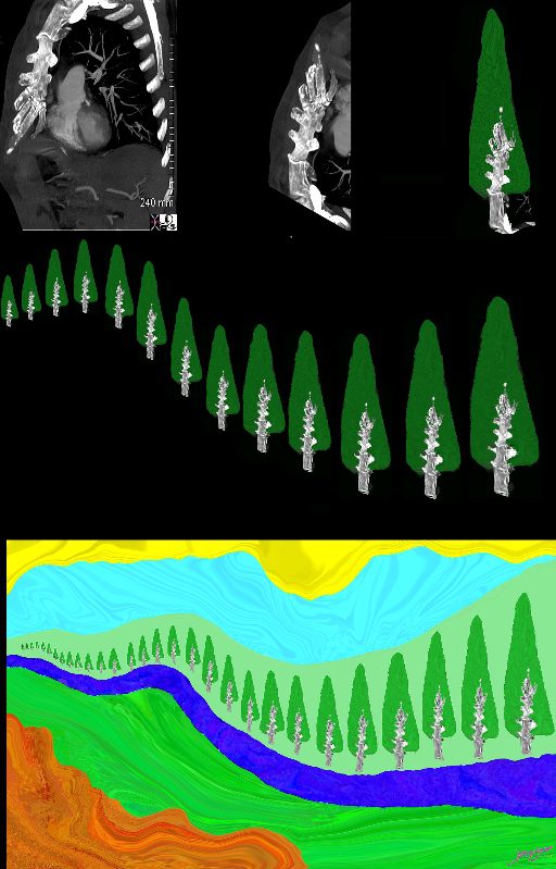
Top left image is sagittal oblique CT scan of the chest and shows a prominent sternum and heavily calcified costochondral junctions. When turned upside down (middle top , a cedar tree is created, then multiplied (middle image) and placed along a river in a mountain under a beautiful summer sun.
Ashley Davidoff MD TheCommonVein.net lungs-0708