
Parts
Size
Normal Size Abnormal Position
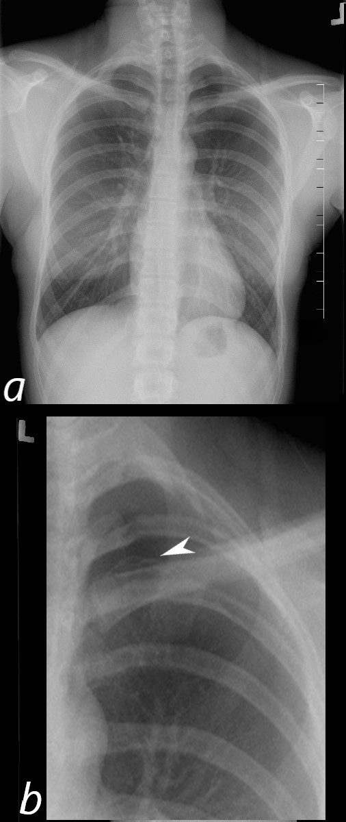
20-year-old female presents with acute left sided chest pain. She has asthenic build which raises the suspicion for a spontaneous pneumothorax. Frontal CXR shows a small subtle pneumothorax characterised by a thin pleural line (b, white arrowhead) and relative lucency of the left apex
Ashley Davidoff MD TheCommonVein.net 117246c01
Mildly Thickened Post Biopsy
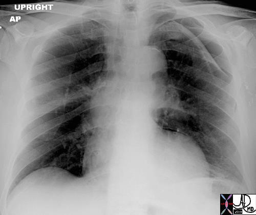
This case shows a chest x-ray of a patient who had a lung biopsy which was complicated by a pneumothorax and a small amount of pleuroparenchymal hemorrhage. The two components of the pleural layer have been separated and the cohesive/adhesive forces have been disrupted by the air which now intervenes and disturbs the physics of the capillary action. The parietal pleura remains attached to the chest wall while the visceral pleura remains attached to the lung. This is a small pneumothorax and had no effect on the patient or the mechanics of lung movement. In this case we followed the patient with a CXR at 2 and 4 hours later. When no progression was demonstrated we allowed the patient to be discharged home with special instructions to limit activities till the next day. In this case there is an abnormal increase in the density of the visceral pleura
Ashley Davidoff MD TheCommonVein.net 42041b01
Mildly Thickened Post Biopsy
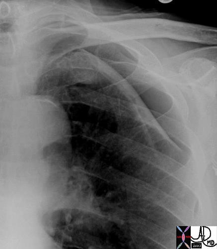
The above image is a magnified view of the pneumothorax.
Courtesy Ashley Davidoff MD TheCommonVein.net 42041b02
Parapneumonic Effusion – Empyema Thickened Pleura
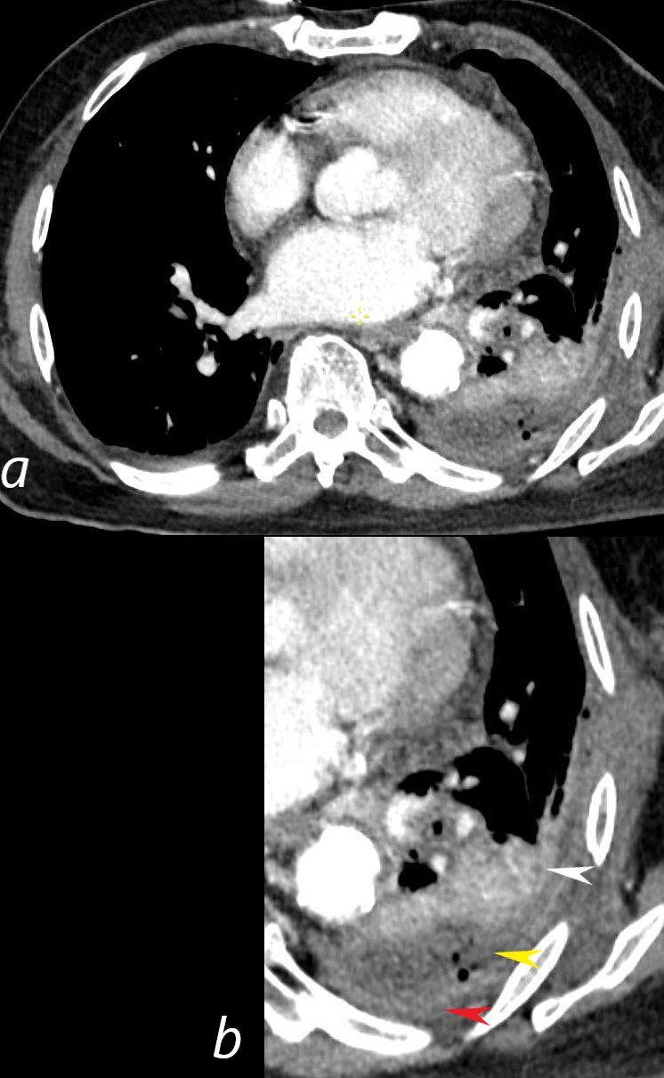
CT scan in a 76-year-old male shows a left lower lobe consolidation (b, white arrowhead) associated with a loculated parapneumonic effusion with trapped air bubbles (b yellow arrowhead) which was culture positive and a thickened enhancing pleura
Ashley Davidoff MD TheCommonVein.net 136194cL
Shape
Position
Normal Size Abnormal Position
20-year-old female presents with acute left sided chest pain. She has asthenic build which raises the suspicion for a spontaneous pneumothorax. Frontal CXR shows a small subtle pneumothorax characterised by a thin pleural line (b, white arrowhead) and relative lucency of the left apex
Ashley Davidoff MD TheCommonVein.net 117246c01