
Pulmonary Hemorrhage and Heterogeneous Changes in the Secondary Lobules
75-year-old man on blood thinners s/p aortic valve replacement, s/p trauma, presents with hemoptysis. He was afebrile and without an elevated white count
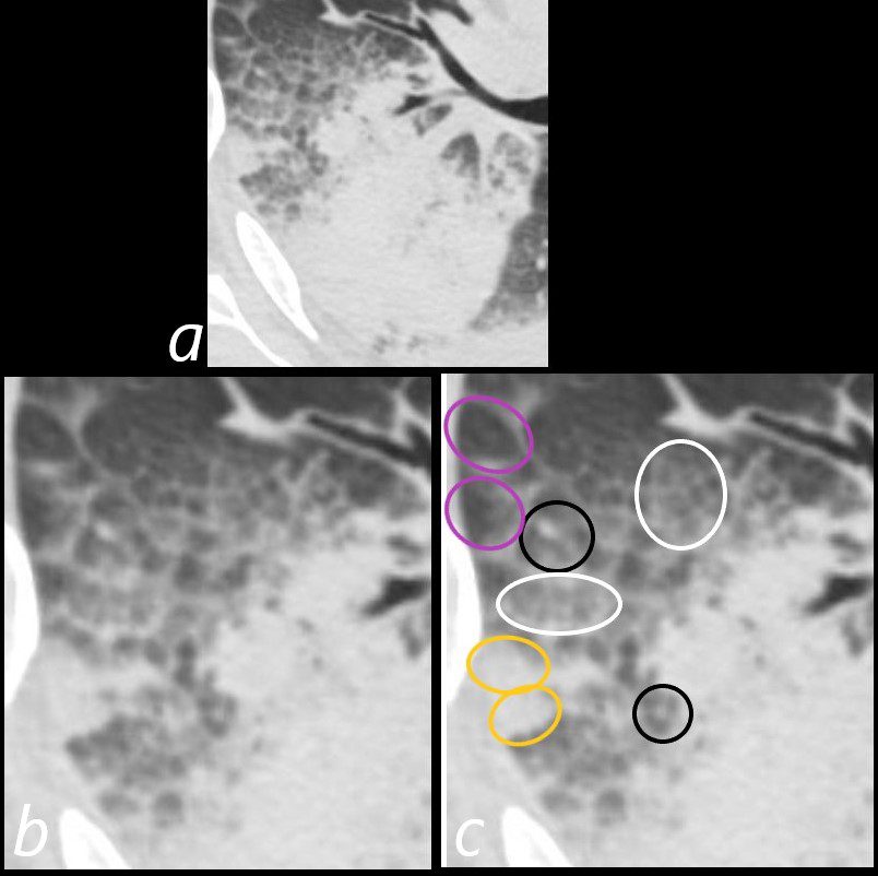
Axial CT at the level below the carina shows heterogenous changes of the secondary lobules. The changes in the posterior segment of the right upper lobe (a magnified below in b and c ) and show secondary lobules with ground glass changes (white rings) It is difficult to distinguish those with normal lung parenchyma and those with mosaic attenuation. It is hypothesized that those with prominent centrilobular nodules (black rings c) more than likely represent mosaic attenuation due to small airway disease. The lower density secondary lobules which do not have a prominent centrilobular nodule (c, purple rings), may either reflect normal parenchyma or mosaic attenuation. The orange rings likely reflect consolidation in side-by-side secondary lobules.
Ashley Davidoff MD TheCommonVein.net 165Lu 135854cL01
Calcification
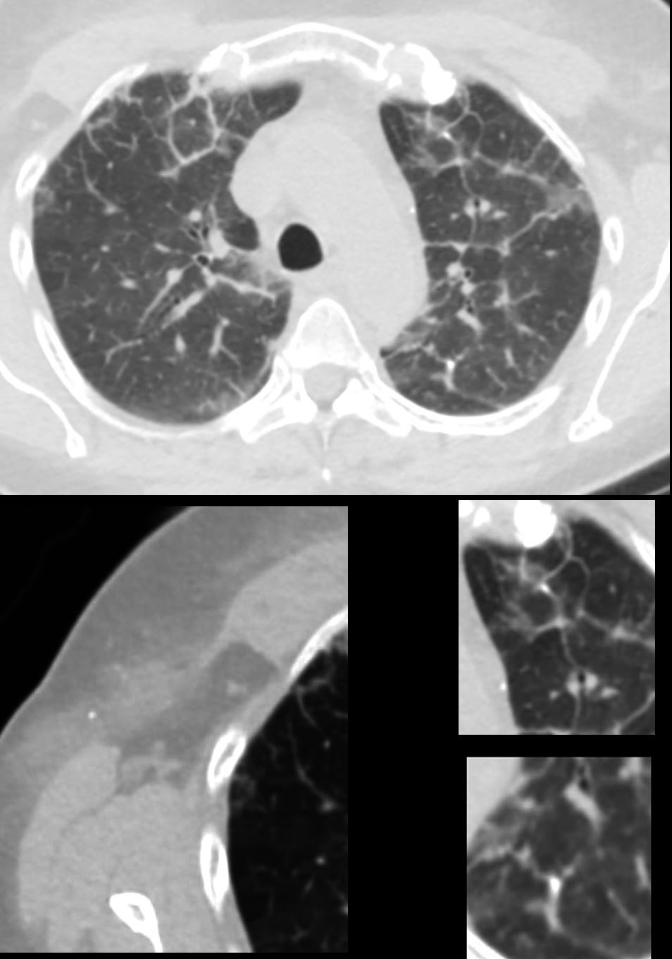
56 -year-old female with a history of amyloidosis (AL) presents for follow up. Axial CT of the chest shows diffuse reticular process best appreciated anteriorly with thickening of the interlobular septa. Punctate, dystrophic calcifications are seen in the interlobular septa (white arrowheads a, c and d), as well as in the pleura abutting the mediastinum (white arrowhead a). Image b shows similar calcifications, likely deposits of amyloid in the axilla with surrounding soft tissue changes
Ashley Davidoff MD TheCommonVein.net 244 Lu 135741d02cL
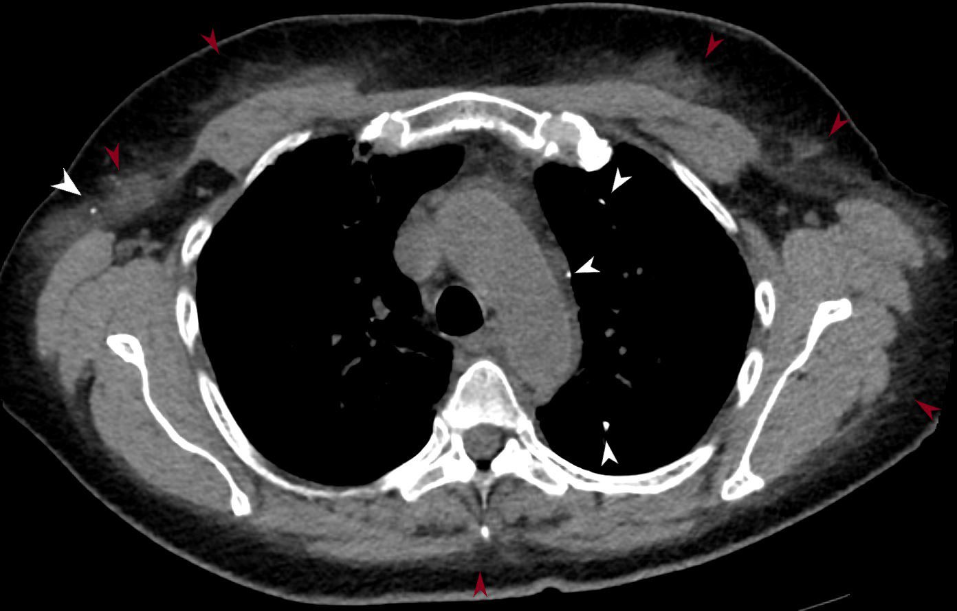
56 -year-old female with a history of amyloidosis (AL) presents for follow up. Axial CT of the chest shows punctate, dystrophic calcifications in the left upper lobe, pleura abutting the mediastinum and in the right axilla (white arrowheads). There is induration in the soft tissues suggesting amyloidosis (maroon arrowheads).
Ashley Davidoff MD TheCommonVein.net 244 Lu 135741d02m05L