
Infection
TB
29-year-old male presents with night sweats
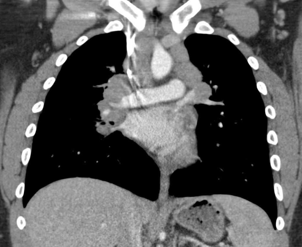
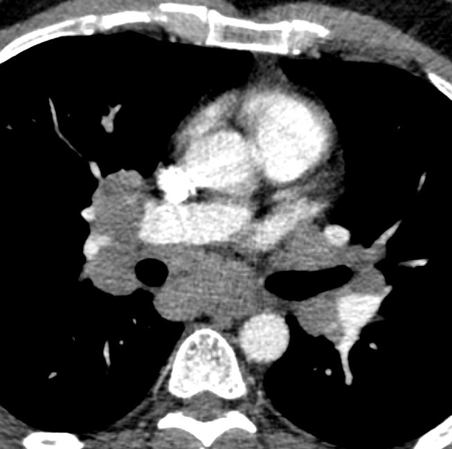
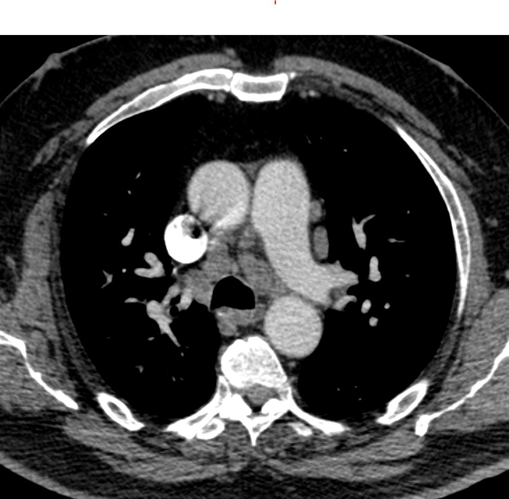
CT at the level of the left atrium shows extensive hilar and mediastinal lymphadenopathy. Wedge biopsy and culture revealed a diagnosis of MAC (mycobacterium avium complex)
Ashley Davidoff MD TheCommonvein.net 135812 247Lu
Ashley Davidoff MD TheCommonVein.net
Inflammation Infection
Allergic Bronchopulmonary Aspergillosis Finger in Glove and Low Density Lymph Nodes
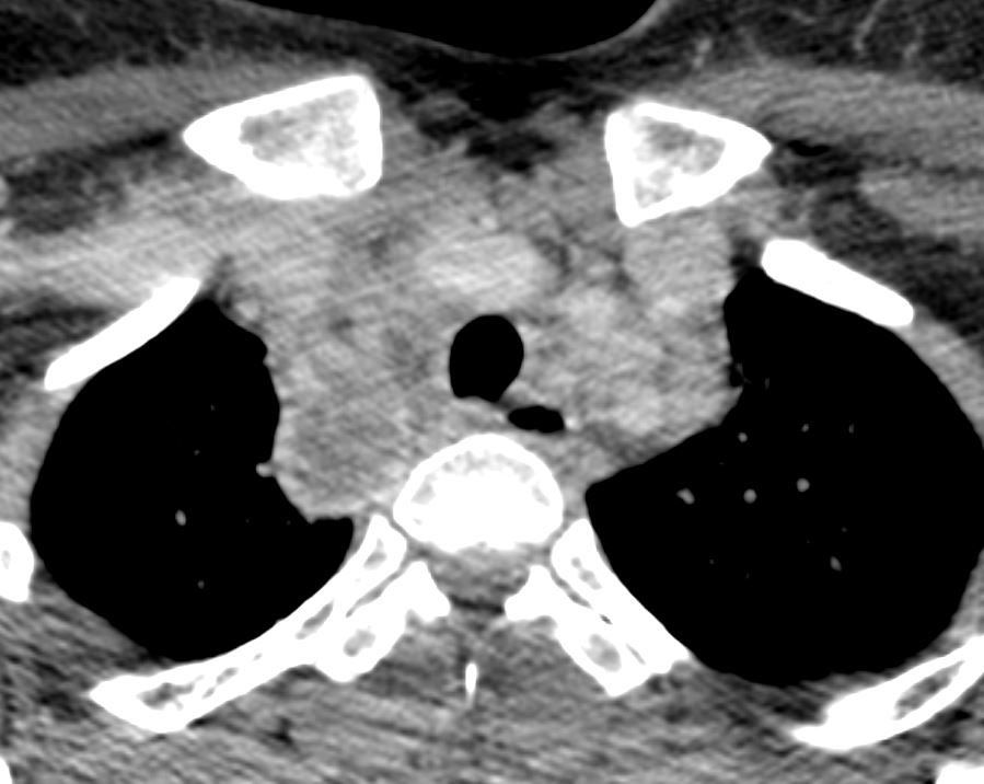
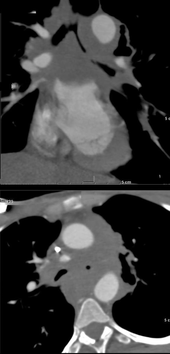
CT scan with contrast shows a cluster of low density lymph nodes in the anterior mediastinum in the retro-clavicular region
Ashley Davidoff MD The CommonVein.net 294Lu 117970
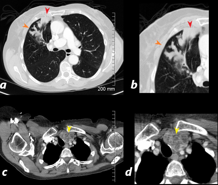
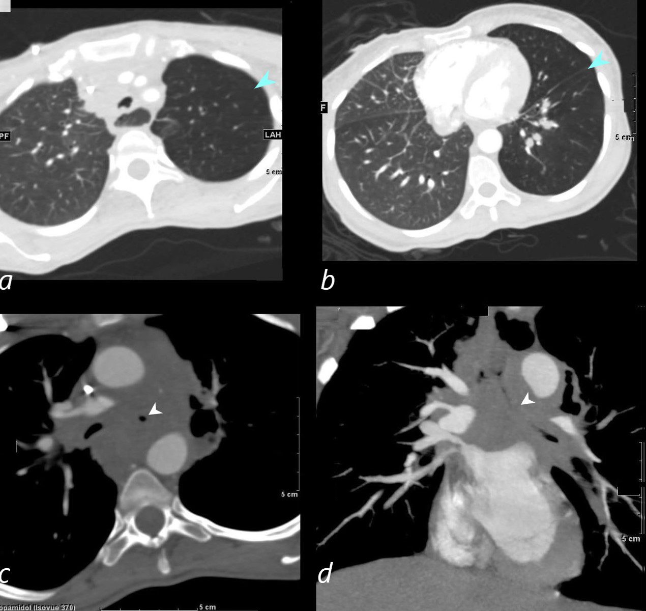
CT scan with contrast shows finger in glove appearance of the anterior segmental airways of the right upper lobe (orange arrowheads a, b) with a focal region of subsegmental atelectasis (red arrow head a,b).
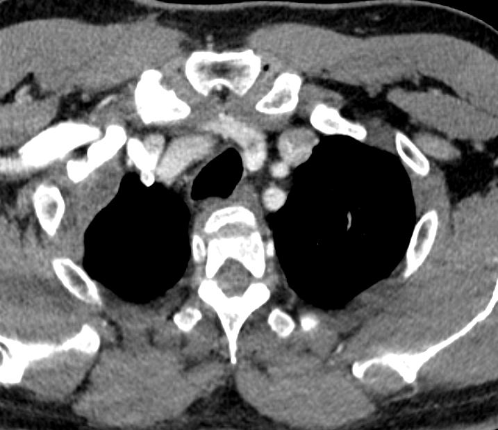
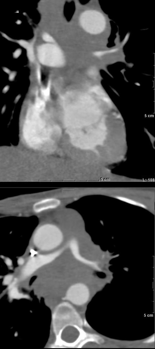
The lower panel shows a cluster of low density lymph nodes in the anterior mediastinum in the retro-clavicular region (yellow arrowheads c,d) .
Ashley Davidoff MD The CommonVein.net
CT scan with contrast shows finger in glove appearance of the anterior segmental airways of the right upper lobe (orange arrowheads a, b) with a focal region of subsegmental atelectasis (red arrow head a,b).
The lower panel shows a cluster of low density lymph nodes in the anterior mediastinum in the retro-clavicular region (yellow arrowheads c,d) .
Ashley Davidoff MD The CommonVein.net 294LU 117972b
Inflammation
Sarcoidosis
CT Hilar and Mediastinal Adenopathy
Ashley Davidoff MD TheCommonVein.net
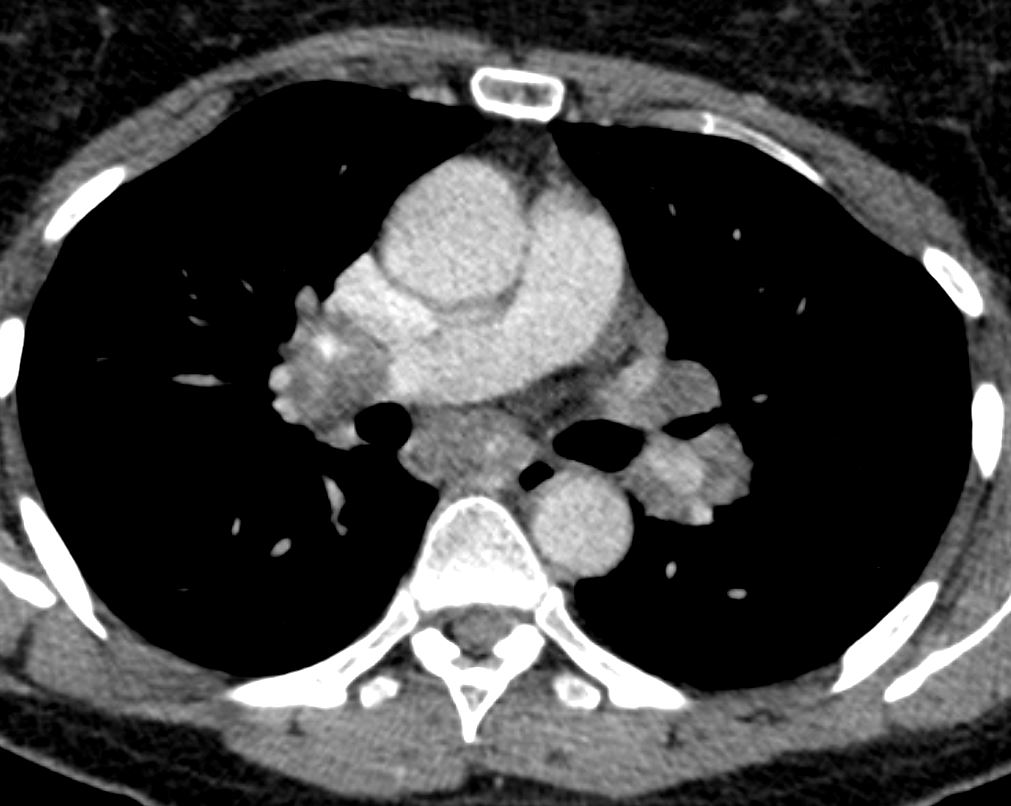
CT Hilar and Mediastinal Adenopathy
Ashley Davidoff MD TheCommonVein.net
Chronic Eosinophilic Pneumonia (CEP)
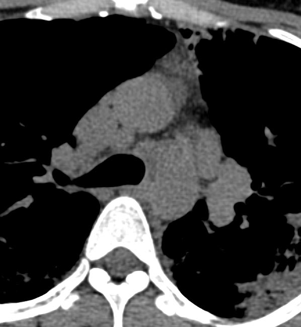
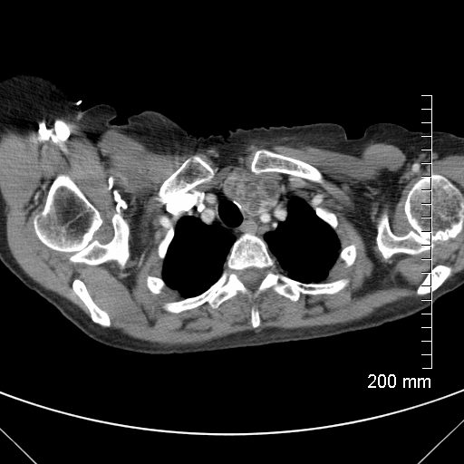
CT scan in the axial plane performed 6 months ago at the time of clinical presentation shows an enlarged A-P window lymph node likely reactive. Subsequent diagnosis by BAL of chronic eosinophilic pneumonia (CEP) was made
Ashley Davidoff TheCommonVein.net
DIP – Mediastinal Adenopathy
60-year-old male smoker with a history of progressive dyspnea. Axial CT through the level of the left pulmonary artery shows borderline enlarged mediastinal lymph nodes.
Pathology of the lung confirmed a diagnosis of DIP
Ashley Davidoff MD TheCommonVein.net 253Lu 136017
Malignancy
Carcinoma of the Cervix Metastatic Mediastinal Adenopathy Encasement of the Airways
33-year-old female with known primary carcinoma of the cervix presents with infiltrating and encasing mediastinal adenopathy with subtotal narrowing of the left main stem bronchus and significant stenosis of the right
Ashley Davidoff MD TheCommonVein.net 254Lu 136048b01
Carcinoma of the Cervix Metastatic Mediastinal Adenopathy Encasement of the Pulmonary Arteries
33-year-old female with known primary carcinoma of the cervix presents with infiltrating and encasing mediastinal adenopathy with severe stenosis of both the left (LPA) and right main pulmonary (RPA) arteries. In the upper panel the severe narrowing of the LPA is appreciated and in the lower panel the bilateral severe proximal stenosis is appreciated.
Ashley Davidoff MD TheCommonVein.net 254Lu 136048b
Carcinoma of the Cervix Metastatic Mediastinal Adenopathy Encasement of the Airways with Ball Valve Effect on the Left Upper Bronchus
33-year-old female with known primary carcinoma of the cervix presents with infiltrating and encasing mediastinal adenopathy with severe stenosis of both the left and right main stem bronchi. The upper panels reveal hyperinflation of the left upper lobe (a, b blue arrow heads) likely secondary to air trapping. The lower panel reveals the severe stenosis of the left main stem bronchus (c white arrowhead) with subtotal occlusion appreciated in d, (white arrowhead). This severe subtotal occlusion probably accounts for the hyperinflation secondary to ball valve mechanism
Ashley Davidoff MD TheCommonVein.net 254Lu 136050b02L
Carcinoma of the Cervix Metastatic Mediastinal Adenopathy – Encasement of the Pulmonary Veins
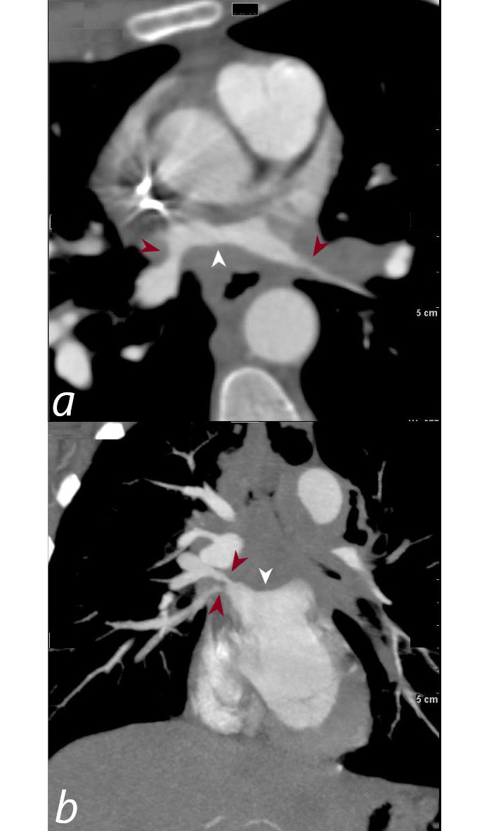
33-year-old female with known primary carcinoma of the cervix presents with infiltrating and encasing mediastinal adenopathy with stenosis of the bilateral pulmonary veins. The upper panel (a) reveals encasement of the right superior pulmonary vein (a, maroon arrowheads) and compression of the left atrium (white arrowhead). The lower panel b, reveals stenosis of both the superior and inferior pulmonary veins on the right,(maroon arrowheads) and mass effect on the superior aspect of the left atrium (LA) (white arrowhead).
Ashley Davidoff MD TheCommonVein.net 254Lu 136050b04L
Mechanical/
Atelectasis
Trauma
Metabolic
Circulatory-
Hemorrhage
Immune Infiltrative Idiopathic Iatrogenic Idiopathic