
- 54 year old female with history of asthma, bronchitis, bronchiectasis, ABPA
- asthma
- bronchitis
- bronchiectasis
- ABPA
- bronchiectasis
Finger in glove in RLL noted 9 years prior
CXR Hyperinflation, Bronchiectasis and
Volume Loss of the Right lung
– Hyperinflation,
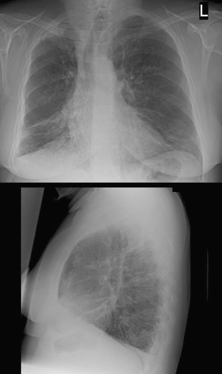
54 year old female with history of asthma, bronchitis, bronchiectasis, ABPA
CXR shows hyperinflation, with flattening of the hemidiaphragm bronchiectasis and volume loss of the right lung with rightward deviation of the trachea and downward displacement of the major fissure)
Ashley Davidoff TheCommonVein.net 220Lu 31258b
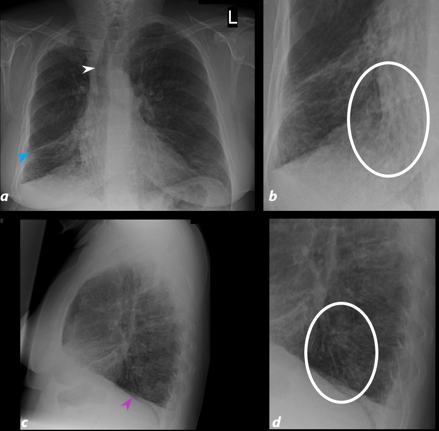
54 year old female with history of asthma, bronchitis, bronchiectasis, ABPA
CXR shows hyperinflation, with flattening of the hemidiaphragm (pink arrowhead c) bronchiectasis (oval white ring b.and d) and volume loss of the right lung with rightward deviation of the trachea (white arrowhead (a) and downward displacement of the major fissure (blue arrowhead, a)
Ashley Davidoff TheCommonVein.net 220Lu 31258aL
Finger in Glove in RLL noted 9 years prior
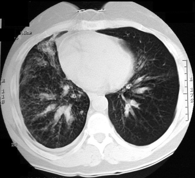
54 year old female with history of asthma, bronchitis, bronchiectasis, ABPA
CT scan shows extensive segmental, subsegmental and small airway disease in the right lower lobe. The ectatic right lower lobe segmental bronchi, and to lesser extent in the left lower lobe. are fluid filled manifesting as “finger in glove” appearance. There are innumerable groundglass micronodules in the right lower lobe and middle lobe, reflecting small airway disease.
Ashley Davidoff TheCommonVein.net 220Lu 31258
Lower Lobe Bronchiectasis and ABPA over 9 years
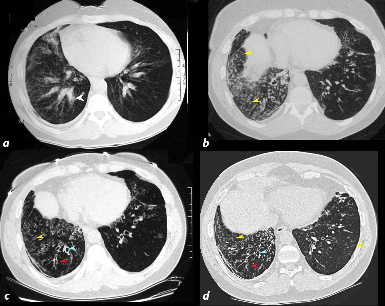
54 year old female with history of asthma, bronchitis, bronchiectasis, ABPA
CT scan shows initial CT (a) of the lower lobes and part of the middle lobe performed 9 years prior with extensive segmental, fluid/soft tissue filled bronchiectasis within the right lower and to lesser extent the left lower lobe reminiscent of finger in glove morphology. Subsequent imaging 2 (b),5 (c) and 9 years(d), reveals ongoing bronchiectasis (teal arrowhead) subsegmental airway thickening (red arrowhead) and ground glass micronodules indicating small airway disease. The middle lobe was also involved- not shown here.
Ashley Davidoff TheCommonVein.net
220Lu 31257
ABPA Bronchiectasis Bronchitis Bronchiolectasis and Small Airway Disease Right Lower Lobe and Middle Lobe 7 Years ago

54 year old female with history of asthma, bronchitis, bronchiectasis, ABPA
Current CT scan shows extensive small airway disease in the right lower lobe, magnified in lower image with centrilobular nodules and thickened interlobular septa characterized by ground glass micronodules.
Ashley Davidoff TheCommonVein.net 220Lu 31246c
Current CT –
ABPA Bronchiectasis Bronchitis Bronchiolectasis and Small Airway Disease Right Lower Lobe and Middle Lobe
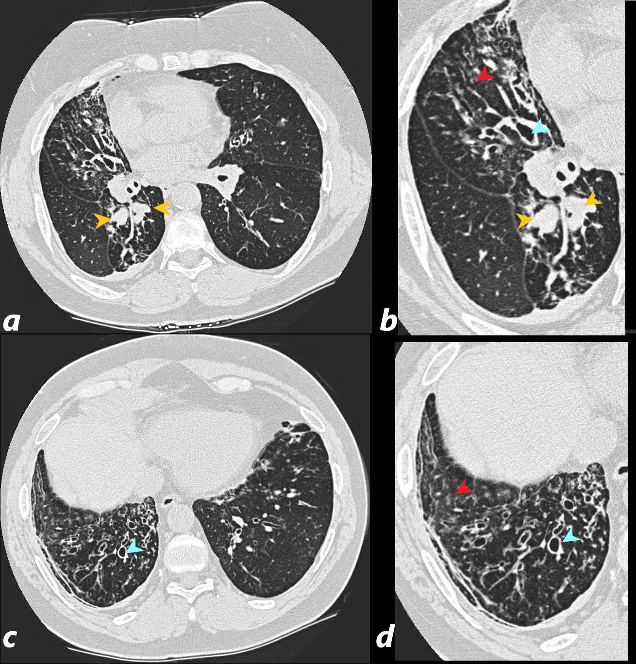
54 year old female with history of asthma, bronchitis, bronchiectasis, ABPA
Current CT scan shows proximal impaction of bronchectatic lower lobe bronchi (orange arrowheads a, and magnified in b) with bronchiectasis in the middle lobe (teal arrowhead in b) and in the right lower lobes (teal arrowheads in c and d ) with suggestion of small airway disease characterized by ground glass micronodules indicating small airway disease (red arrowheads b and d).
Ashley Davidoff TheCommonVein.net 220Lu 31258b03c
Current CT – ABPA Bronchiectasis Bronchitis Bronchiolectasis and Small Airway Disease Right Lower Lobe and Middle Lobe
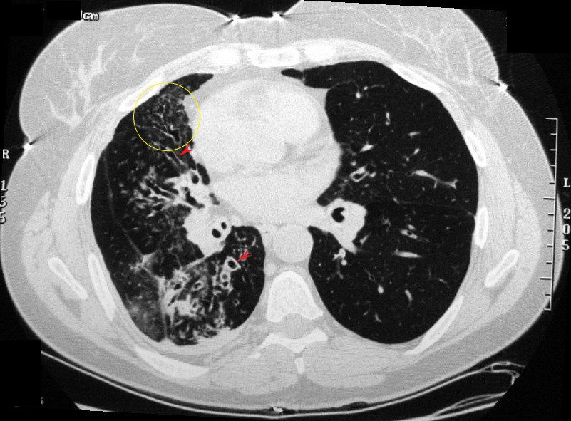
54 year old female with history of asthma, bronchitis, bronchiectasis, ABPA
Current CT scan shows suggestion of small airway disease (yellow circle) characterized by ground glass micronodules and bronchial wall thickening in segmental and subsegmental airways (red arrowheads)
Ashley Davidoff TheCommonVein.net 220Lu 31250