
74-year-old man presents with dyspnea and orthopnea.
CT Enlarged Left Atrium
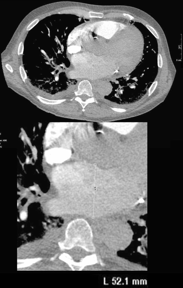
74-year-old man presents with dyspnea and orthopnea. CT through the chest at the level of the left atrium shows an enlarged left atrium measuring 5.2cms (normal up to 4cms). Additionally, there are small bilateral effusions right greater than left. The shape of the right effusion suggests it is partially loculated. Peribronchial thickening is suggested. These findings suggest congestive heart failure.
Ashley Davidoff MD TheCommonVein.net 135770c
CT Enlarged Right Atrium
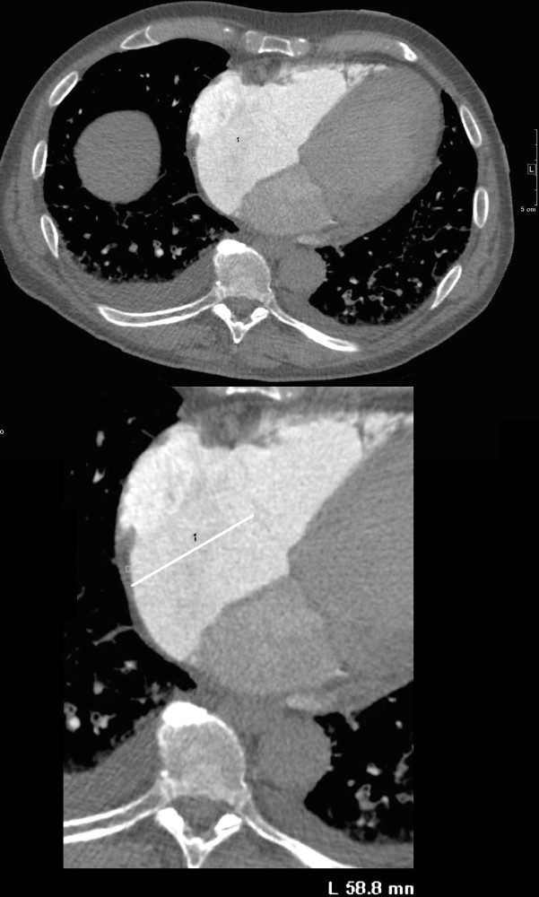
74-year-old man presents with dyspnea and orthopnea. CT through the chest at the level of the right atrium shows an enlarged right atrium measuring 5.9cms (normal up to 5cms). Additionally, there are small bilateral effusions right greater than left. The mild irregular shape of the effusions suggests that they are partially loculated. Peribronchial thickening is suggested. These findings suggest congestive heart failure.
Ashley Davidoff MD TheCommonVein.net 135771cL
Tricuspid Regurgitation
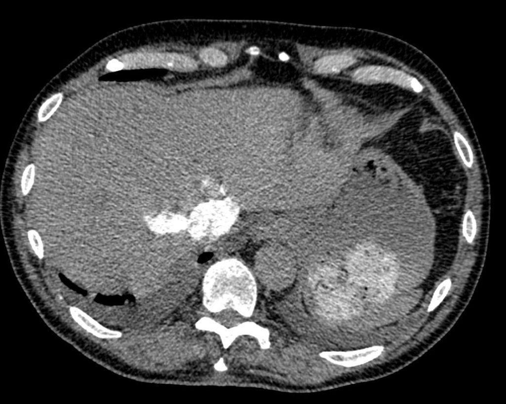
74-year-old man presents with dyspnea and orthopnea. CT through the upper abdomen at the level of the hepatic veins shows reflux of contrast into the hepatic veins indicating tricuspid regurgitation. Additionally, there are small bilateral effusions right greater than left. The mild irregular shape of the effusions suggests that they are partially loculated. Peribronchial thickening is suggested. These findings suggest congestive heart failure with both right and left sided involvement.
Ashley Davidoff MD TheCommonVein.net 135772
CHF with Interstitial Edema
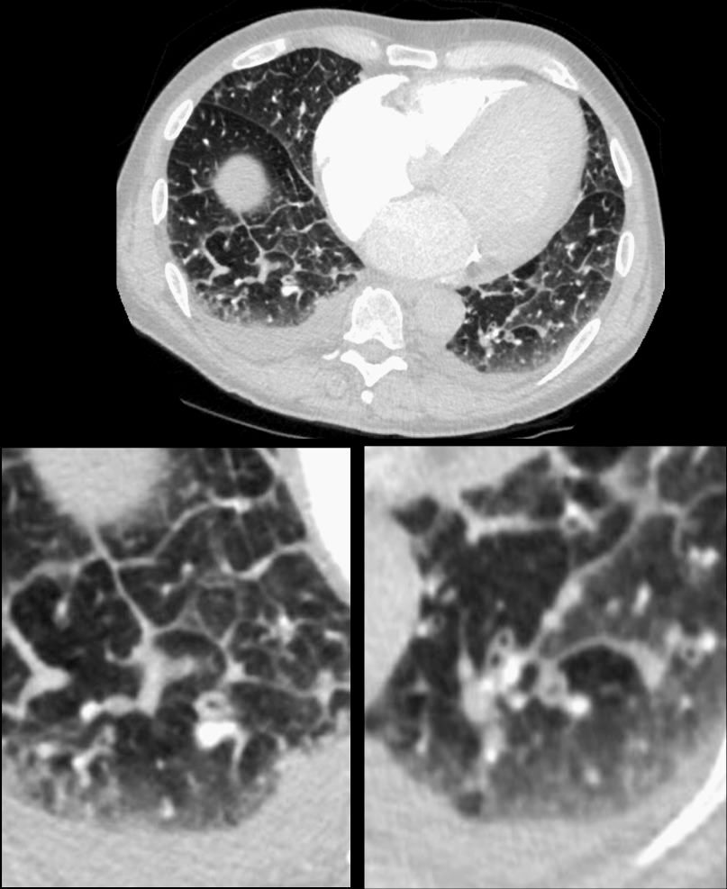
74-year-old man presents with dyspnea and orthopnea. CT shows thickening of the interlobular septa (Kerley B lines), peribronchial cuffing, and enlargement of the lobular arteriole in the right lower lobe. There is a suggestion of vasoconstriction of the arteriole as it enters the secondary lobule. ground glass changes in the some of the secondary lobules on the left and perhaps mosaic attenuation vs normal secondary lobule at the right base are noted. Additionally, there are small bilateral effusions right greater than left. The mild irregular shape of the effusions suggests that they are partially loculated. These findings indicate moderate congestive heart failure with interstitial edema.
Ashley Davidoff MD TheCommonVein.net 135775c01