
Total Lung Compressive Atelectasis
Effusion
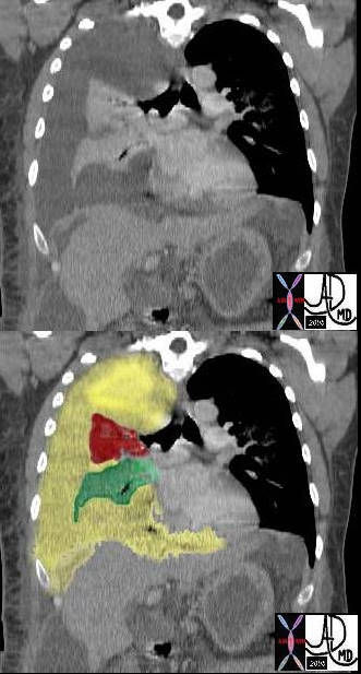
In this case there a large right sided pleural effusion (yellow) with secondary atelectasis of the right lung. (red and green) This coronal CT of the chest at the level of the left ventricle shows a large right pleural effusion which lies between the visceral and parietal pleura. Once the effusion is large enough to weaken the capillary forces that hold the parietal and visceral pleura together, it fail, and the lung collapses which is what is noted on this image – ie total lung collapse because of loss of cohesive adhesive forces.
Courtesy of: Ashley Davidoff, M.D. TheCommonvein.net 42558c
White Out of the CXR with Passive Compressive Atelectasis of the Left Lung
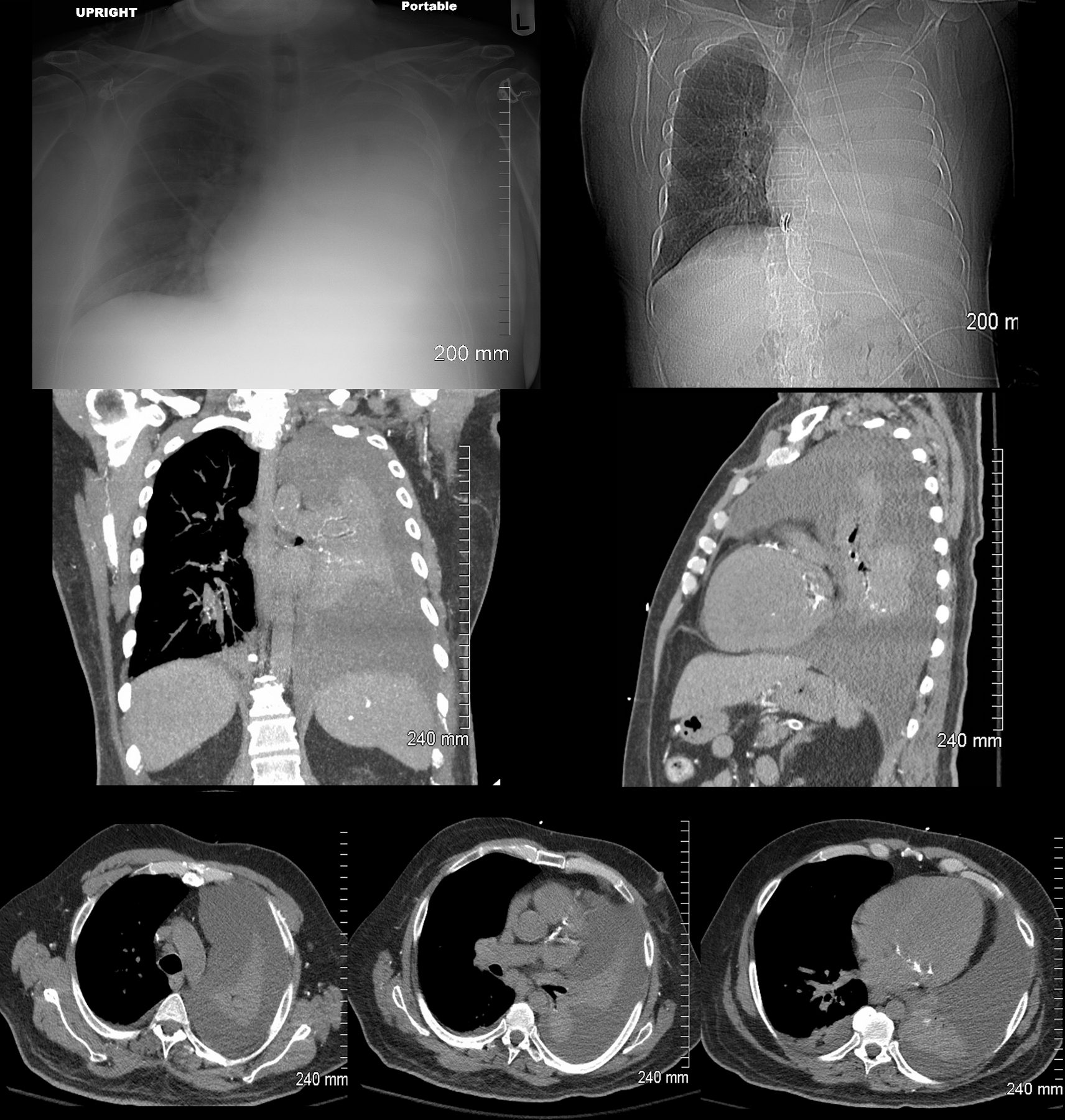
48 year-old male presents with a dyspnea. CXR shows a total white out of the left chest with pulmonary congestion. CT scan shows a large left pleural effusion with total atelectasis of the left lung. Incidental note is made of premature calcific coronary artery disease.
Ashley Davidoff MD TheCommonVein.net
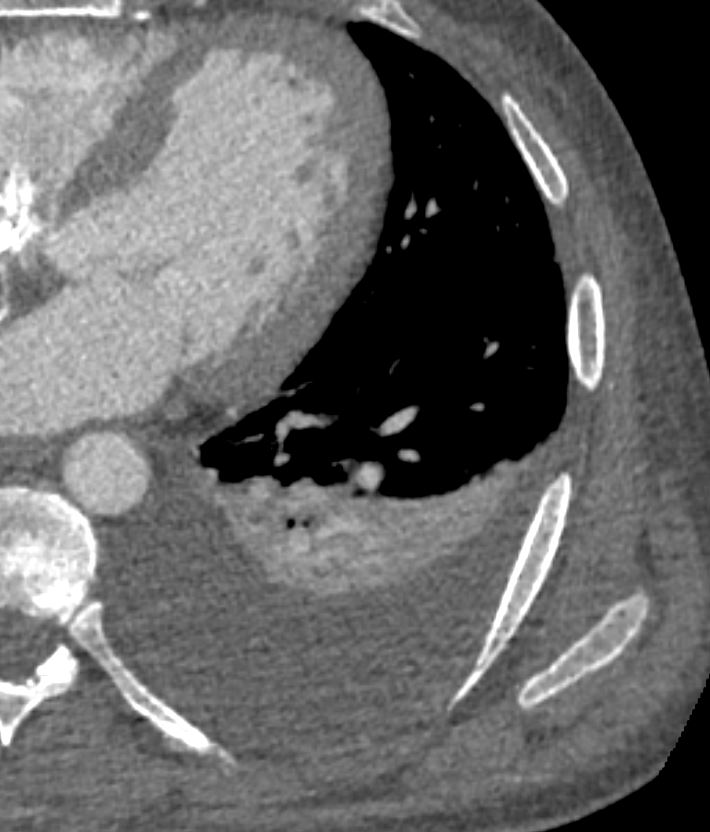
Left Lower Lobe Collapse ,
Partial Left Upper and Right Lower Collapse
88 year old male with bilateral effusions shown on the CXR. Axial CT shows thickened pleura on the left with compressive atelectasis of the lower lobe and a smaller region of crescentic compressive atelectasis on the right. 3D reconstruction shows atelectasis of the left lower lobe and portion of the lingula. The left effusion is complex.
Ashley Davidoff MD TheCommonVein.net
Normal vs Lobar Atelectasis
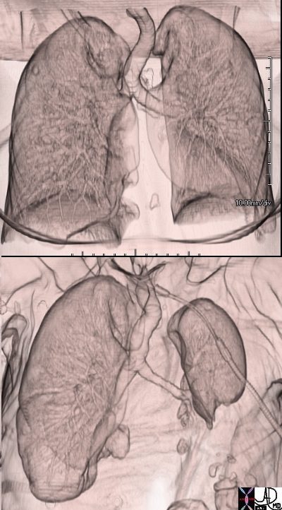
3D reconstruction of a normal patient (above) and of a patient with compressive atelectasis (below) The image below is from an 88 year old male with bilateral complex effusions with compressive atelectasis of the lower lobe and portion of the lingula
Ashley Davidoff MD TheCommonVein.net
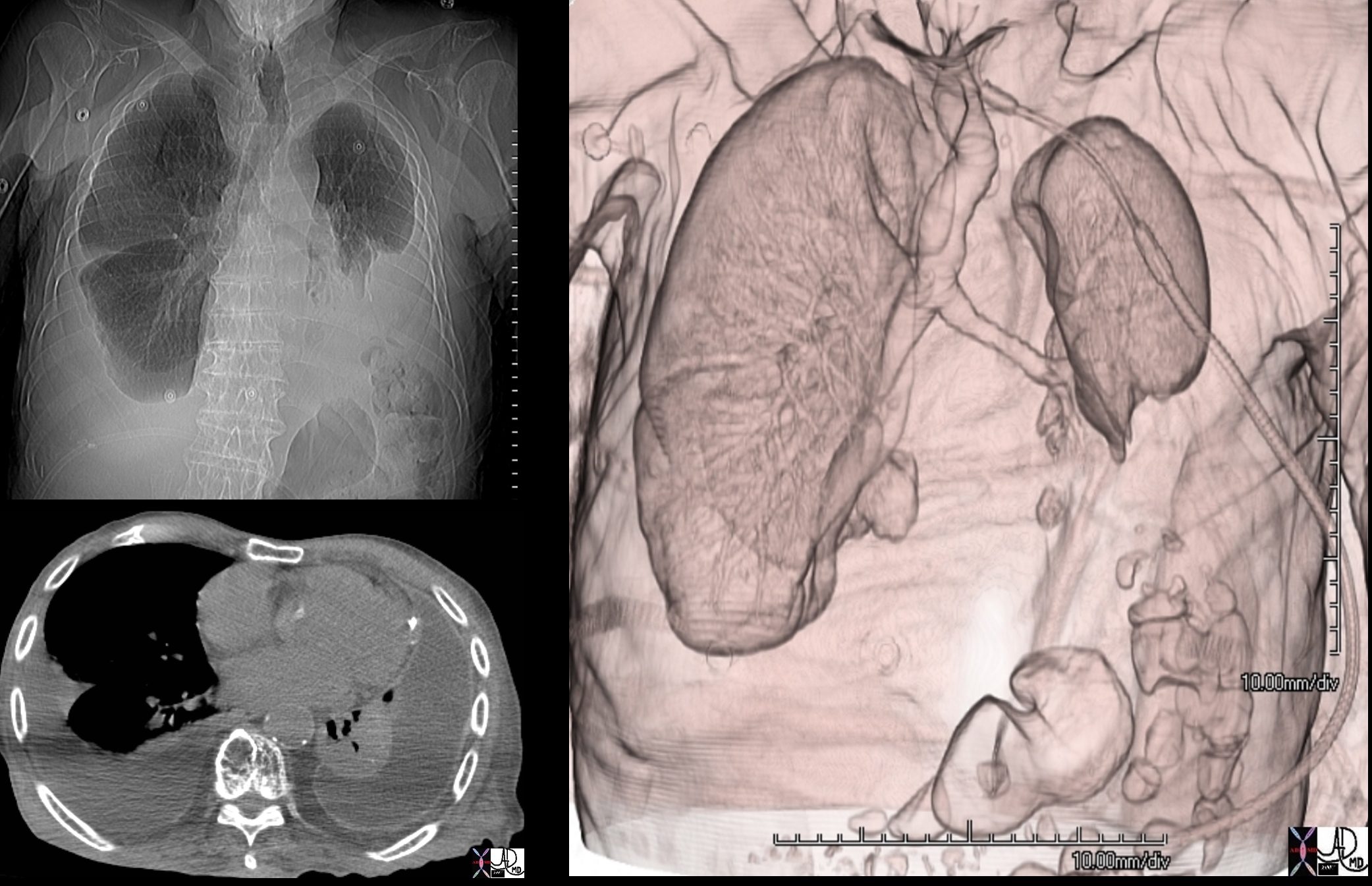
Total Right Lung Collapse
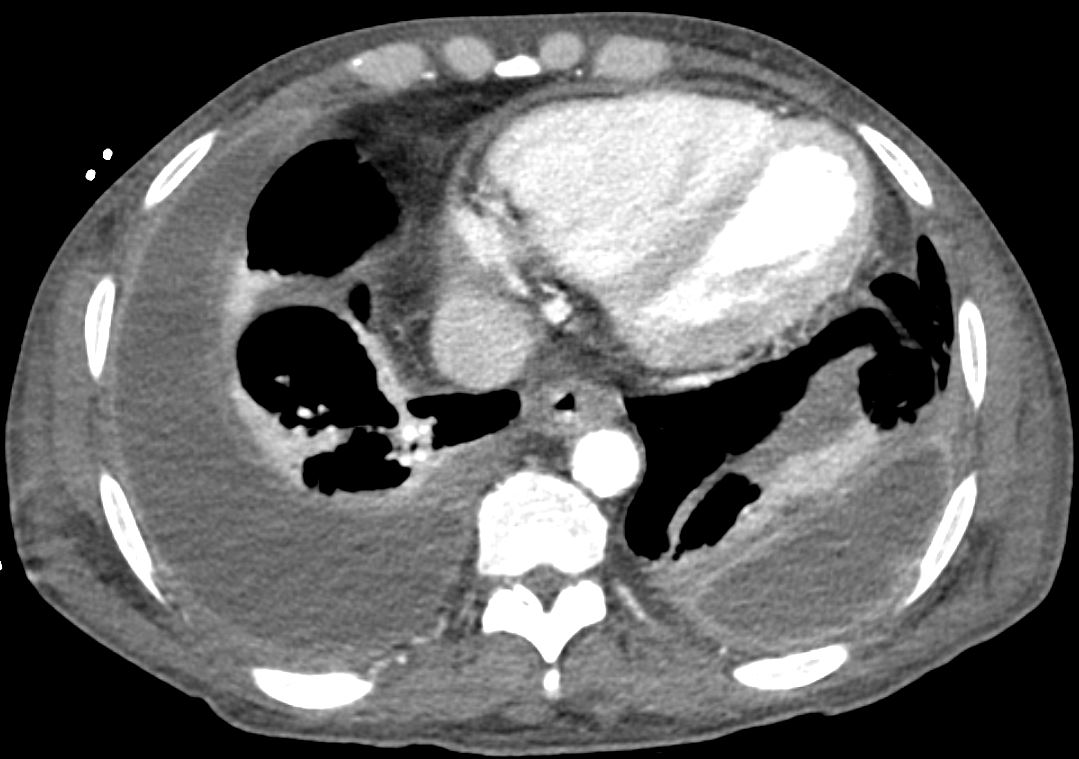
Tension Hydrothorax

85-year-old female with a history of lung cancer, presents with a dyspnea and hypotension. CT scan shows a large right pleural effusion under pressure, with mediastinal shift to the right. In addition, there is compression of the heart with back up of venous return due the pressure effect on the heart and vascular structures. Among the structures showing venous distension are the SVC (blue arrowhead, a) right sided upper limb veins (blue arrowhead b) and the left upper pulmonary veins (red arrowhead, b. The effusion in the right pleural cavity with atelectatic lung herniates into the left hemithorax, (white arrowhead, c). There is a dense sediment in the pleural fluid (red arrowhead, d) suggesting blood in the pleural cavity. The left atrium is compressed (maroon arrowhead, d)
Ashley Davidoff MD TheCommonVein.net
Ashley Davidoff MD TheCommonVein.net
Bilateral Loculated Effusion – Primary Effusion Lymphoma (PEL)
Axial CT scan with contrast of 71-year-old male shows bilateral complex and loculated effusions with thickened enhancing pleura. Pleural tap of the left effusion revealed evidence of lymphoma likely related to the patients underlying herpes virus infection. An entity called primary effusion lymphoma (PEL) is a rapidly progressing non-Hodgkin’s B-cell lymphoma that develops in body cavities
Ashley Davidoff MD TheCommonVein.net 135684