
Ashley Davidoff, M.D. TheCommonVein.net 42540b06

Ashley Davidoff MD TheCommonVein.net lungs-0701
Ashley Davidoff MD TheCommonVein.net lungs-0702
Ashley Davidoff MD TheCommonVein.net lungs-0011
by Ashley Davidoff MD TheCommonVein.net lungs-0016

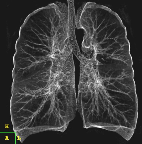
42474b18.800 lung trachea bronchi tracheobronchial tree
Ashley Davidoff MD
TheCommonVein.net 42474b18.800

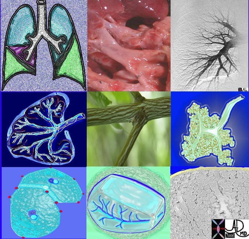
Tracheobronchial Tree
Tree, flower, tracheobronchial tree, trachea bronchi lung
Ashley Davidoff TheCommonVein.net 32620b14.800b02p
82738p chest lung connective tissue pulmonary artery pulmonary vein axial interstitial tissue secondary lobule lobes segments trachea bronchi interlobular septa polygonal Ashley Davidoff MD
TheCommonVein.net
TheCommonVein.net 32679

Tracheobronchial Tree
Tree, flower, tracheobronchial tree, trachea bronchi lung
Ashley Davidoff TheCommonVein.net lungs-0010
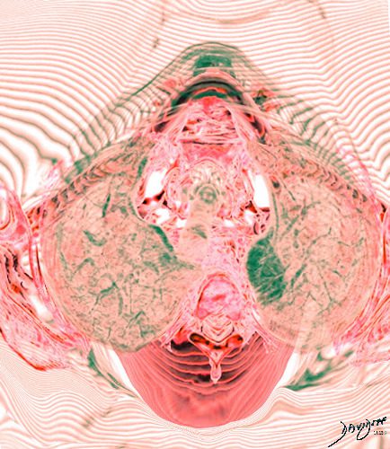
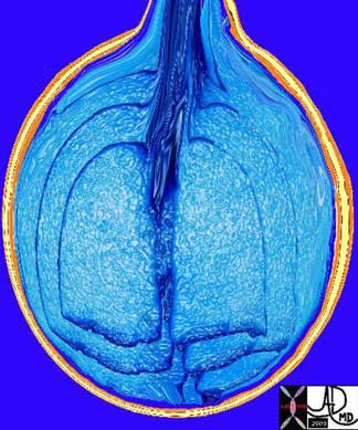
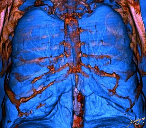
3 D rendering looking down the trachea with the lungs on either side showing the waves of respiratory motion and its effect on the surrounding air. Newtons 3rd law For every action there is an equal and opposite reaction
Ashley Davidoff TheCommonvein.net lungs-0005
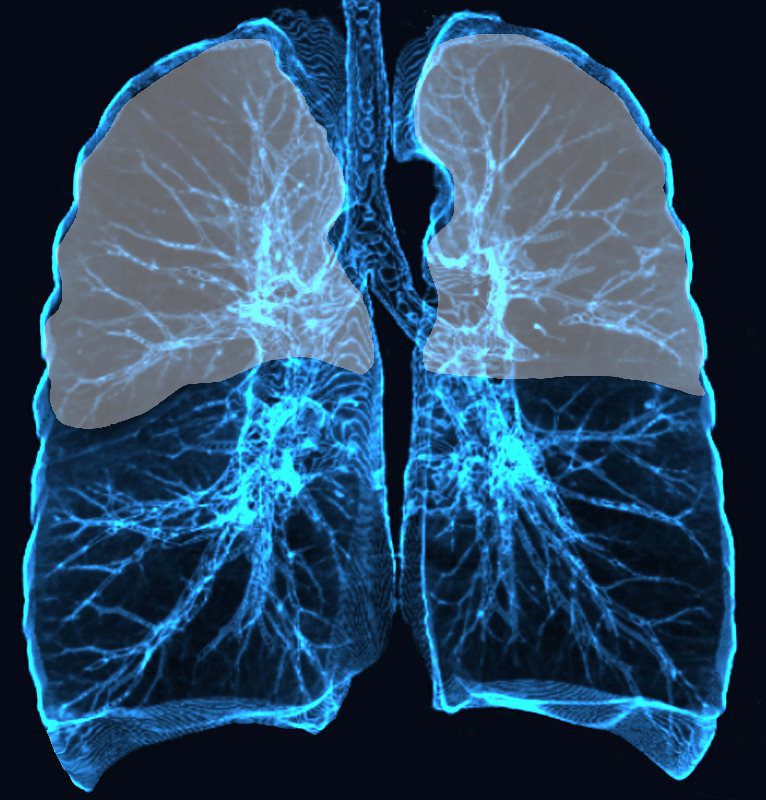
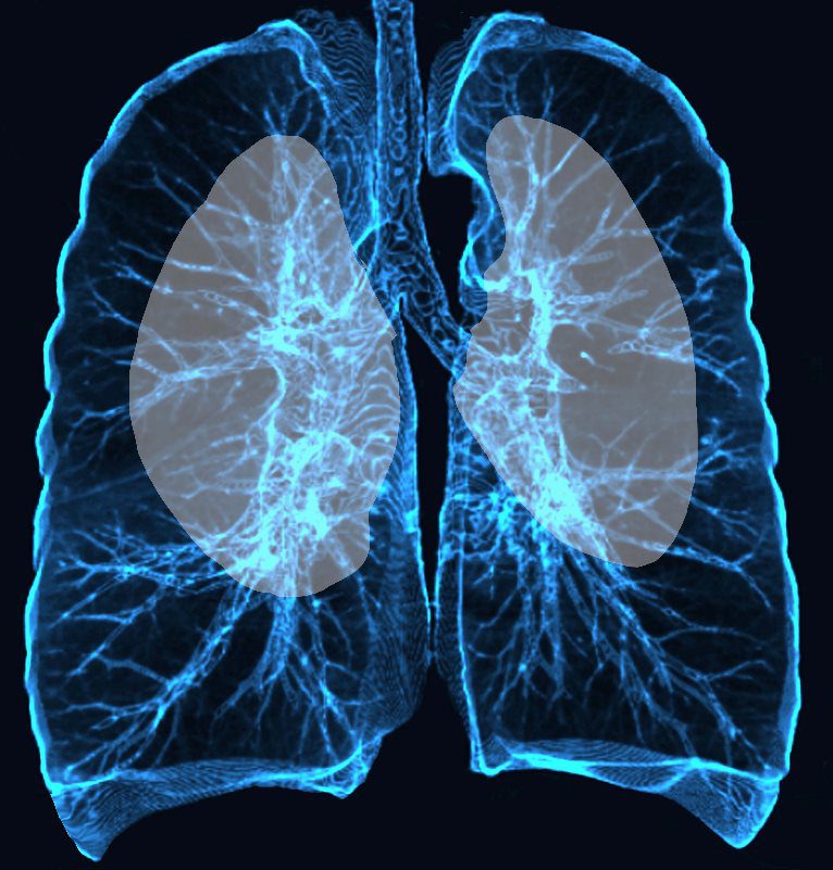
Upper and mid lung field distribution
Ashley Davidoff MD TheCommonvein.net lungs-0772
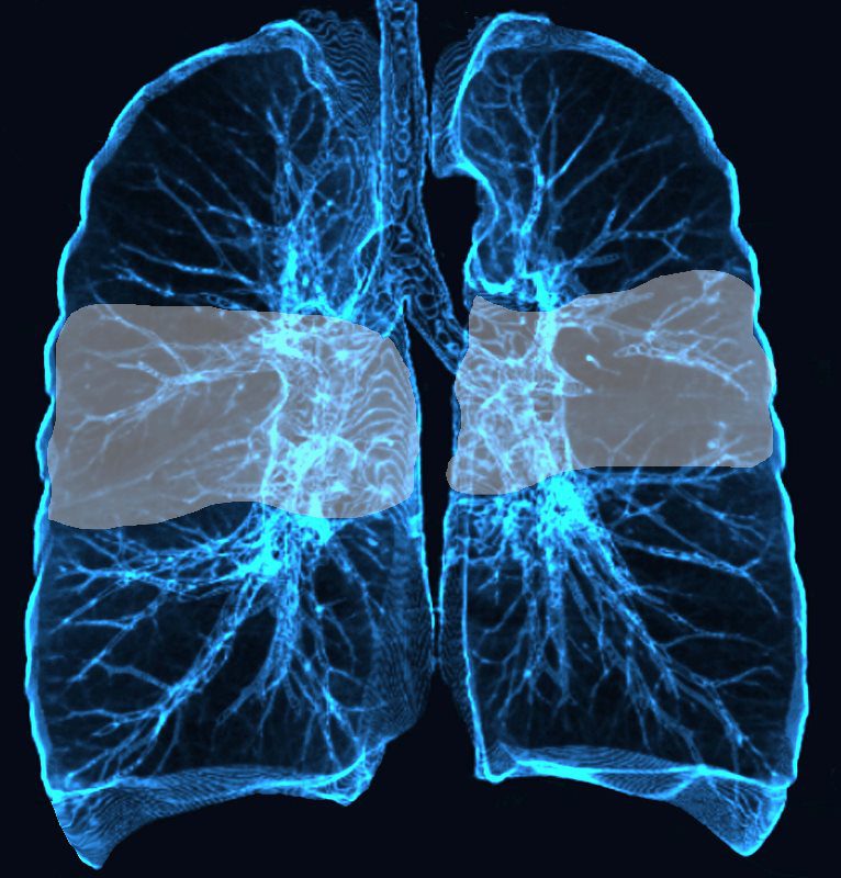
Mid lung field distribution
Ashley Davidoff MD TheCommonvein.net lungs-0773
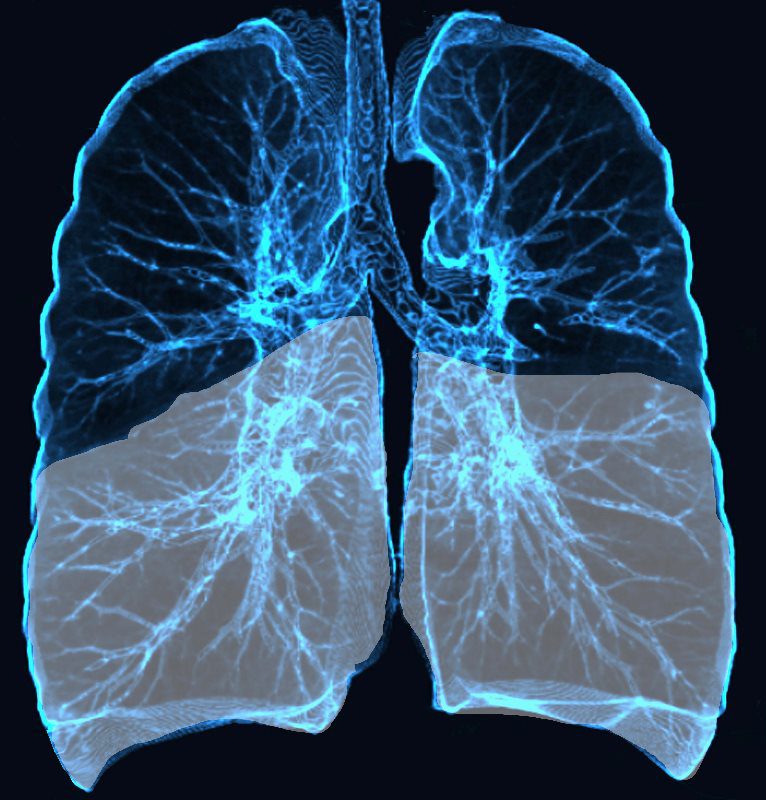
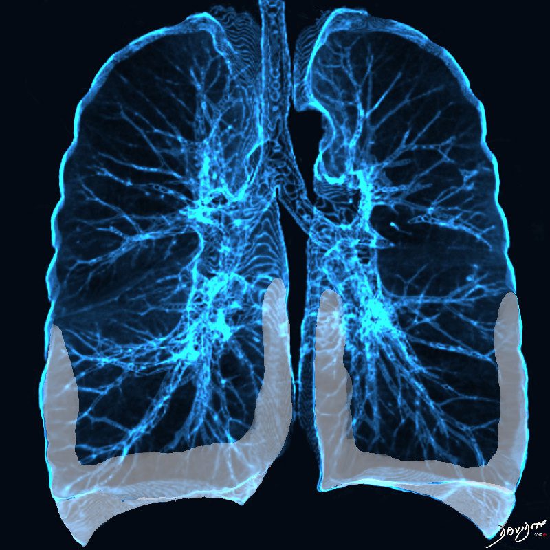
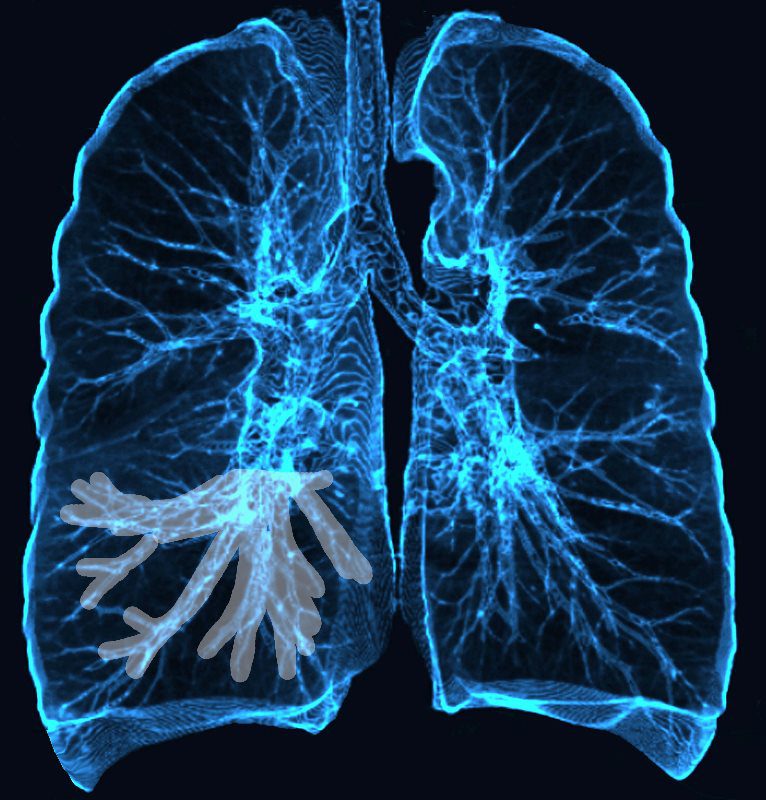
Lower Lobe distribution
Ashley Davidoff MD TheCommonvein.net lungs-0771
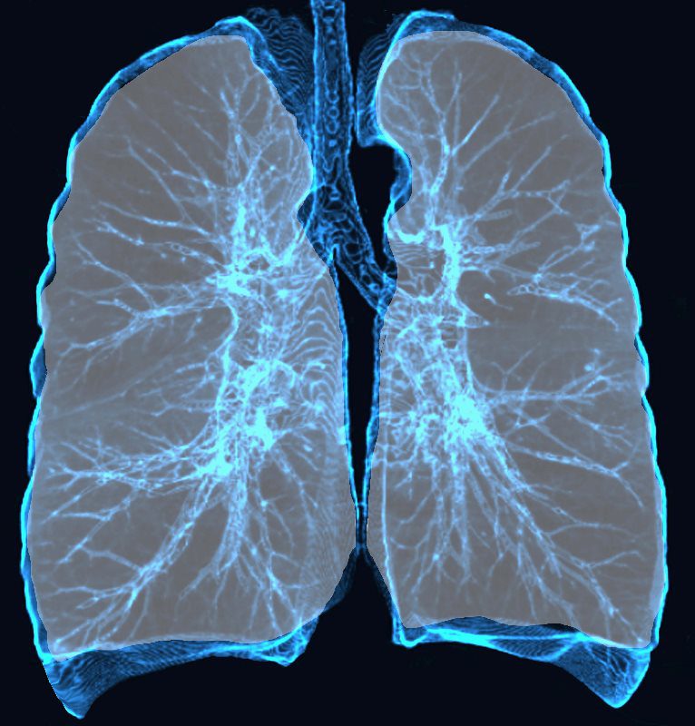
Diffuse Lung Disease
Ashley Davidoff MD TheCommonvein.net lungs-0775
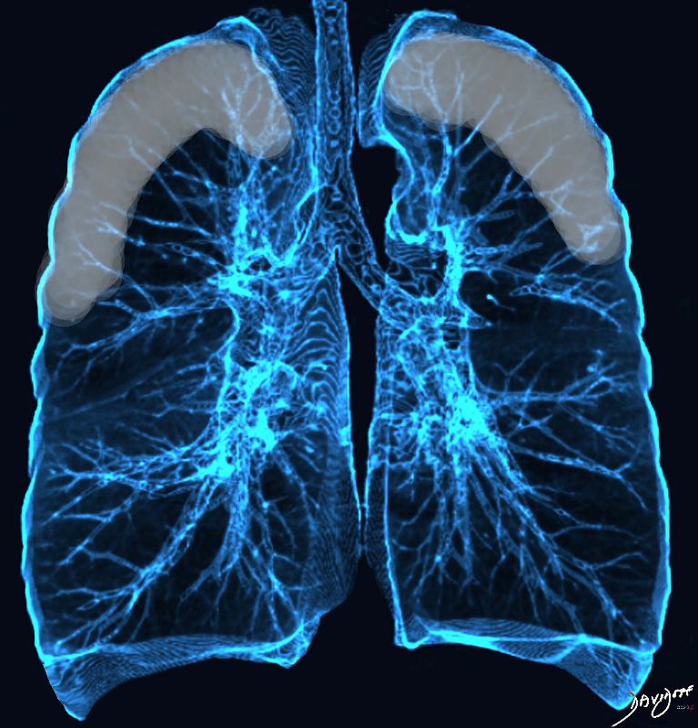
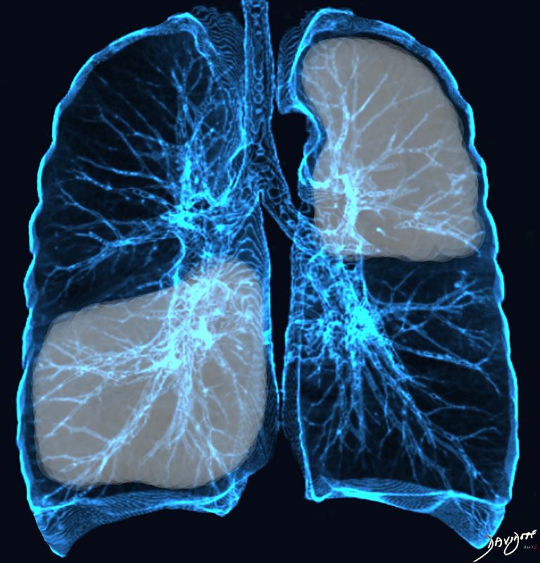
Perihilar distribution
Ashley Davidoff MD TheCommonvein.net lungs-0770
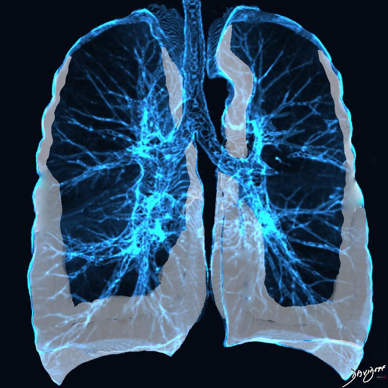
Basilar and peripheral distribution
Ashley Davidoff MD TheCommonvein.net lungs-0769b
As the disease progresses the lower disease becomes more extensive and the disease progresses into the periphery of the upper lobes as well
Ashley Davidoff MD TheCommonvein.net lungs-0769c
Chronic eosinophilia is characterised by alveolar filling with eosinophils and inflammatory exudates(a) and interalveolar interstitial thickening, (overlaid in red in b). The infiltrates are classically peripherally positioned, usually upper lobes, more commonly bilateral but can be unilateral, and manifest as consolidation and or ground glass opacities. The CT shows bilateral peripheral consolidations in the upper lobes
Ashley Davidoff MD The CommonVein.net lungs-0775e
Subpleural Sparing
Ashley Davidoff MD TheCommonvein.net lungs-0775 0775-lo res subpleural sparing
Broncho vascular distribution
Ashley Davidoff MD TheCommonvein.net lungs-0769
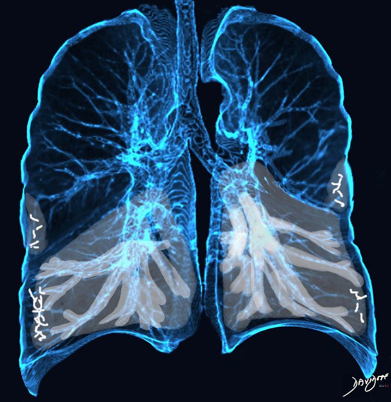
Broncho vascular distribution associated with peripheral sparing, ground glass changes, reticulations, and volume loss, dominantly in the lower lobes but to some extent in the middle lobe and upper lobes
Ashley Davidoff MD TheCommonvein.net lungs-0771b
Broncho vascular distribution associated with peripheral sparing, ground glass changes, reticulations, and volume loss, dominantly in the lower lobes but to some extent in the middle lobe and upper lobes
Ashley Davidoff MD TheCommonvein.net lungs-0771b
Broncho vascular distribution associated with increased reticular changes, more prominent traction bronchiectasis, decreased lung volumes , and decreased lung volumes, dominantly in the lower lobes but to some extent in the middle lobe and upper lobes. Pulmonary hypertension becomes more common.
Ashley Davidoff MD TheCommonvein.net lungs-0771d
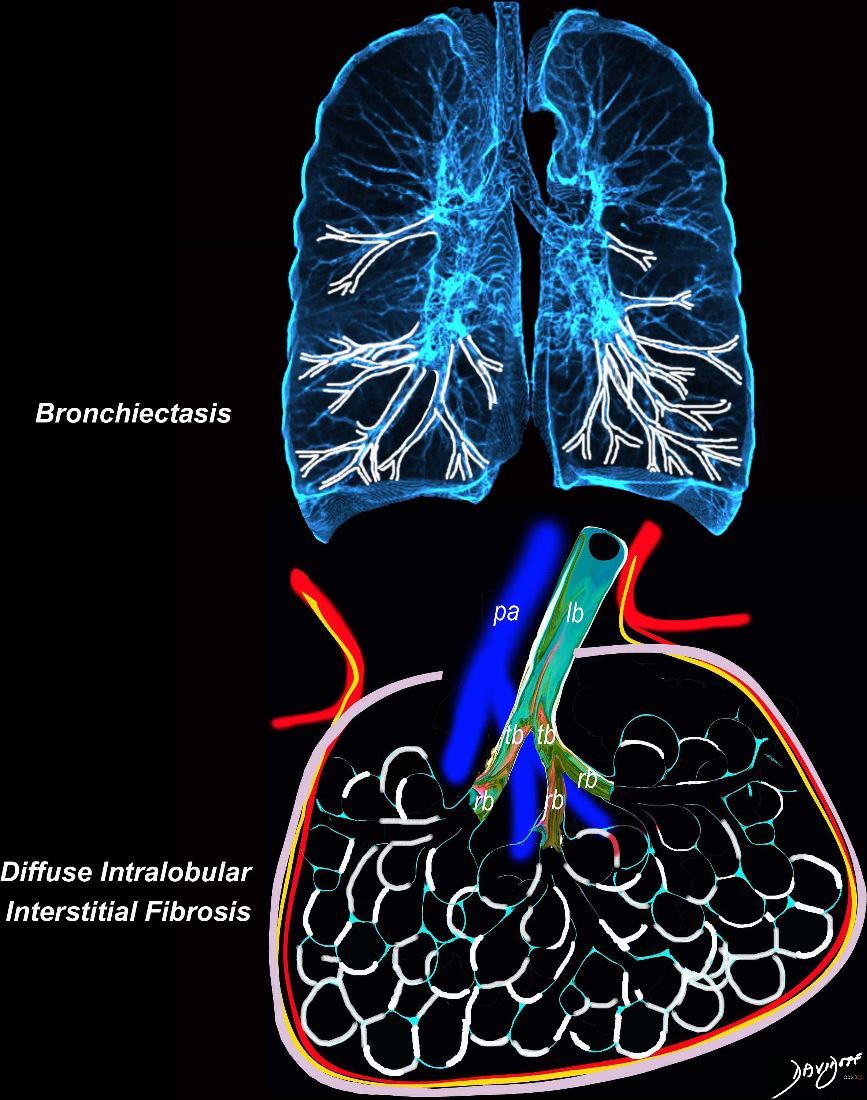
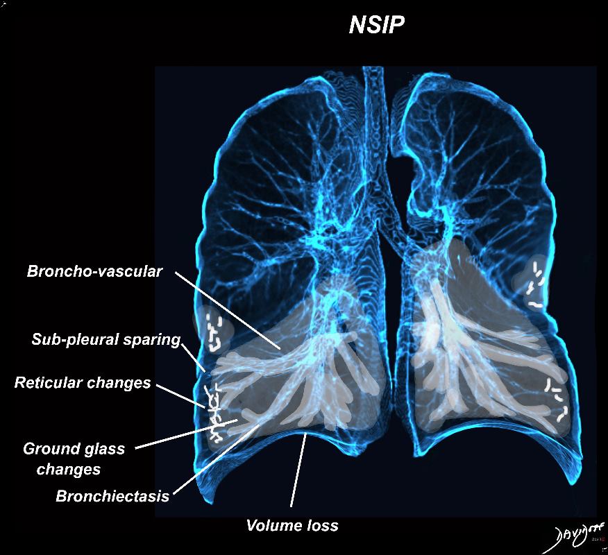
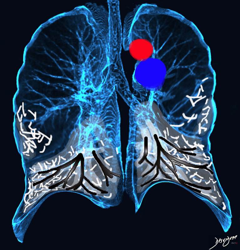
Broncho vascular and inter- alveolar interstitial fibrosis dominantly in the lower lobes but affecting the middle and upper lobes to lesser extent resulting in bronchiectasis and reticulations. The overall increase in density results in ground glass changes
Ashley Davidoff MD TheCommonvein.net lungs-0738 NSIP