
he Mycobacterium tuberculosis complex (MTC or MTBC) is a genetically related group of Mycobacterium species that can cause tuberculosis in humans or other animals. It includes: Mycobacterium tuberculosis. Mycobacterium africanum.
M. tuberculosis is a member of the M. tuberculosis complex; other members include Mycobacterium africanum and Mycobacterium bovis.
- Patient is a 68-year-old female, with history of seropositive RA (on rituximab, who presents for follow-up.
- exposure to TB as a child in Russia where she was apparently treated with an unknown regimen.
- She was first seen
- 1 year prior fo
- PNA,
- weight loss,
- night sweats,
- quantiferon gold+, and
- lymphocyte dominant pleural effusion
- Imaging showed
- diffuse tree-in-bud opacities
- Microbiology was
- negative at that time,
- rx
- empirically began on
- Rifampin 600 mg po daily,
- Isoniazid 300 mg daily,
- Pyridoxine 50 mg daily, and
- Ethambutol 1600 mg daily
- due to severe nausea and vomiting.
- empirically began on
- TB confirmed on bronchoscopy
- positive for Mycobacterium Tuberculosis Complex
- Rx
- TB medications on including INH, rifampin and ethambutol
- Rising eosinophil count and elevated LFTs concerning for DRESS.
- Drug reaction with eosinophilia and systemic symptoms (DRESS) is a
- severe adverse drug reaction
- characterized by an
- extensive skin rash
- visceral organ involvement,
- lymphadenopathy,
- atypical lymphocytosis.
- Drug reaction with eosinophilia and systemic symptoms (DRESS) is a
CXR Micronodules Right Upper Lobe
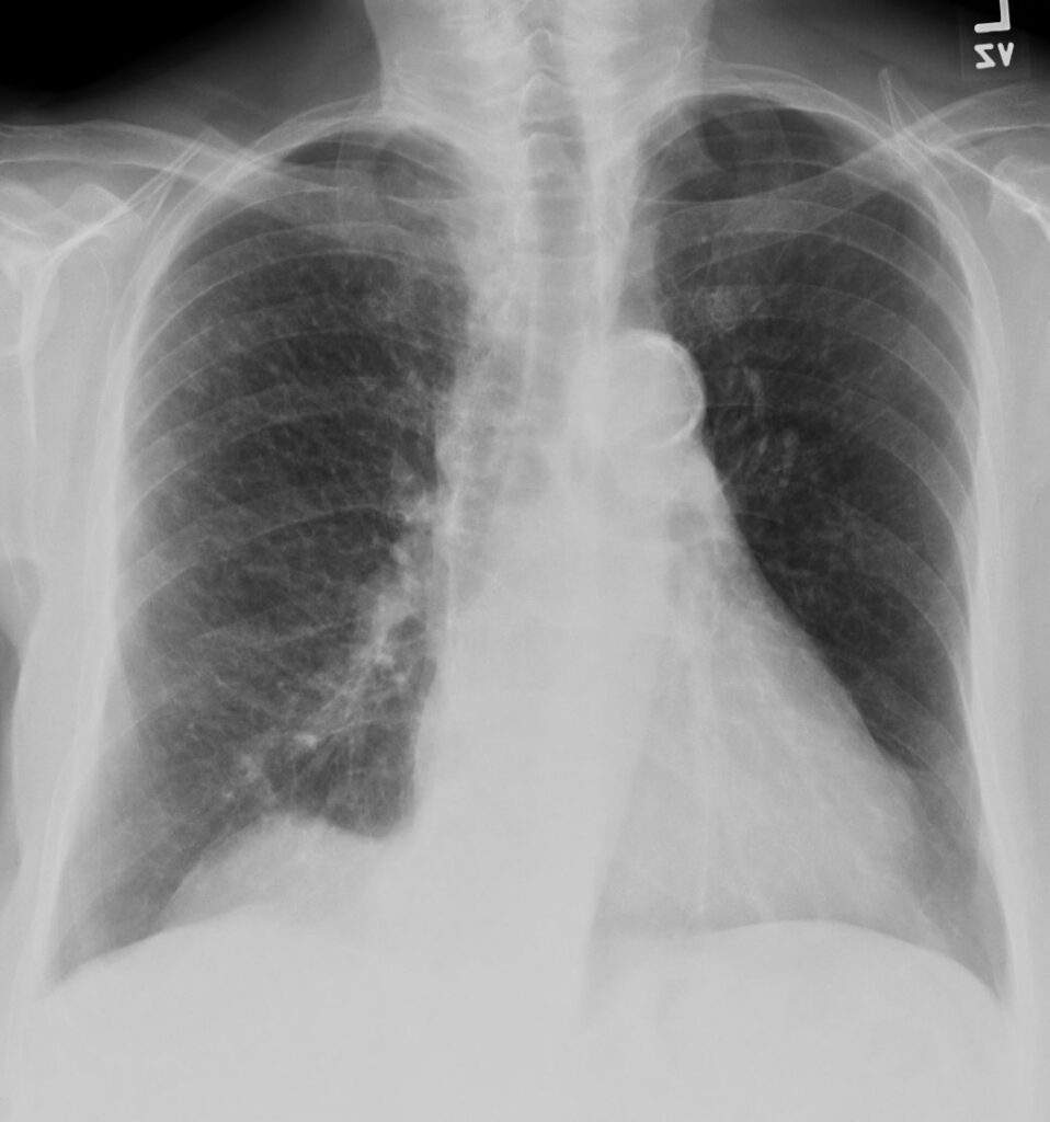
68 year old female presented with malaise night sweats weight loss Quantiferon gold positive, with a past history of treated TB in her native country as a child. Frontal CXR shows a nodular pattern of disease affecting the right upper and right lower lobe. Bronchoscopy isolated Mycobacterium complex. She was treated with good result
Ashley Davidoff MD TheCommonVein.net 135820 192Lu
Subsegmental and Small Airway Disease Right Upper Lobe Active TB
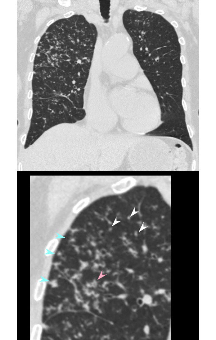
68 year old female presented with malaise night sweats weight loss QuantiFeron gold positive, with a past history of treated TB in her native country as a child. CT shows subsegmental and small airway disease dominant in the right upper lobe characterized by airway thickening (pink arrowhead),ground glass micronodules (white arrowheads) and centrilobular nodules (teal blue arrowheads). There are lesser changes in the right lower lobe, and left upper lobe. Bronchoscopy isolated Mycobacterium complex. She was treated with good result
Ashley Davidoff MD TheCommonVein.net 135822cL 192Lu
Subsegmental and Small Airway Disease Right Upper Lobe
Active TB
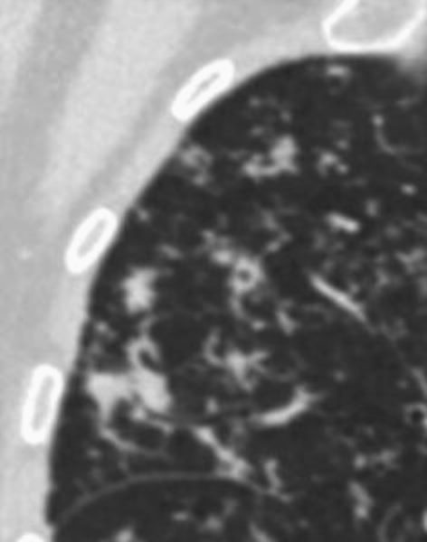
CT shows subsegmental and small airway disease dominant in the right upper lobe characterized by airway thickening, ground glass micronodules and centrilobular nodules
Ashley Davidoff MD TheCommonVein.net 135823 192Lu
Small Airway Disease and Interlobular Septal Changes
Right Upper Lobe
ActiveTB
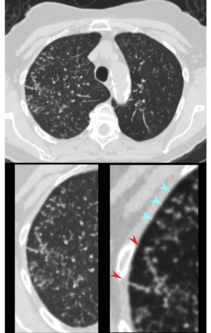
CT in the axial plane shows extensive small airway disease dominant in the right upper lobe characterized by innumerable, ground glass micronodules, centrilobular nodules (teal blue arrowheads) and nodular thickening of the interlobular septa likely reflecting lymphatic involvement (red arrowheads)
Ashley Davidoff MD TheCommonVein.net 135825cL 192Lu
Small Airway Disease and Tree in Bud Changes
Favors Transbronchial Spread
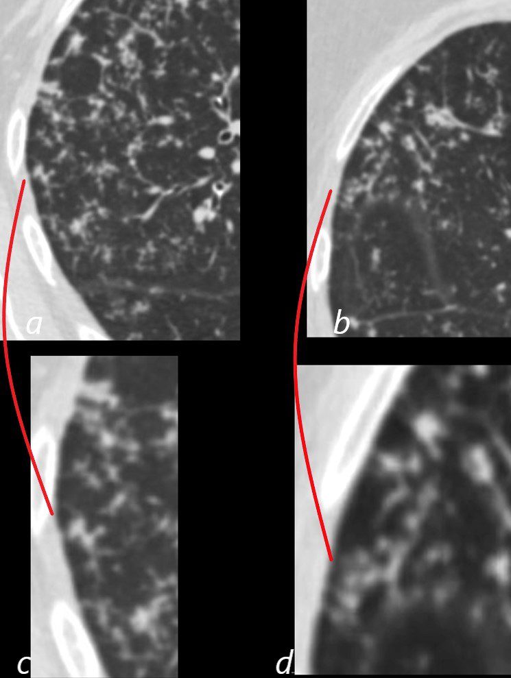
CT in the axial plane shows extensive small airway disease dominant in the right upper lobe characterized by innumerable, ground glass micronodules, and tree in bud changes. (a magnified in c and b magnified in d)
Ashley Davidoff MD TheCommonVein.net 135827aL 192Lu
Small Calcified Granulomatous Lymph Node
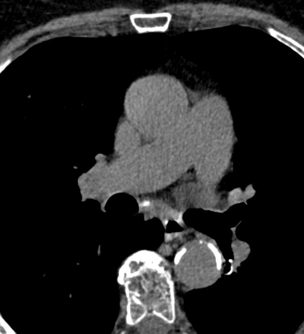
68 year old female presented with malaise night sweats weight loss QuantiFeron gold positive,
CT at the level of the main pulmonary artery shows a small calcified subcarinal node
Ashley Davidoff MD TheCommonVein.net mycobacterium-complex-135829 192Lu
2 years Later Following Treatment
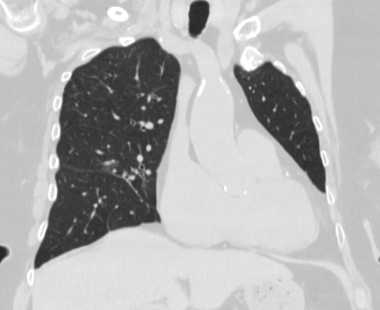
Coronal CT shows significantly improved small airway disease with no evidence of micronodules
Ashley Davidoff MD TheCommonVein.net 135830 192Lu