
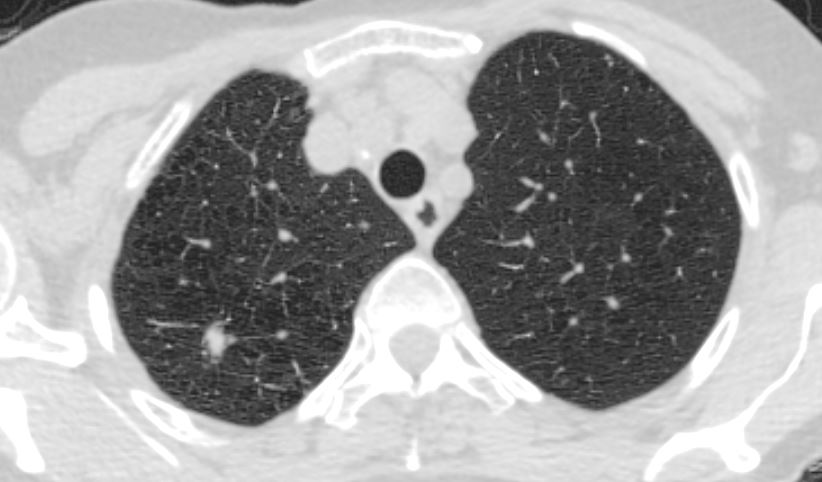
The PET scan showed faint FDG uptake which was below blood pool
Pathology revealed inflammatory pseudotumor
Ashley Davidoff MD TheCommonVein.net
PET Scan
. The 1 x 0.6 cm right upper lobe nodule showed howed faint FDG uptake which was below blood pool
It was stated that the nodule could be hypocellular and the true metabolic activity was likely underestimated. Findings
were still suggestive of malignant lung nodule.
2. No hypermetabolic mediastinal or hilar lymph nodes. No scan
evidence of distant metastasis.
Pathology
Benign alveolar lung parenchyma with moderate, predominantly plasmacytic, mononuclear chronic inflammatory cell infiltrate consistent with inflammatory pseudotumor.
No granulomas, vasculitis, viral cytopathic changes or carcinoma seen.
Unremarkable bronchial, vascular and parenchymal resection margins.
Smaller lung segment with mild emphysematous changes.