
Source
Signs in Thoracic Imaging
Journal of Thoracic Imaging 21(1):76-90, March 2006.
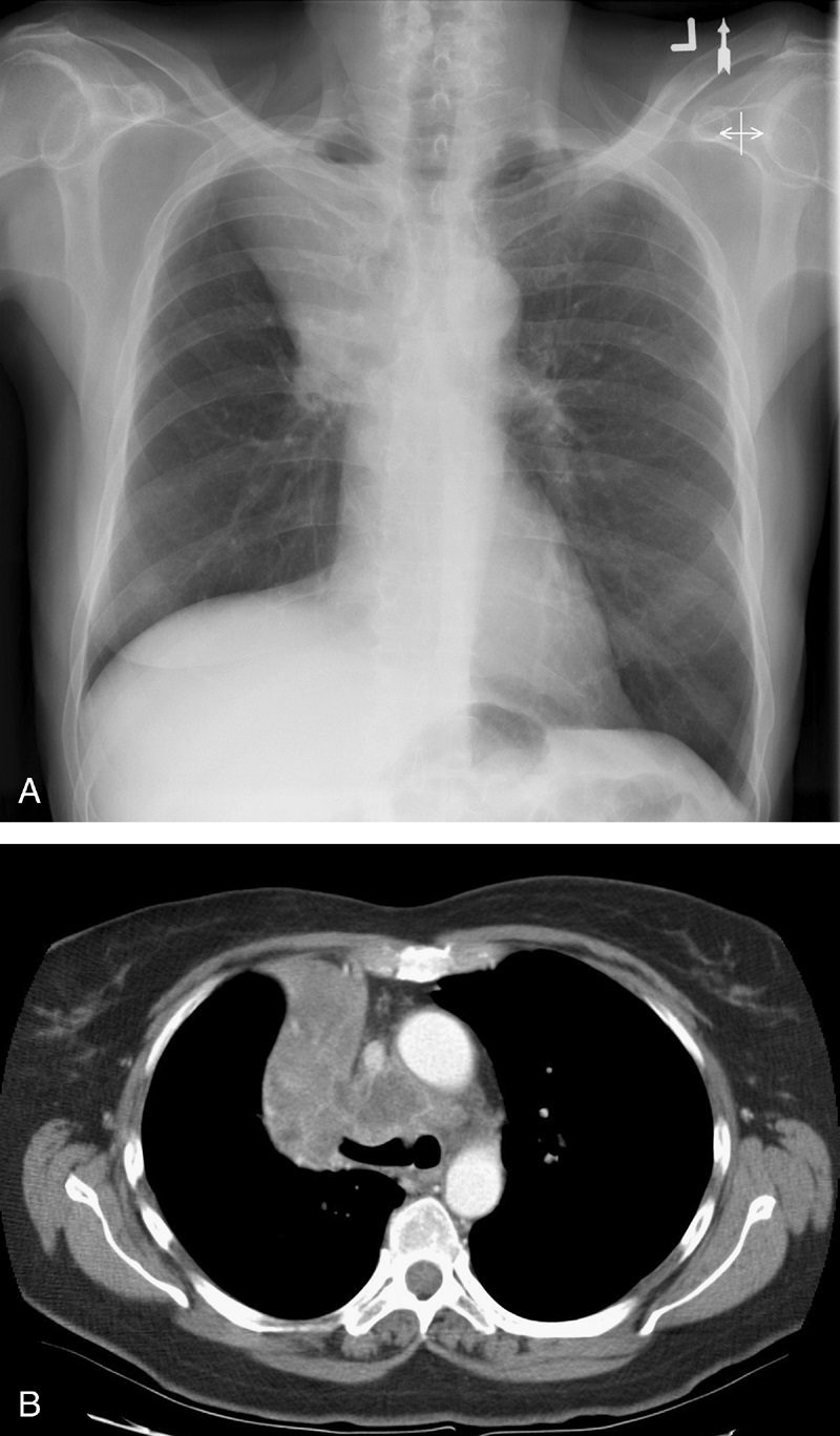
Dr Ross Golden was an American-born radiologist who originally described this sign as a form of right upper lobe collapse associated with a central mass.45 When the right upper lobe bronchus is obstructed by an endobronchial lesion, there is elevation and medial displacement of the minor fissure with proximal convexity of the fissure due to the mass (Fig. 13). This creates the “reverse S” characteristic of central obstructing bronchogenic carcinomas.3,4,45 Given the same presentation of a proximal, obstructing endobronchial lesion within the left upper lobe bronchus with associated left upper lobe collapse, the upwardly retracted major fissure will follow an S-shaped contour along its length. Although initially used to describe signs of right upper lobe collapse, the Golden S sign can be applicable to atelectasis involving any lobe.46