
Ashley Davidoff MD TheCommonVein.net
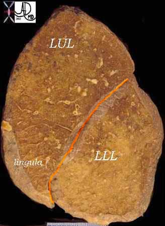
Lingula Left Side in Pink
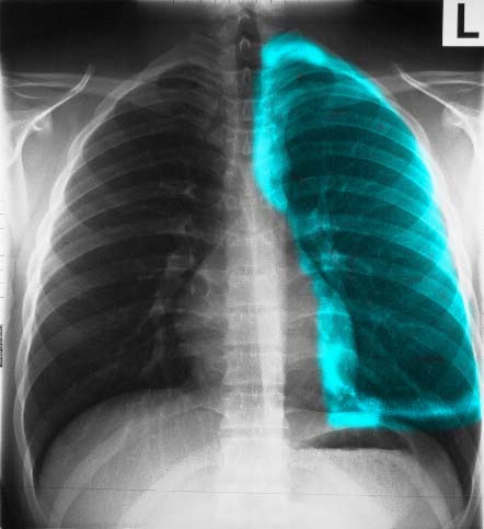
The A-P examination of the chest shows the LUL, with the teal overlay representing the upper lobe and lingula.. Note how the lingula hugs the left heart border. Ashley Davidoff MD TheCommonVein.net 30397b05
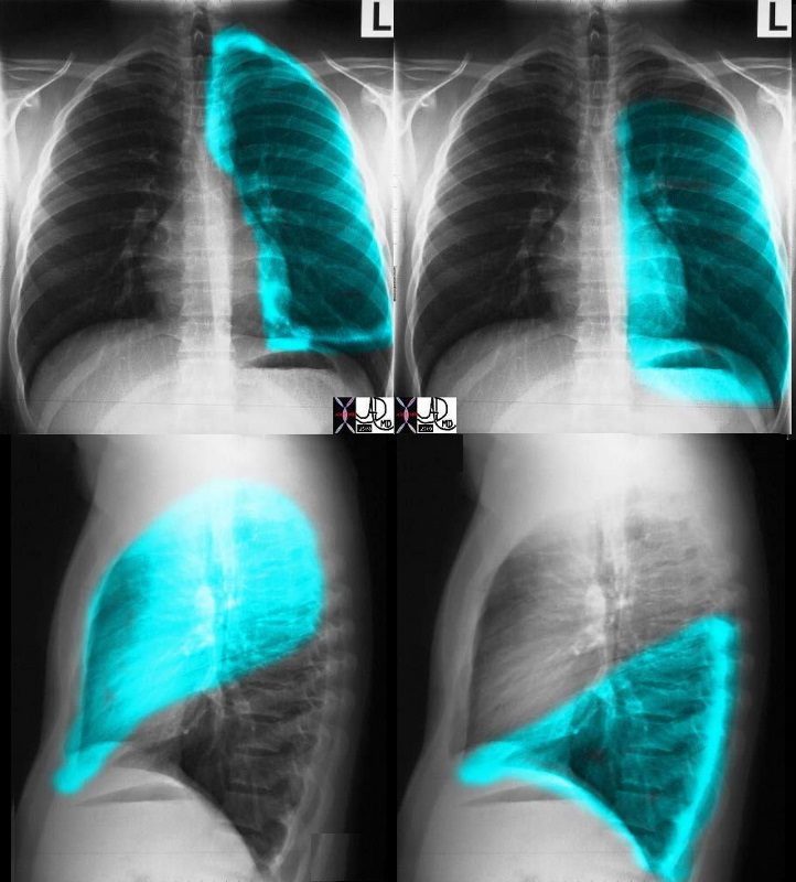
In left sided images (top and bottom on the left), the overlay represents the upper lobe and lingula. Note how the lingula hugs the left heart border. The top and bottom on the right represent the left lower lobe. The volume of the LUL and LLL is about equal. Courtesy Ashley Davidoff MD. 30398c02.8
Lingular Segments Superior and Inferior
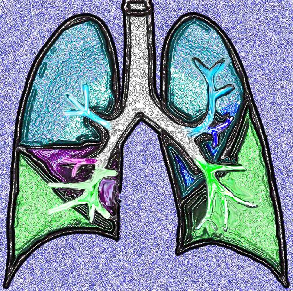
This diagram shows the basic division of the tracheobronchial tree into lobes. The right lung is divided into right upper (RUL) (teal) right middle, (RML pink) and right lower lobe (RLL green). The left lung is divided into left upper (LUL teal), which includes the lingula(dark blue), and left lower lobe (LLL= green). Note that the two mainstem bronchi are of unequal length and size. The right mainstem is short and fat while the left is long and thin. This irregular dichotomous branching pattern is characteristic of the branching pattern of all the conducting systems within the lungs.
Ashley Davidoff
TheCommonVein.net 32686b05
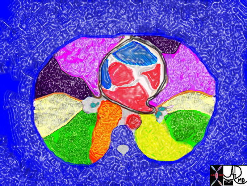
The axial CT through the level of the heart shows a few of the right and left pulmonary segments including parts of the middle lobe, lingula and of the lung bases
Ashley Davidoff MD TheCommonVein.net 32557bb03.8s
Inferior Segment Lingula Infiltrate
Inferior segment
Ashley Davidoff MD TheCommonVein.net 130903c.8
52 year old male presents with a cough and fever
Lingular Pneumonia

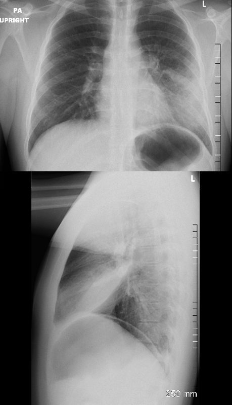
52 year old male presents with a cough and fever
Frontal CXR shows a lingular infiltrate with a positive silhouette sign. Both the superior and inferior lingular segments appear to be involved
Ashley Davidoff MD TheCommonVein.net
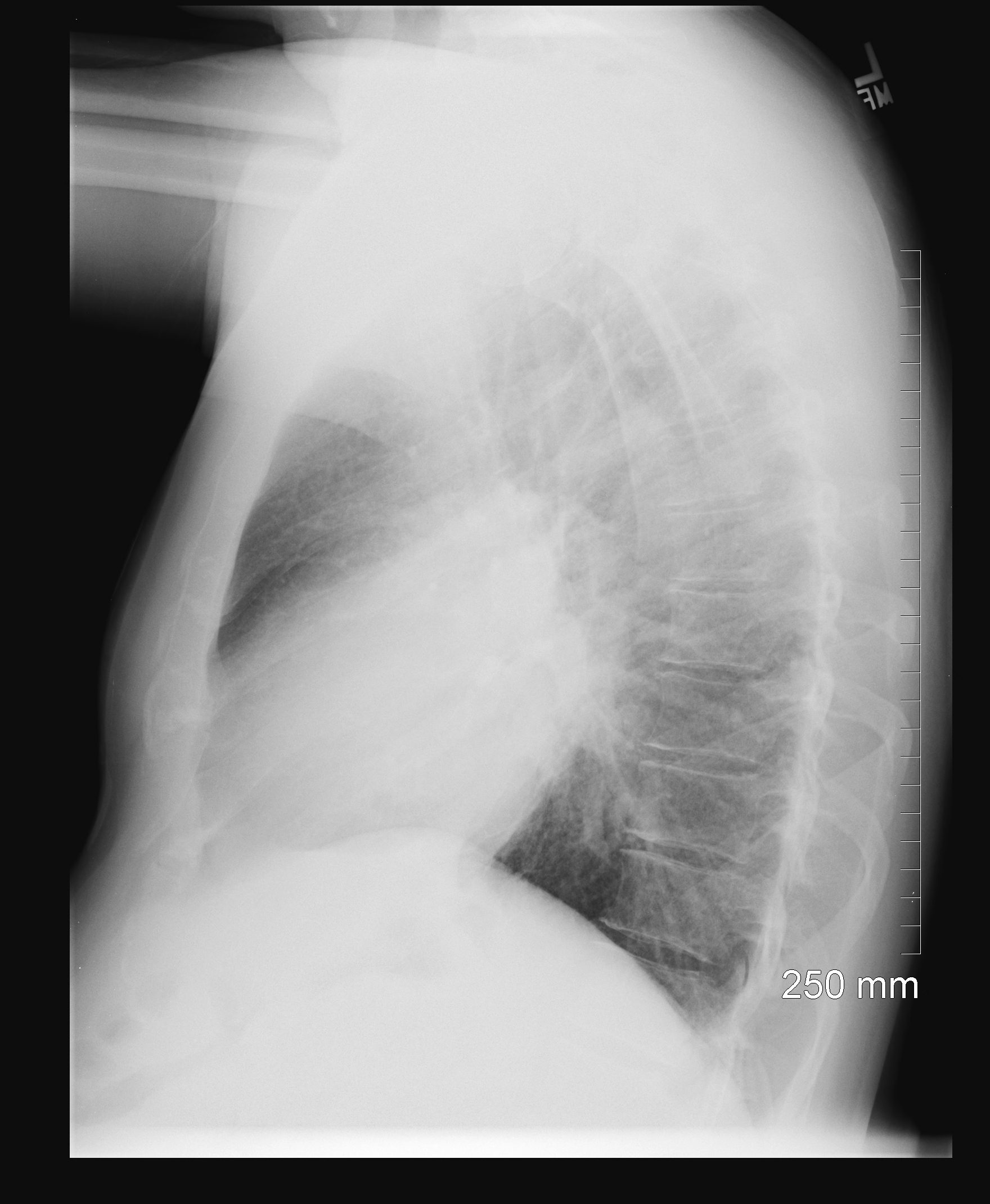
52 year old male presents with a cough and fever
Lateral CXR shows a dense lingular infiltrate overlying the heart. Both the superior and inferior lingular segments appear to be involved
Ashley Davidoff MD TheCommonVein.net
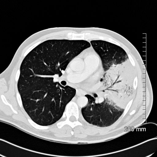
52 year old male presents with a cough and fever
CT scan in the axial plane shows a lingular consolidation with air bronchograms and a positive silhouette sign. Both the superior and inferior lingular segments are involved
Ashley Davidoff MD TheCommonVein.net
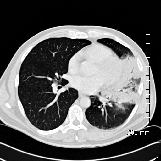
52 year old male presents with a cough and fever
CT scsn in the axial plane shows a lingular consolidation with air bronchograms and a positive silhouette sign. Both the superior and inferior lingular segments are involved
Ashley Davidoff MD TheCommonVein.net
CXR Silhouetting Left Heart Border Lingula Atelectasis
Central Obstructing Carcinoid Tumor
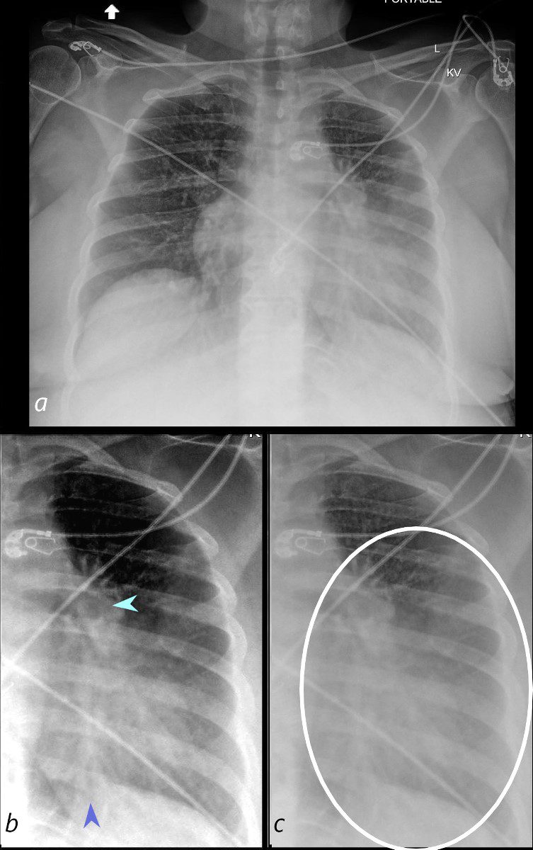
58-year-old female presents with a cough Frontal CXR shows silhouetting of the left heart border with hazy or veiling opacity extending out from the left hilum and fading out inferiorly (white circle c). The left hilum is pulled superiorly (teal arrowhead b) , resulting in an almost horizontal course of the left main bronchus and vertical orientation of the left lower lobe bronchovascular bundle (dark blue arrowhead b)
Ashley Davidoff MD TheCommonVein.net 257Lu 136109cL01
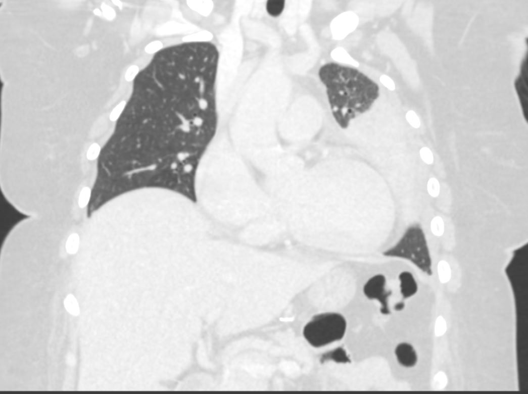
58-year-old female presents with a cough. CT in the coronal plane shows post obstructive atelectasis of the lingula which silhouettes the left heart border. A small portion of aerated left upper lobe is noted in the left apex.
Pathology revealed findings consistent with a carcinoid tumor of the left bronchus.
Ashley Davidoff MD TheCommonVein.net 257Lu 136115
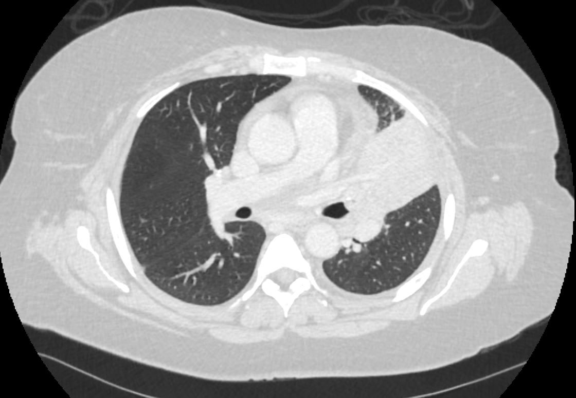
58-year-old female presents with a cough. CT in the axial plane shows an obstructing lesion in the left mainstem bronchus of the lung with post obstructive atelectasis of the lingula and a small portion of aerated left upper lobe anteriorly. T he major fissure is displaced anteriorly.
Pathology revealed findings consistent with a carcinoid tumor of the left bronchus.
Ashley Davidoff MD TheCommonVein.net 257Lu 136110
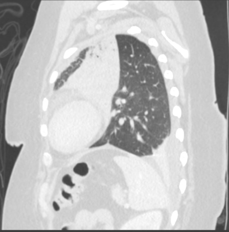
58-year-old female presents with a cough. CT in the sagittal plane shows post obstructive atelectasis of the lingula, hyperinflation of the left lower lobe, superior and anterior migration of the left major fissure, and a small portion of aerated left upper lobe anteriorly. There is a loculated effusion with subsegmental compressive atelectasis of the left lower lobe.
Pathology revealed findings consistent with a carcinoid tumor in the left mainstem bronchus
Ashley Davidoff MD TheCommonVein.net 257Lu 136121