162Lu Pulmonary Langerhans Cell Histiocytosis and Treated TB
43 year old female w/PMH of OSA on cpap, +ppd s/p rx 2004, allergic rhinitis, pulmonary Langerhans cell histiocytosis diagnosed in 2008 which resolved with smoking cessation.
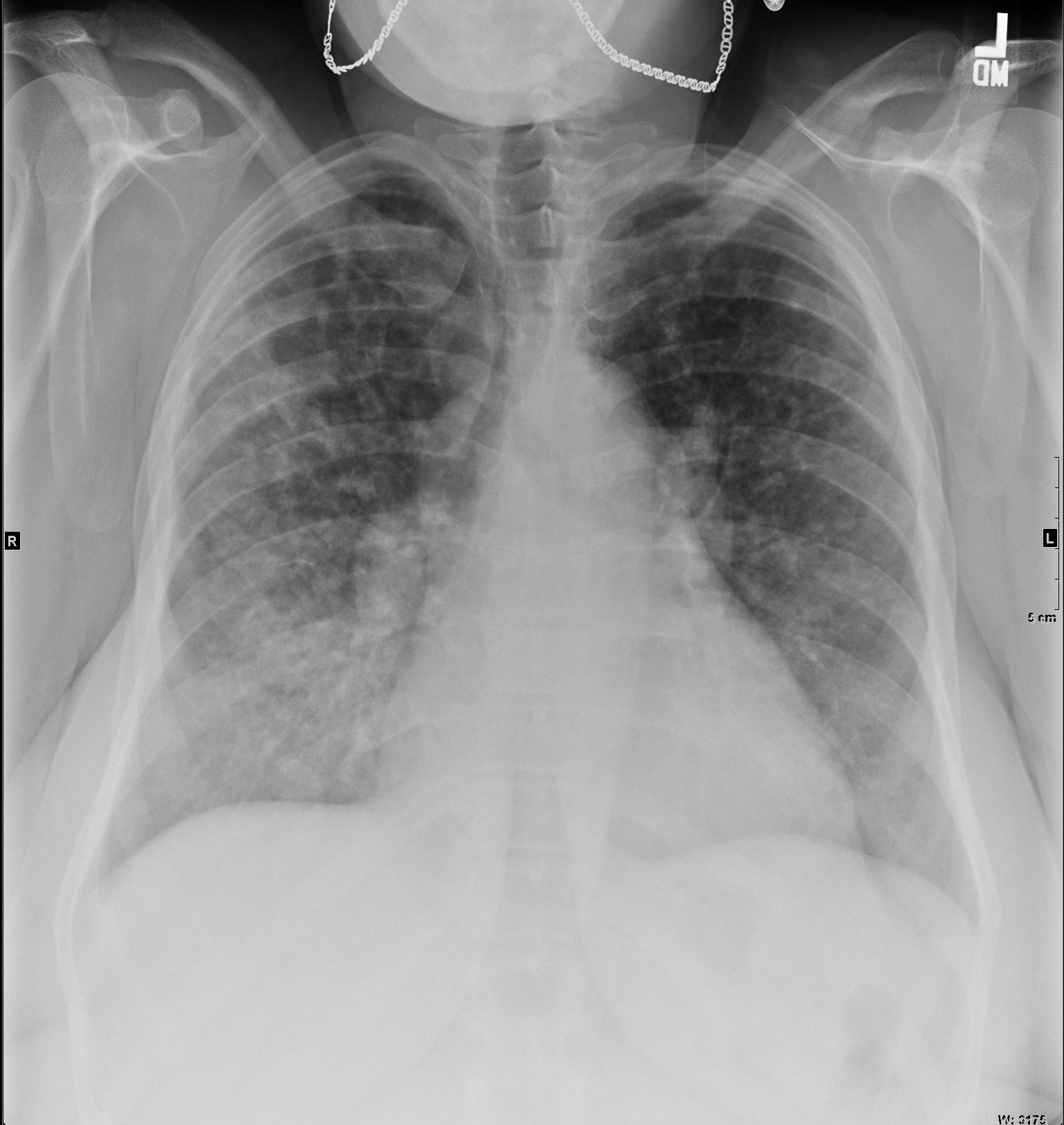
50 year old obese female heavy smoker presents with a cough 15 and half years ago CXR is normal Ashley Davidoff MD TheCommonVein.net 50F 0012 months later she is in respiratory distress and CXR shows bilateral diffuse nodular infiltrates Ashley Davidoff MD TheCommonVein.net 50F 001 0
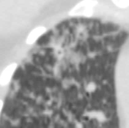
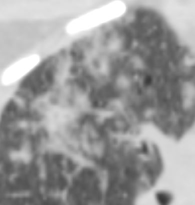
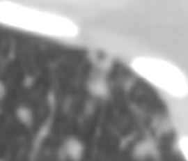
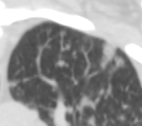
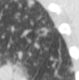
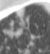
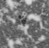
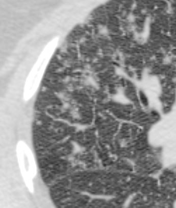
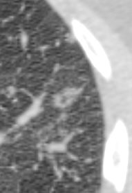
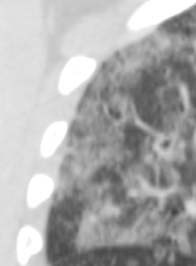
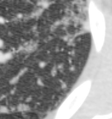
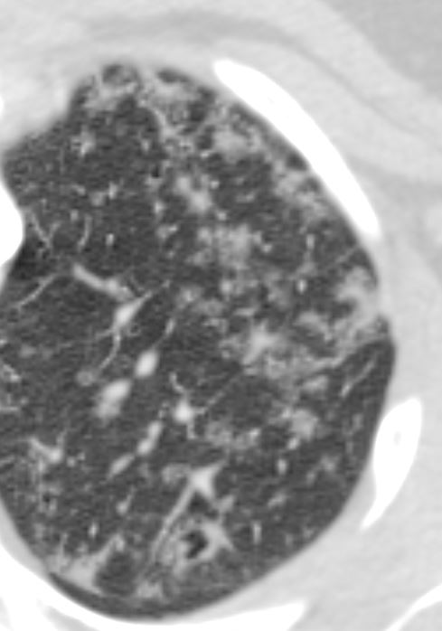
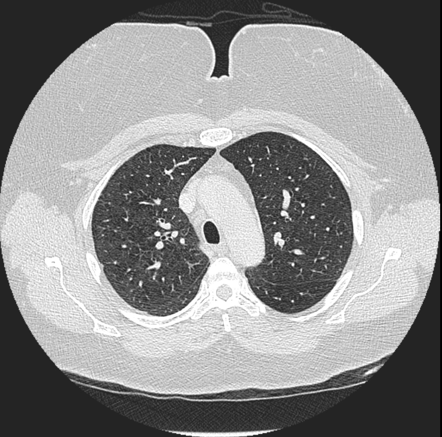
CT shows extensive diseae that appears to be centered around the bronchioles and small airways associated with centrilobular nodules, bronchiolectasis and thick walled cysts, more prominent in the upper lobes and mid lung fields but also involving the bases
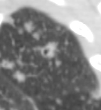
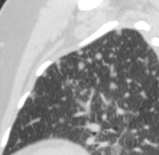
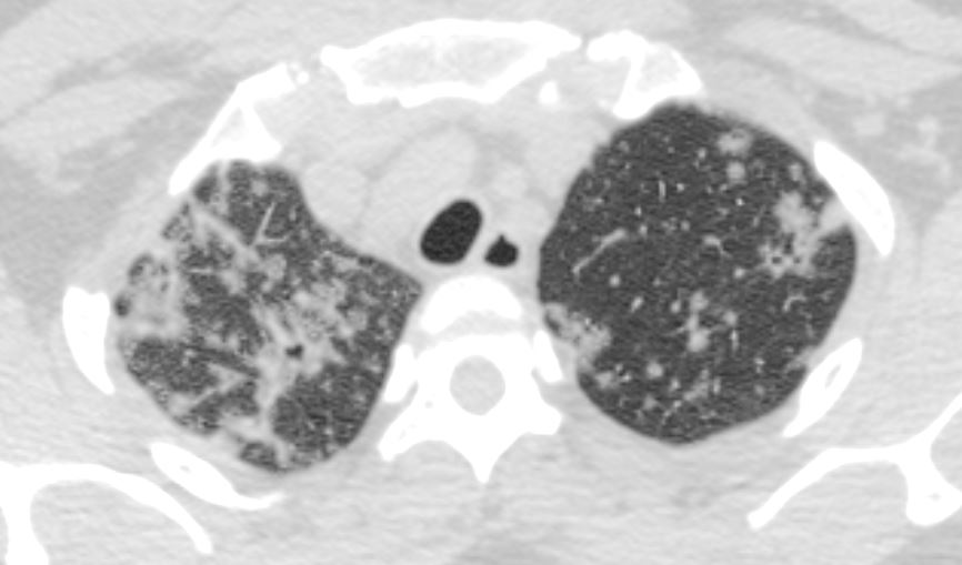
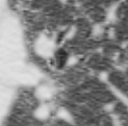
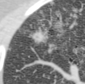
Seen throughout the lungs are nodular opacities with somewhat of an upper lobe predominance and areas of cavitation seen. This is a nonspecific imaging finding. Differential considerations do include infection (bacterial, fungal, or tuberculosis) or possible connective tissue disorder (Wegener’s granulomatosis or rheumatoid disease). Septic emboli or diffuse metastases are possible, although less likely.
CT shows extensive diseae that appears to be centered around the bronchioles and small airways associated with centrilobular nodules, bronchiolectasis and thick walled cysts, more prominent in the upper lobes and mid lung fields but also involving the bases Ashley Davidoff MD TheCommonVein.net 50F 01aCT shows extensive diseae that appears to be centered around the bronchioles and small airways associated with centrilobular nodules, bronchiolectasis and thick walled cysts, more prominent in the upper lobes and mid lung fields but also involving the bases Ashley Davidoff MD TheCommonVein.net 50F 001bCT shows extensive diseae that appears to be centered around the bronchioles and small airways associated with centrilobular nodules, bronchiolectasis and thick walled cysts, more prominent in the upper lobes and mid lung fields but also involving the bases Ashley Davidoff MD TheCommonVein.net 50F 001cCT shows extensive diseae that appears to be centered around the bronchioles and small airways associated with centrilobular nodules, bronchiolectasis and thick walled cysts, more prominent in the upper lobes and mid lung fields but also involving the bases Ashley Davidoff MD TheCommonVein.net 50F 001CT shows extensive diseae that appears to be centered around the bronchioles and small airways associated with centrilobular nodules, bronchiolectasis and thick walled cysts, more prominent in the upper lobes and mid lung fields but also involving the bases Ashley Davidoff MD TheCommonVein.net 50F 001CT shows extensive diseae that appears to be centered around the bronchioles and small airways associated with centrilobular nodules, bronchiolectasis and thick walled cysts, more prominent in the upper lobes and mid lung fields but also involving the bases Ashley Davidoff MD TheCommonVein.net 50F 001CT shows extensive diseae that appears to be centered around the bronchioles and small airways associated with centrilobular nodules, bronchiolectasis and thick walled cysts, more prominent in the upper lobes and mid lung fields but also involving the bases Ashley Davidoff MD TheCommonVein.net 50F 001CT shows extensive diseae that appears to be centered around the bronchioles and small airways associated with centrilobular nodules, bronchiolectasis and thick walled cysts, more prominent in the upper lobes and mid lung fields but also involving the bases Ashley Davidoff MD TheCommonVein.net 50F 001CT shows extensive diseae that appears to be centered around the bronchioles and small airways associated with centrilobular nodules, bronchiolectasis and thick walled cysts, more prominent in the upper lobes and mid lung fields but also involving the bases Ashley Davidoff MD TheCommonVein.net 50F 001CT shows extensive diseae that appears to be centered around the bronchioles and small airways associated with centrilobular nodules, bronchiolectasis and thick walled cysts, more prominent in the upper lobes and mid lung fields but also involving the bases Ashley Davidoff MD TheCommonVein.net 50F 001CT shows extensive diseae that appears to be centered around the bronchioles and small airways associated with centrilobular nodules, bronchiolectasis and thick walled cysts, more prominent in the upper lobes and mid lung fields but also involving the bases Ashley Davidoff MD TheCommonVein.net 50F 001CT shows extensive diseae that appears to be centered around the bronchioles and small airways associated with centrilobular nodules, bronchiolectasis and thick walled cysts, more prominent in the upper lobes and mid lung fields but also involving the bases Ashley Davidoff MD TheCommonVein.net 50F 001CT shows extensive diseae that appears to be centered around the bronchioles and small airways associated with centrilobular nodules, bronchiolectasis and thick walled cysts, more prominent in the upper lobes and mid lung fields but also involving the bases Ashley Davidoff MD TheCommonVein.net 50F 001CT shows extensive diseae that appears to be centered around the bronchioles and small airways associated with centrilobular nodules, bronchiolectasis and thick walled cysts, more prominent in the upper lobes and mid lung fields but also involving the bases Ashley Davidoff MD TheCommonVein.net 50F 001CT shows extensive diseae that appears to be centered around the bronchioles and small airways associated with centrilobular nodules, bronchiolectasis and thick walled cysts, more prominent in the upper lobes and mid lung fields but also involving the bases Ashley Davidoff MD TheCommonVein.net 50F 001CT shows extensive diseae that appears to be centered around the bronchioles and small airways associated with centrilobular nodules, bronchiolectasis and thick walled cysts, more prominent in the upper lobes and mid lung fields but also involving the bases Ashley Davidoff MD TheCommonVein.net 50F 001CT shows extensive diseae that appears to be centered around the bronchioles and small airways associated with centrilobular nodules, bronchiolectasis and thick walled cysts, more prominent in the upper lobes and mid lung fields but also involving the bases Ashley Davidoff MD TheCommonVein.net 50F 001CT shows extensive diseae that appears to be centered around the bronchioles and small airways associated with centrilobular nodules, bronchiolectasis and thick walled cysts, more prominent in the upper lobes and mid lung fields but also involving the bases Ashley Davidoff MD TheCommonVein.net 50F 001CT shows extensive diseae that appears to be centered around the bronchioles and small airways associated with centrilobular nodules, bronchiolectasis and thick walled cysts, more prominent in the upper lobes and mid lung fields but also involving the bases Ashley Davidoff MD TheCommonVein.net 50F 001CT shows extensive diseae that appears to be centered around the bronchioles and small airways associated with centrilobular nodules, bronchiolectasis and thick walled cysts, more prominent in the upper lobes and mid lung fields but also involving the bases Ashley Davidoff MD TheCommonVein.net 50F 001CT shows extensive diseae that appears to be centered around the bronchioles and small airways associated with centrilobular nodules, bronchiolectasis and thick walled cysts, more prominent in the upper lobes and mid lung fields but also involving the bases Ashley Davidoff MD TheCommonVein.net 50F 001CT shows extensive diseae that appears to be centered around the bronchioles and small airways associated with centrilobular nodules, bronchiolectasis and thick walled cysts, more prominent in the upper lobes and mid lung fields but also involving the bases Ashley Davidoff MD TheCommonVein.net 50F 001CT shows extensive diseae that appears to be centered around the bronchioles and small airways associated with centrilobular nodules, bronchiolectasis and thick walled cysts, more prominent in the upper lobes and mid lung fields but also involving the bases Ashley Davidoff MD TheCommonVein.net 50F 001CT shows extensive diseae that appears to be centered around the bronchioles and small airways associated with centrilobular nodules, bronchiolectasis and thick walled cysts, more prominent in the upper lobes and mid lung fields but also involving the bases Ashley Davidoff MD TheCommonVein.net 50F 001CT shows extensive diseae that appears to be centered around the bronchioles and small airways associated with centrilobular nodules, bronchiolectasis and thick walled cysts, more prominent in the upper lobes and mid lung fields but also involving the bases Ashley Davidoff MD TheCommonVein.net 50F 001CT shows extensive diseae that appears to be centered around the bronchioles and small airways associated with centrilobular nodules, bronchiolectasis and thick walled cysts, more prominent in the upper lobes and mid lung fields but also involving the bases Ashley Davidoff MD TheCommonVein.net 50F 001CT shows extensive diseae that appears to be centered around the bronchioles and small airways associated with centrilobular nodules, bronchiolectasis and thick walled cysts, more prominent in the upper lobes and mid lung fields but also involving the bases Ashley Davidoff MD TheCommonVein.net 50F 001CT shows extensive diseae that appears to be centered around the bronchioles and small airways associated with centrilobular nodules, bronchiolectasis and thick walled cysts, more prominent in the upper lobes and mid lung fields but also involving the bases Ashley Davidoff MD TheCommonVein.net 50F 001
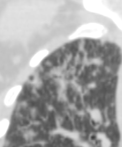
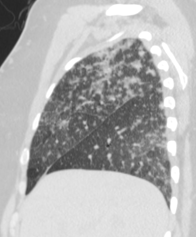
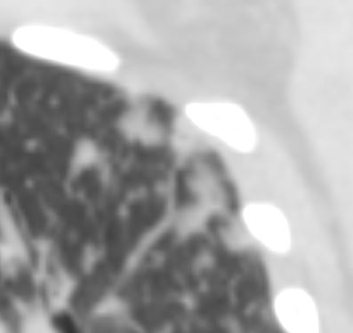
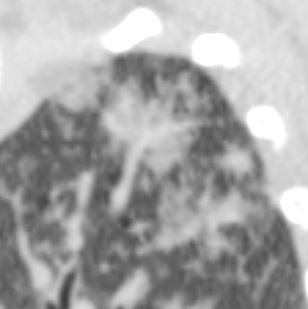
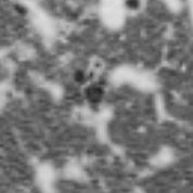
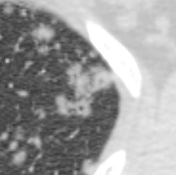
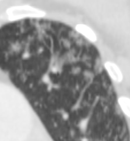
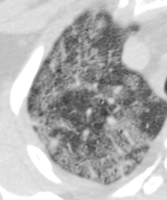
Bilateral nodular opacities, centered around bronchioles and also in a centrilobular distribution. Infiltrates have become more confluent and interspersed with ground glass attenuation , with small areas of cavitation and or thick walled cyst formation. The differential again includes infectious etiologies, including bacterial, fungal or mycobacterial; COP; connective tissue disorders such as Wegener’s or rheumatoid arthritis, or possibly sarcoidosis. Septic emboli or metastasis are much less likely given the interval changes.
Bilateral nodular opacities, centered around bronchioles and also in a centrilobular distribution. Infiltrates have become more confluent and interspersed with ground glass attenuation , with small areas of cavitation and or thick walled cyst formation.
Bilateral nodular opacities, centered around bronchioles and also in a centrilobular distribution. Infiltrates have become more confluent and interspersed with ground glass attenuation , with small areas of cavitation and or thick walled cyst formation. Ashley Davidoff MD TheCommonVein.net 50F 002Bilateral nodular opacities, centered around bronchioles and also in a centrilobular distribution. Infiltrates have become more confluent and interspersed with ground glass attenuation , with small areas of cavitation and or thick walled cyst formation. Ashley Davidoff MD TheCommonVein.net 50F 00Bilateral nodular opacities, centered around bronchioles and also in a centrilobular distribution. Infiltrates have become more confluent and interspersed with ground glass attenuation , with small areas of cavitation and or thick walled cyst formation. Ashley Davidoff MD TheCommonVein.net 50F 00Bilateral nodular opacities, centered around bronchioles and also in a centrilobular distribution. Infiltrates have become more confluent and interspersed with ground glass attenuation , with small areas of cavitation and or thick walled cyst formation. Ashley Davidoff MD TheCommonVein.net 50F 00Bilateral nodular opacities, centered around bronchioles and also in a centrilobular distribution. Infiltrates have become more confluent and interspersed with ground glass attenuation , with small areas of cavitation and or thick walled cyst formation. Ashley Davidoff MD TheCommonVein.net 50F 00Bilateral nodular opacities, centered around bronchioles and also in a centrilobular distribution. Infiltrates have become more confluent and interspersed with ground glass attenuation , with small areas of cavitation and or thick walled cyst formation. Ashley Davidoff MD TheCommonVein.net 50F 00Bilateral nodular opacities, centered around bronchioles and also in a centrilobular distribution. Infiltrates have become more confluent and interspersed with ground glass attenuation , with small areas of cavitation and or thick walled cyst formation. Ashley Davidoff MD TheCommonVein.net 50F 00Bilateral nodular opacities, centered around bronchioles and also in a centrilobular distribution. Infiltrates have become more confluent and interspersed with ground glass attenuation , with small areas of cavitation and or thick walled cyst formation. Ashley Davidoff MD TheCommonVein.net 50F 00Bilateral nodular opacities, centered around bronchioles and also in a centrilobular distribution. Infiltrates have become more confluent and interspersed with ground glass attenuation , with small areas of cavitation and or thick walled cyst formation. Ashley Davidoff MD TheCommonVein.net 50F 00Bilateral nodular opacities, centered around bronchioles and also in a centrilobular distribution. Infiltrates have become more confluent and interspersed with ground glass attenuation , with small areas of cavitation and or thick walled cyst formation. Ashley Davidoff MD TheCommonVein.net 50F 00Bilateral nodular opacities, centered around bronchioles and also in a centrilobular distribution. Infiltrates have become more confluent and interspersed with ground glass attenuation , with small areas of cavitation and or thick walled cyst formation. Ashley Davidoff MD TheCommonVein.net 50F 00Bilateral nodular opacities, centered around bronchioles and also in a centrilobular distribution. Infiltrates have become more confluent and interspersed with ground glass attenuation , with small areas of cavitation and or thick walled cyst formation. Ashley Davidoff MD TheCommonVein.net 50F 00Bilateral nodular opacities, centered around bronchioles and also in a centrilobular distribution. Infiltrates have become more confluent and interspersed with ground glass attenuation , with small areas of cavitation and or thick walled cyst formation. Ashley Davidoff MD TheCommonVein.net 50F 00
Biopsy performed from right middle and lower lobes showed
Pulmonary Langerhans Cell Histiocytosis
Patient stopped smoking and improved significantly
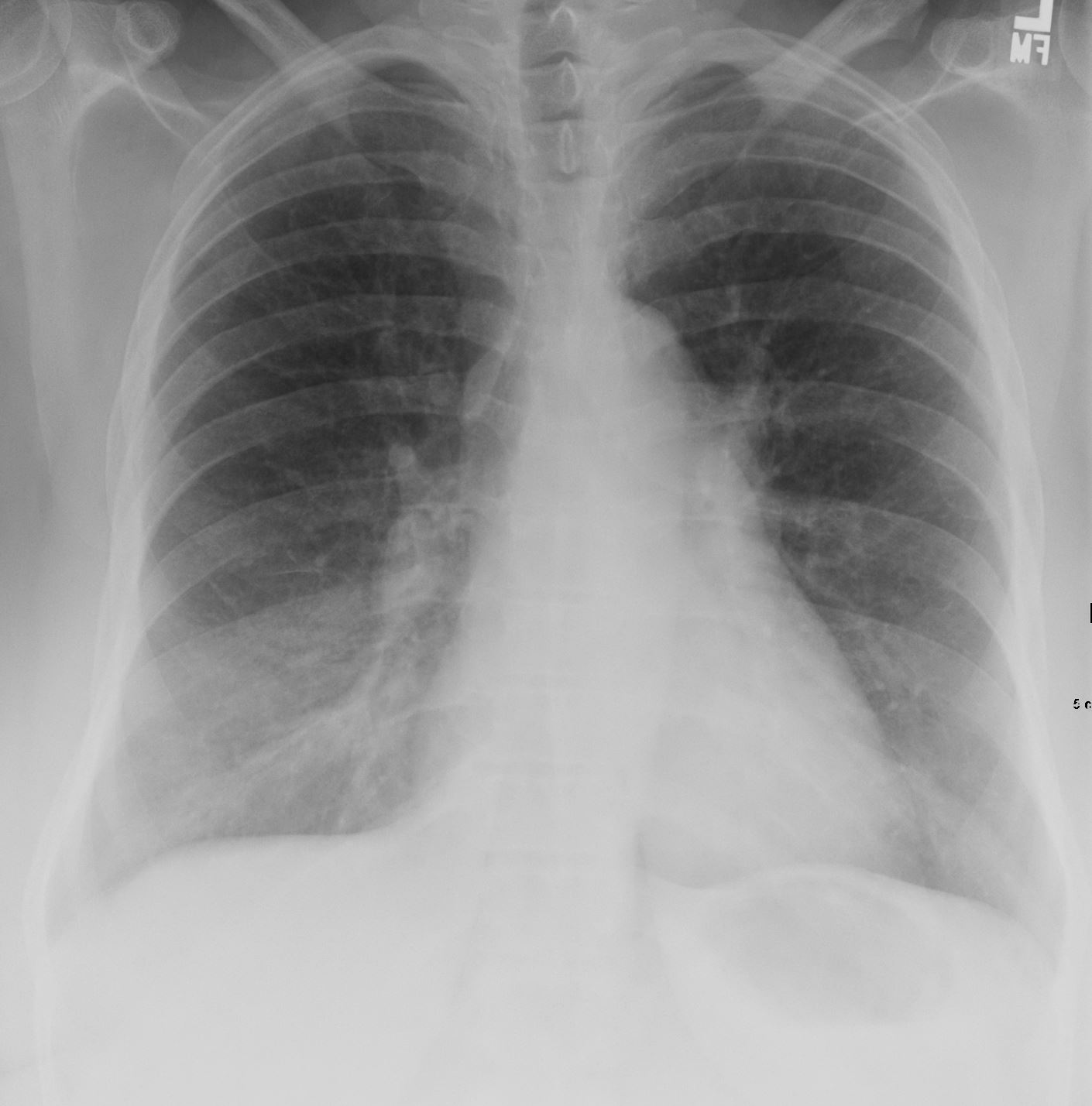
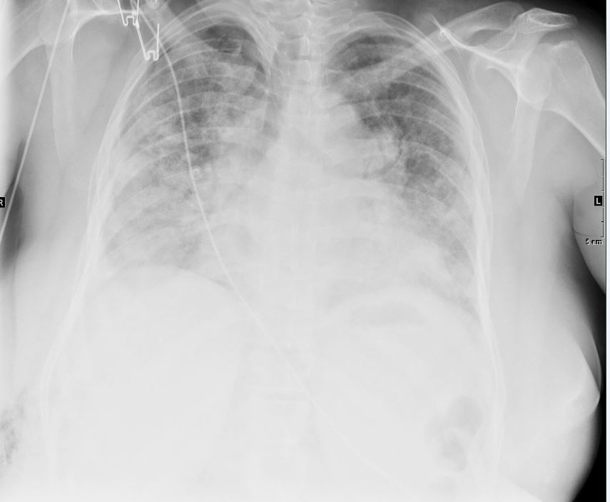
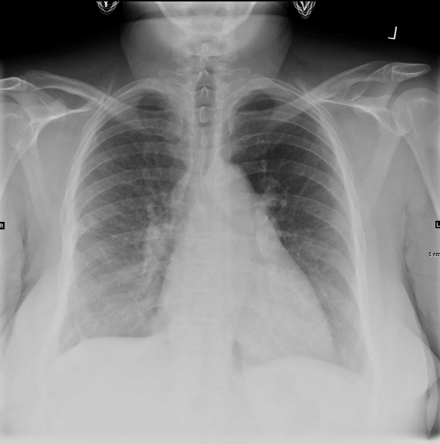
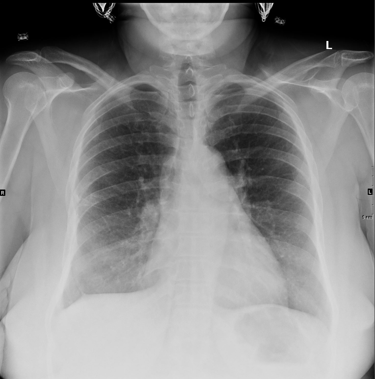
CXR performed 10 months later shows significant improvement Ashley Davidoff MD TheCommonVein.net 50F 014
CXR 2.5 years later
CXR 2.5 years later shows contuinued improvement Ashley Davidoff MD TheCommonVein.net 50F-015
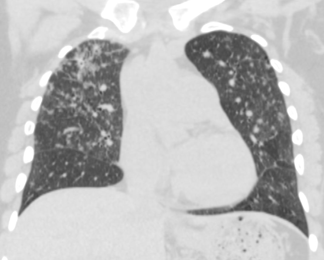
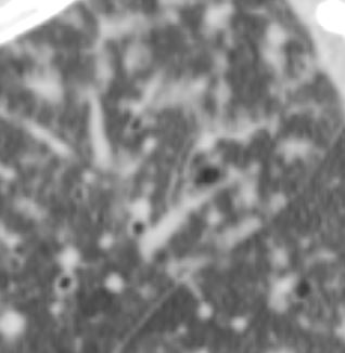
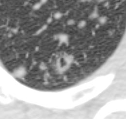
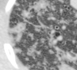
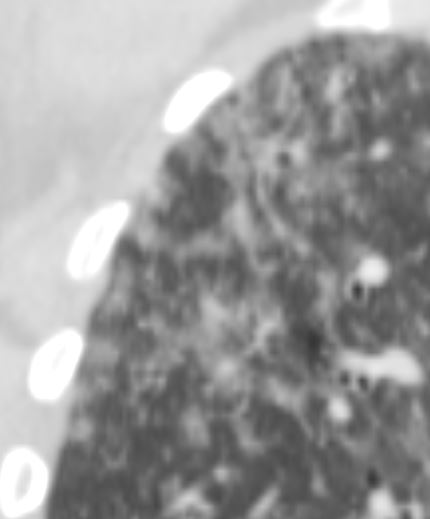
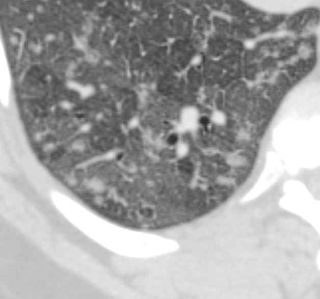
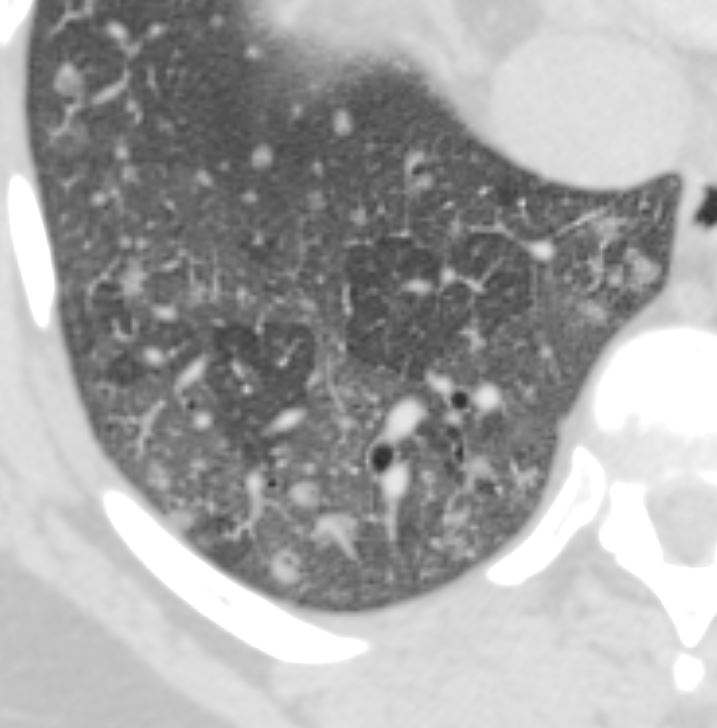
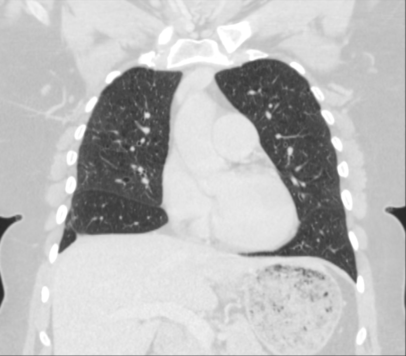
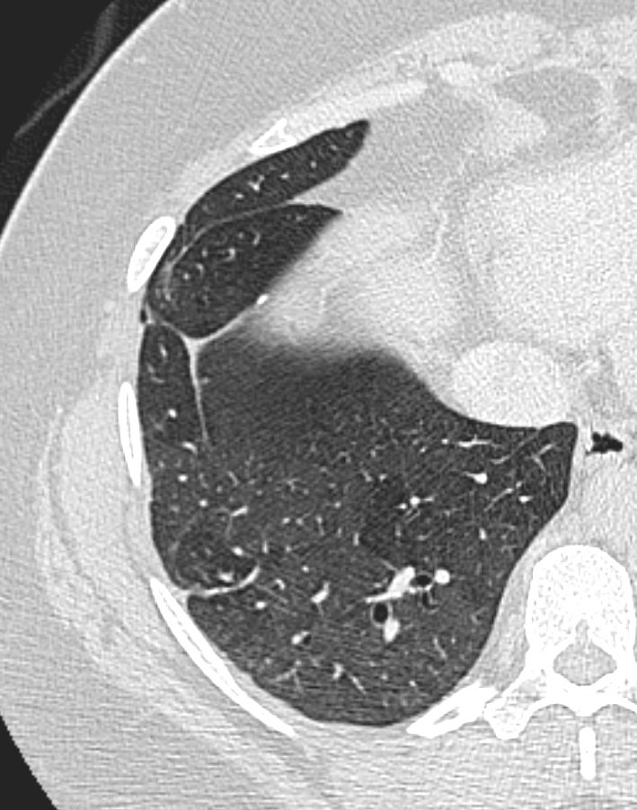
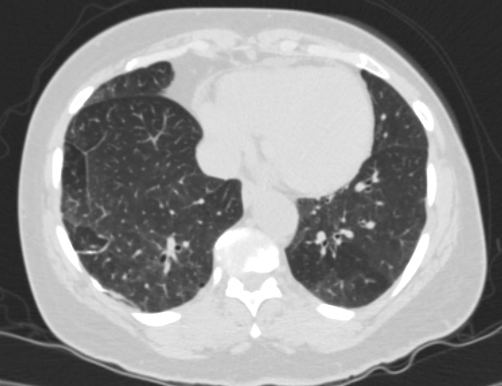
CT 5 years later
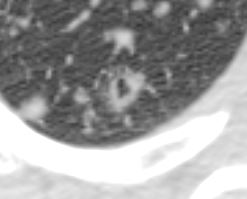
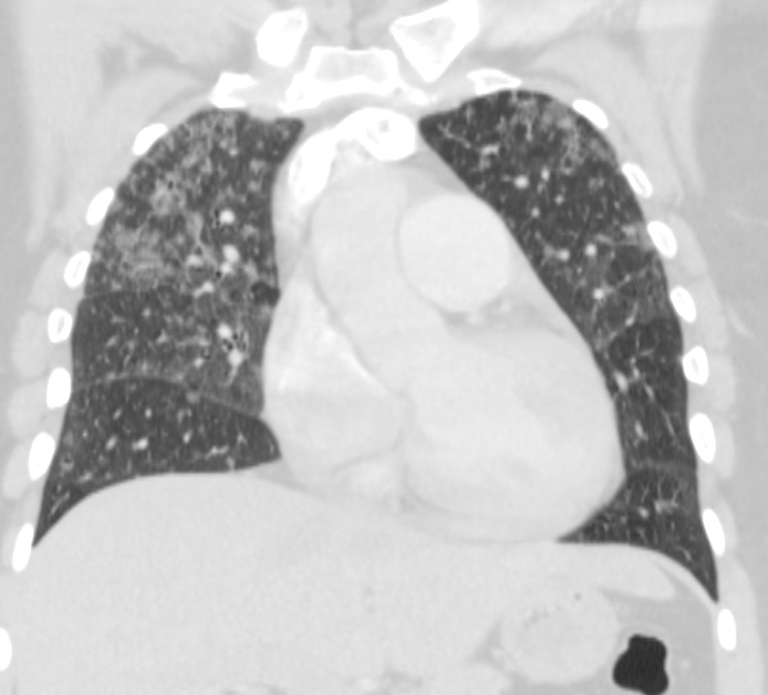
CT 5 years later shows evidence of emphysema , post biopsy changes in the right middle and lower lobes and pulmonary hypertension Ashley Davidoff MD TheCommonVein.net 50F-016CT 5 years later shows evidence of emphysema , post biopsy changes in the right middle and lower lobes and pulmonary hypertension Ashley Davidoff MD TheCommonVein.net 50F-016CT 5 years later shows evidence of emphysema , post biopsy changes in the right middle and lower lobes and pulmonary hypertension Ashley Davidoff MD TheCommonVein.net 50F-017CT 5 years later shows evidence of emphysema , post biopsy changes in the right middle and lower lobes and pulmonary hypertension Ashley Davidoff MD TheCommonVein.net 50F-018
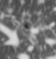
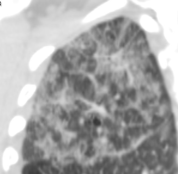
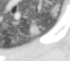
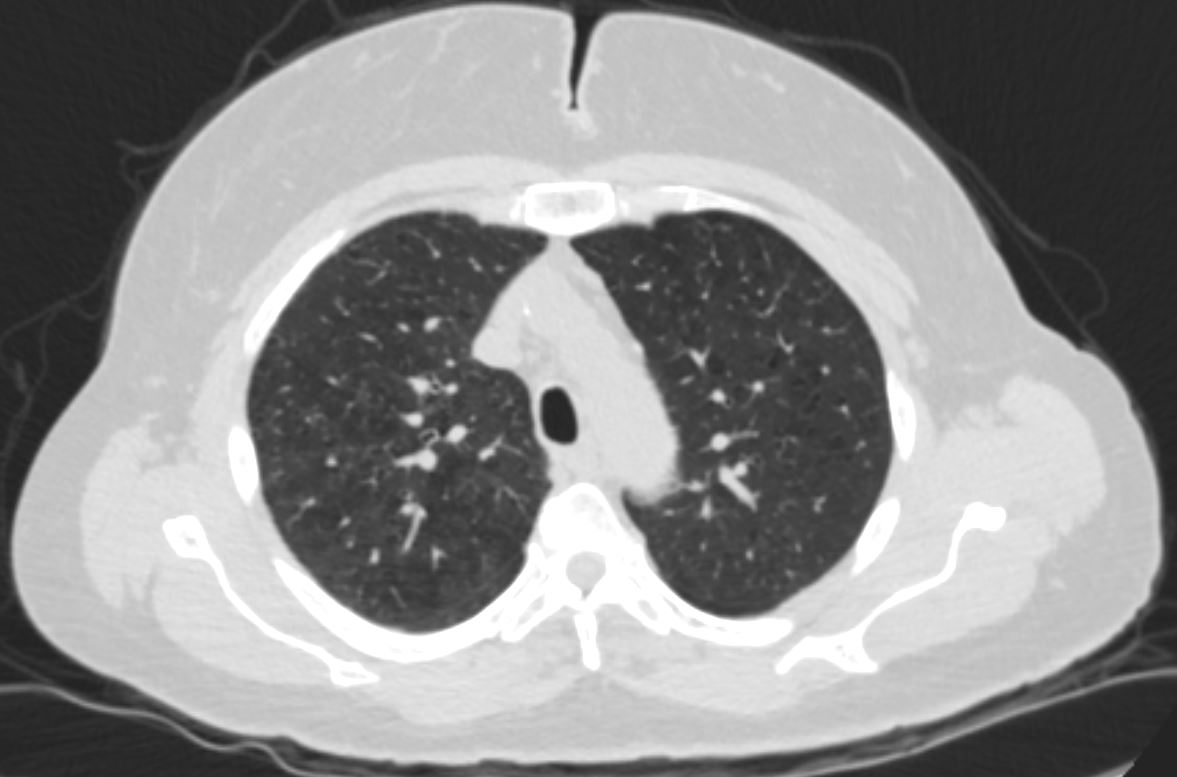
Mosaic Attenuation at the Lung Bases
CT 5 years later shows evidence of emphysema , post biopsy changes in the right middle and lower lobes and pulmonary hypertension Ashley Davidoff MD TheCommonVein.net 50F-019
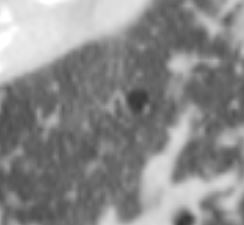
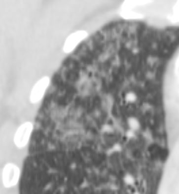
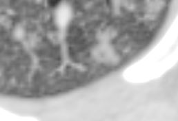
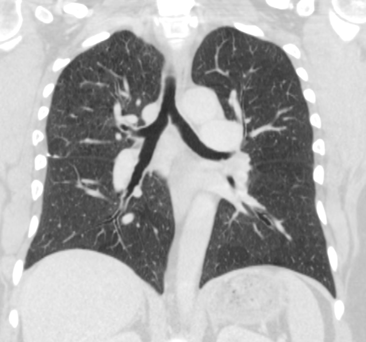
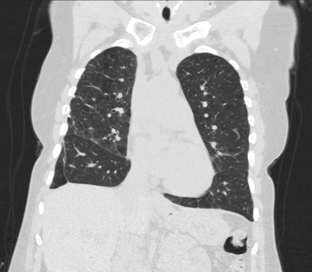
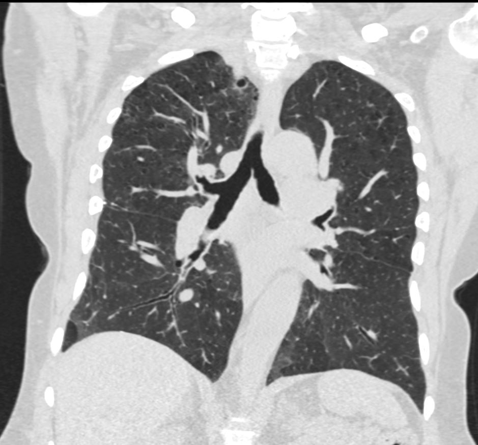
Current CT – 12 years since Most Recent CT and 18 years after the Acute Event
CT 5 years later, and 18 years after the acute event shows evidence of stable emphysema and mosaic attenuation at the lung bases Ashley Davidoff MD TheCommonVein.net 50F-020CT 5 years later, and 18 years after the acute event shows evidence of stable emphysema and mosaic attenuation at the lung bases Ashley Davidoff MD TheCommonVein.net 50F-021CT 5 years later, and 18 years after the acute event shows evidence of stable emphysema and mosaic attenuation at the lung bases Ashley Davidoff MD TheCommonVein.net 50F-022CT 5 years later, and 18 years after the acute event shows evidence of stable emphysema and mosaic attenuation at the lung bases Ashley Davidoff MD TheCommonVein.net 50F-023