
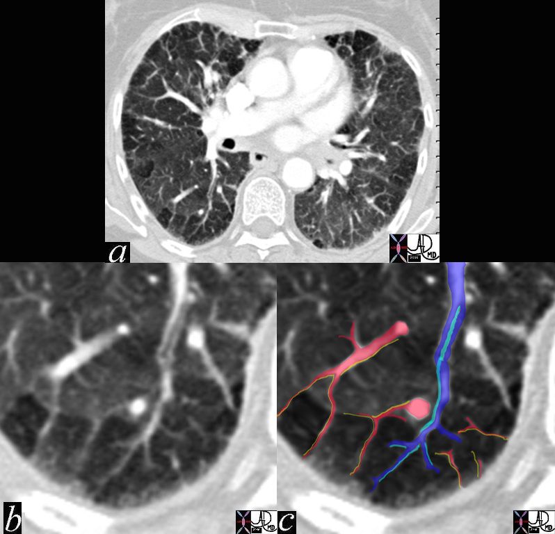
CT scan of the chest shows mosaic attenuation peripheral pulmonary lobules best exemplified in the posterior aspects of the superior segments of the lower lobes bilaterally
A lobular bronchiole is overlaid in teal in c, while the accompanying lobular arteriole is seen in royal blue entering the lobule and then branching. The peripheral venules (red) in the interlobular septa join to form the lobular vein. The accompanying lymphatics (yellow) accompany the venules in the interlobular septa. These are not visualized but are implied
In this remarkable CT we were able to identify a few secondary lobules at the periphery of the lung that have a rectangular shape in this instance. The branching structure that enters the lobule (blue in c), is characterised as an arteriole for two reasons. Firstly, it is paired with a tubular airway seen in (b) in its most proximal portion as a lucent tubule, and subsequently interpolated in light blue in c. Secondly it branches in the center of the lobule. It is distinct from the border forming interlobular septum which surrounds it. A second relatively large vessel colored in red receives a branch from the interlobular septum and by virtue of its size and position it has to be a pulmonary venule. We know that the lymphatic vessel accompanies the venule, and so the yellow lymphatic has been implied but not visualised. We also know that connective tissue surrounds these two structures. In this instance the matrix of the lobule that consists of the alveoli is less dense than it should be and is surrounded by normal lung parenchyma. Lucency, or mosaic attenuation implies either small vessel disease or small airway disease. In the latter instance air trapping from small airway disease is implied.
Ashley Davidoff MD TheCommonVein.net 47152c02 RnD