
67-year-old patient with history of right lower lobe proven kappa AL
amyloidosis presents for follow-up.
2 years earlier Thick Walled Stomach with Early Satiety
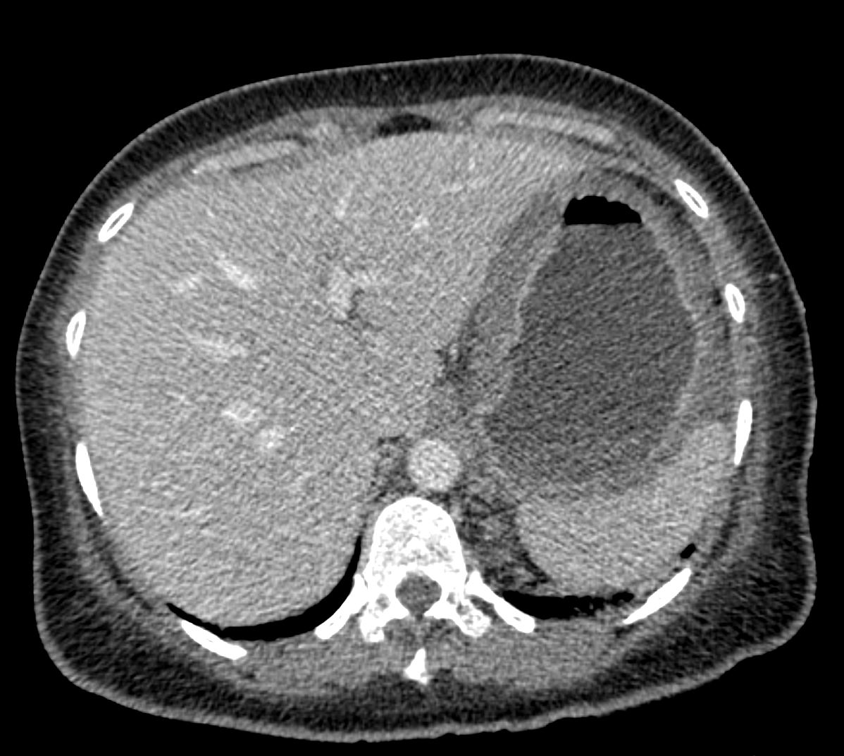
Axial CT 56 -year-old female with a history of amyloidosis presenting with early satiety and dyspnea. CT in the axial plane shows a fluid filled thick-walled stomach with enhancing mucosa. Amyloidosis of the stomach was suspected
Ashley Davidoff MD TheCommonVein.net 244Lu 135741e03
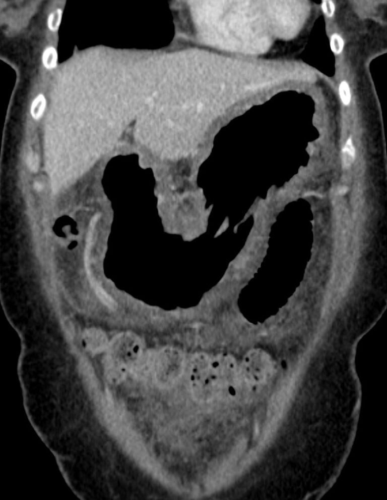
Coronal CT 56 -year-old female with a history of amyloidosis presenting with early satiety and dyspnea. CT in the coronal plane shows an air distended, thick-walled stomach. Amyloidosis of the stomach was suspected
Ashley Davidoff MD TheCommonVein.net 244 Lu 135741e04
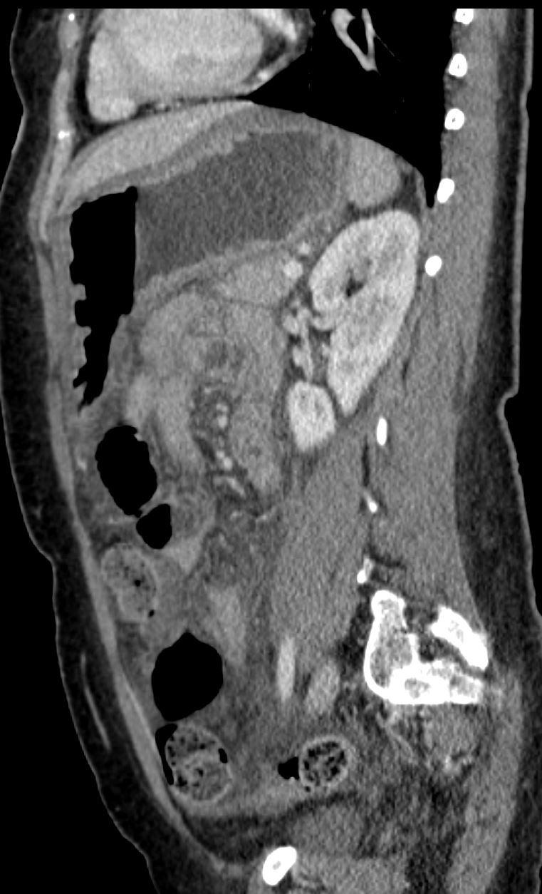
56 -year-old female with a history of amyloidosis presenting with early satiety and dyspnea. CT in the sagittal plane shows a thick-walled stomach with an air-fluid level. Amyloidosis of the stomach was suspected
Ashley Davidoff MD TheCommonVein.net 244 Lu 135741e05
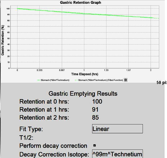
IMPRESSION:
Abnormal gastric emptying that is slow. Findings consistent with delayed gastric emptying.
6 Months Prior
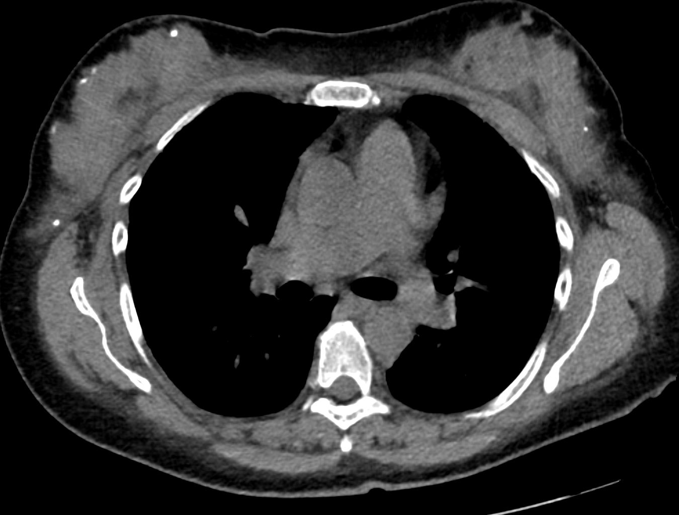
Axial CT – Pulmonary Embolus Left Lower Lobe
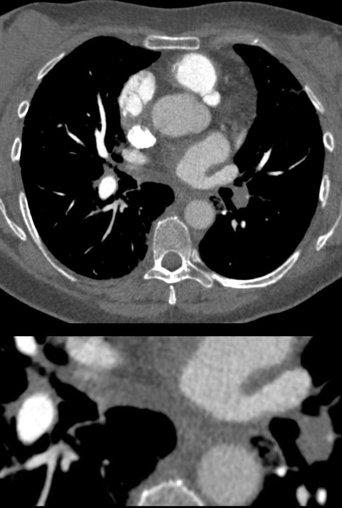
56 -year-old female with a history of amyloidosis presenting with tachycardia and dyspnea. CTPA shows an occlusive embolus (PE) in the left lower lobe pulmonary artery.
Ashley Davidoff MD TheCommonVein.net 135738c
al CT – Pulmonary Embolus Left Lower Lobe (PE)
56 -year-old female with a history of amyloidosis presenting with tachycardia and dyspnea. CTPA shows no contrast enhancement of the pulmonary arteries subtending the left lower lobe compared to the right and a subsegmental wedge shaped defect (Hampton’s hump) in the lateral segment of the left lower lobe
Ashley Davidoff MD TheCommonVein.net 135739c
Perfusion Defect of the Left Lower Lobe from Occlusive Pulmonary Embolus
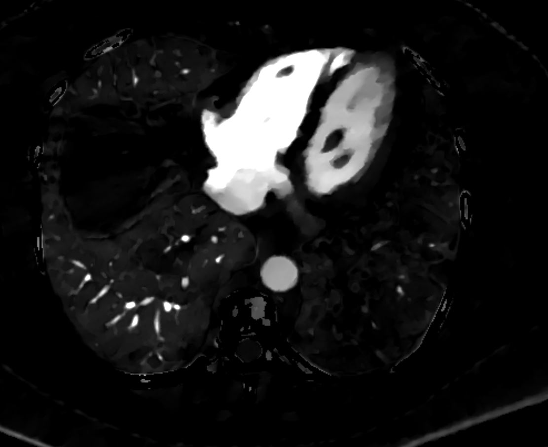
Perfusion Defect of the Left Lower Lobe from Occlusive Pulmonary Embolus
56 -year-old female with a history of amyloidosis presenting with tachycardia and dyspnea. Dual energy CT with an iodine map shows shows an almost lobar perfusion defect of the left lower lobe compared
Ashley Davidoff MD TheCommonVein.net 135740
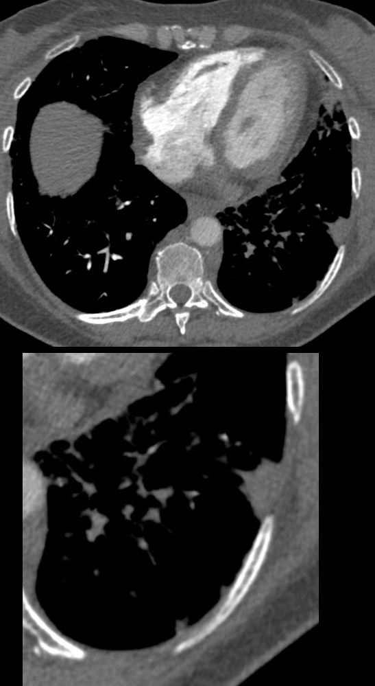
Hampton’s Hump Pulmonary Embolus Left Lower Lobe (PE)
56 -year-old female with a history of amyloidosis presenting with tachycardia and dyspnea. Lung windows from the CTPA a subsegmental wedge shaped defect (Hampton’s hump) in the lateral segment of the left lower lobe. The lung parenchyma is intact in the left lower lobe in the region of lack of lobar perfusion on the dual energy iodine maps, indicating that despite the lack of perfusion there is no necrosis
Ashley Davidoff MD TheCommonVein.net 135742
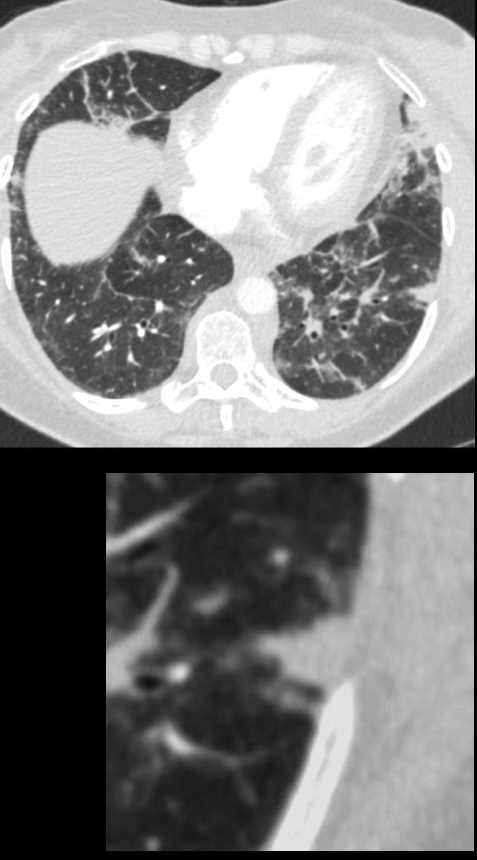
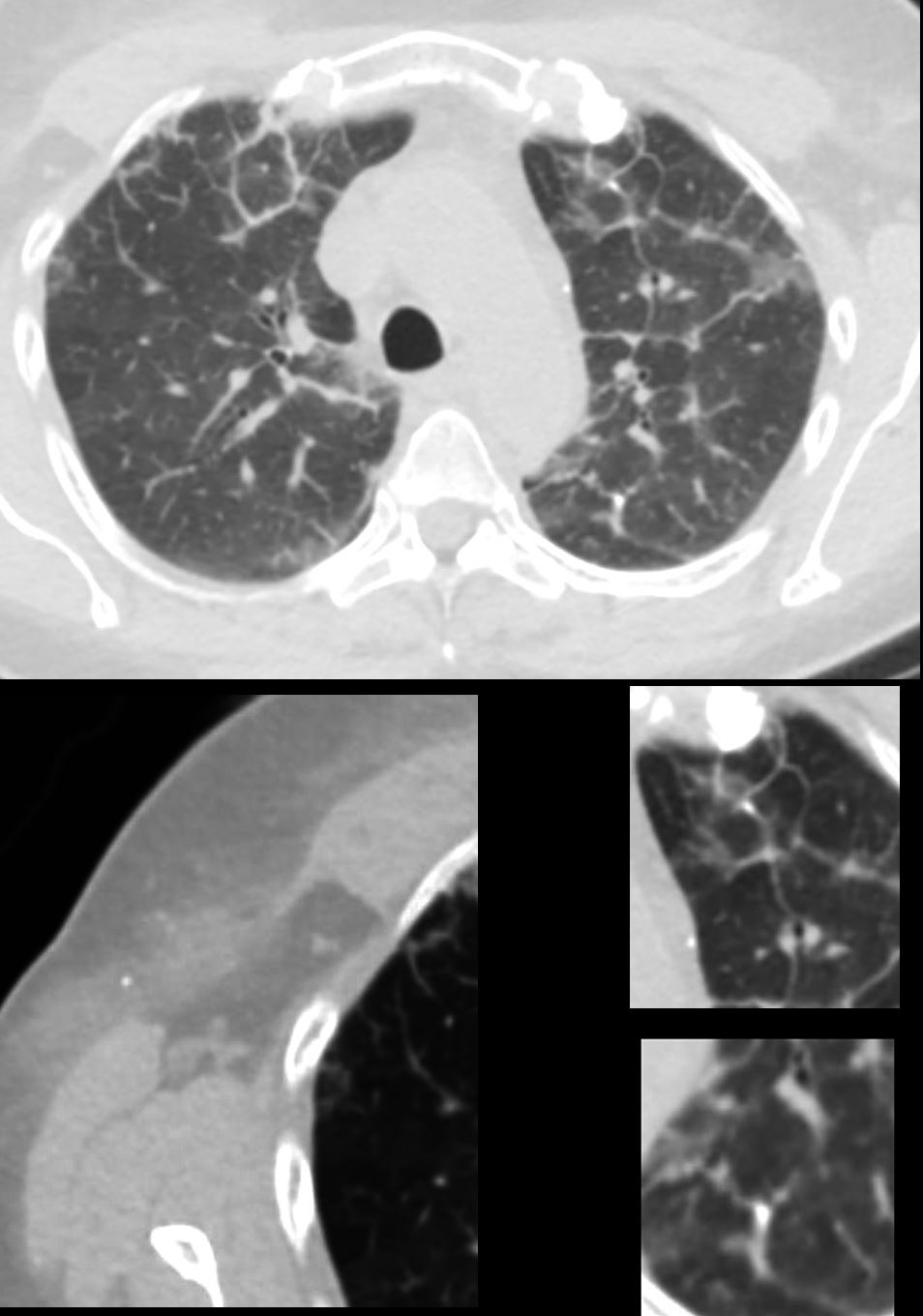
CT Axial Lung Windows Hamptons Hump and Alveolar Septal Amyloidosis
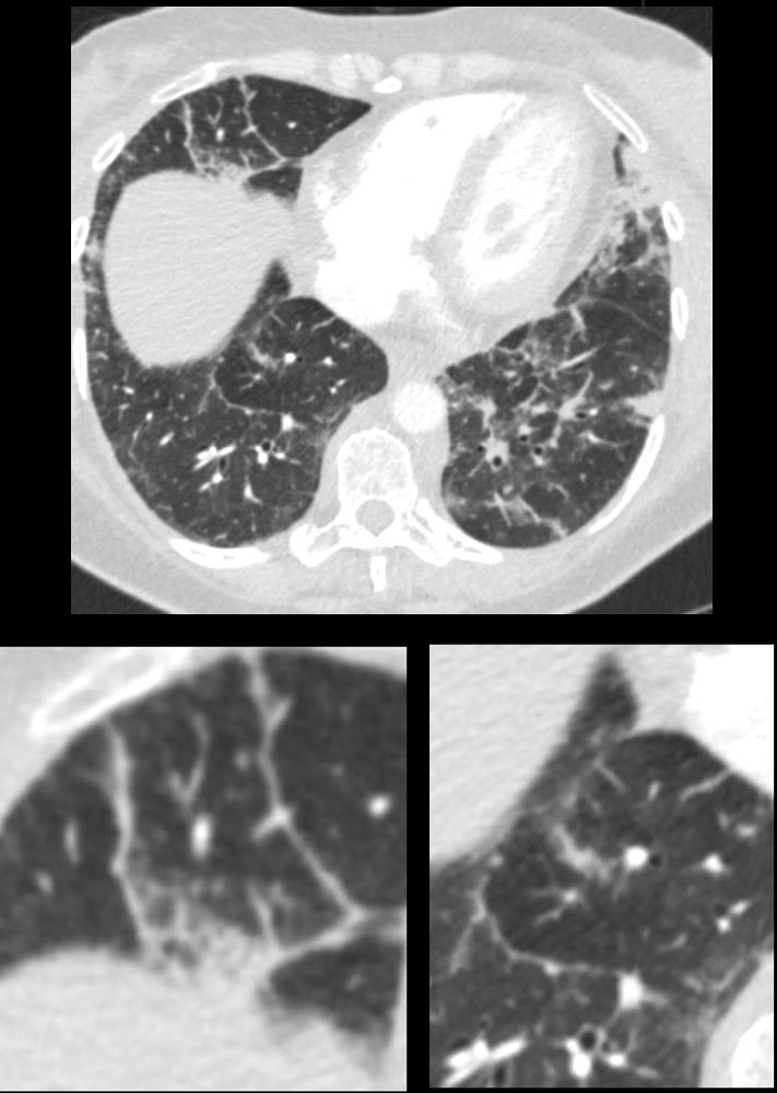
56 -year-old female with a history of amyloidosis presenting with tachycardia and dyspnea. Lung windows show intact parenchyma in the left lower lobe in the region of lack of perfusion on the dual energy iodine maps indicating that despite the lack of perfusion there is no necrosis. There is evidence of thickening of the interlobular septa (lower set of images suggesting a diagnosis of alveolar septal amyloidosis
Ashley Davidoff MD TheCommonVein.net 135741c
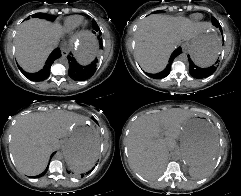
Gastric Changes 18 Months Later –
New Dystrophic Calcifications in the Wall
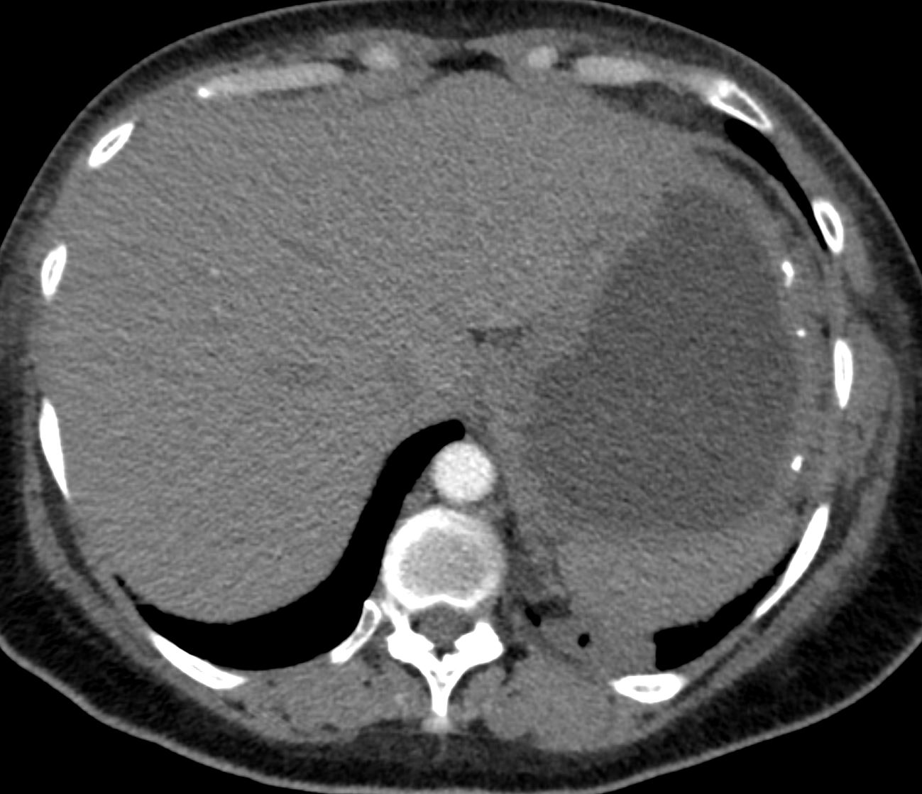
56 -year-old female with a history of amyloidosis (AL) presents for follow up. Axial CT of the upper abdomen shows the fluid filled stomach with dystrophic amyloid calcifications in the gastric
Ashley Davidoff MD TheCommonVein.net 244 Lu 135741e01
56 -year-old female with a history of amyloidosis (AL) presents for follow up. Axial CT of the upper abdomen shows the fluid filled stomach with dystrophic amyloid calcifications in the gastric
Ashley Davidoff MD TheCommonVein.net 244 Lu 135741e02
1. Occlusion of proximal segmental/distalmost lobar left lower lobe pulmonary artery consistent with thrombosis, age indeterminate. Hypoperfusion involving the left lower lung parenchyma. No evidence of right heart strain.
2. Multifocal nodular consolidative opacities primarily within the left lower lobe may reflect any combination of rounded atelectasis, aspiration, and/or infectious process. Infarct cannot be excluded.
3. Diffuse interlobular septal thickening bilaterally which is nonspecific but can be seen in the setting of volume overload.
6 Months Later CXR Septal Thickening
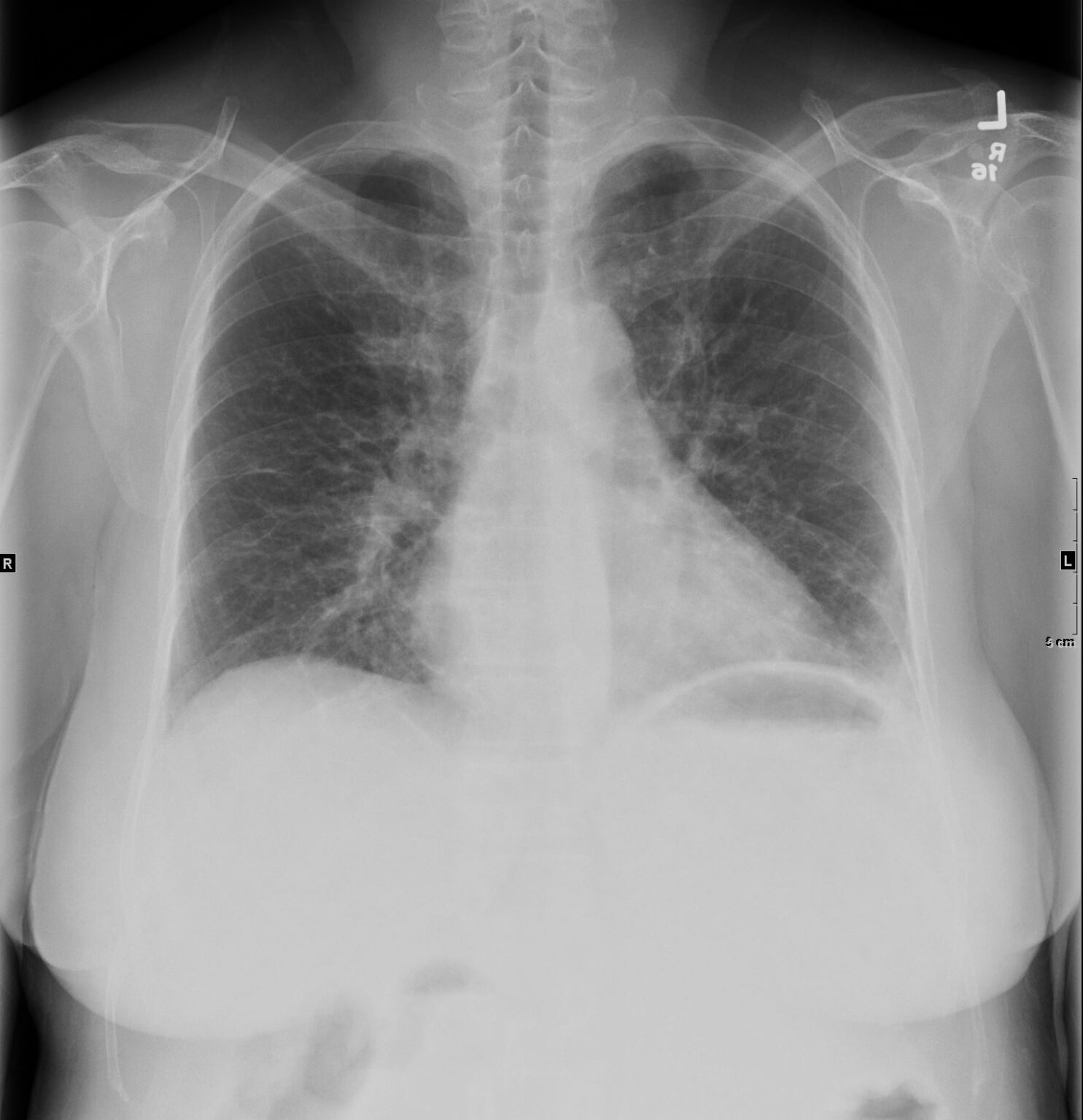
56 -year-old female with a history of amyloidosis (AL) presents for follow up following a pulmonary embolus. Frontal view of the chest shows diffuse reticular process best appreciated at the left base suggesting Kerley b lines. In the appropriate clinical setting these findings are compatible with the diagnosis of alveolar septal amyloidosis. Note the air fluid level in the stomach in this patient with known amyloidosis of the stomach with delayed gastric emptying.
Ashley Davidoff MD TheCommonVein.net 244 Lu 135741c05
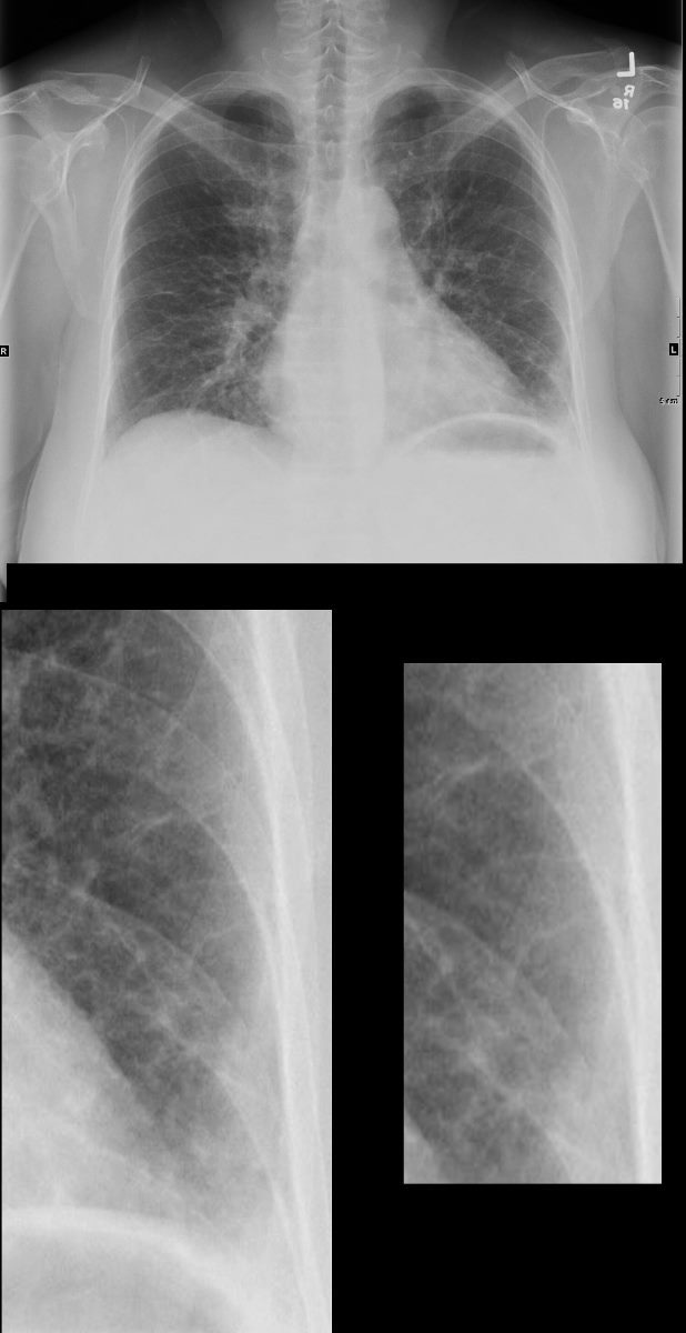
56 -year-old female with a history of amyloidosis (AL) presents for follow up following a pulmonary embolus. Frontal view of the chest shows diffuse reticular process best appreciated at the left base suggesting Kerley b lines (magnified in the lower panels). In the appropriate clinical setting these findings are compatible with the diagnosis of alveolar septal amyloidosis. Note the air fluid level in the stomach in this patient with known amyloidosis of the stomach with delayed gastric emptying.
Ashley Davidoff MD TheCommonVein.net 244 Lu 135741c05c
Ashley Davidoff MD TheCommonVein.net 244 Lu 135741c07
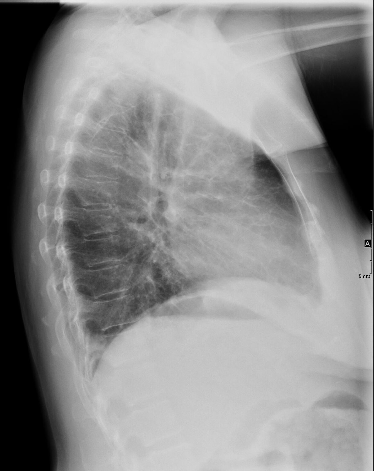
56 -year-old female with a history of amyloidosis (AL) presents for follow up following a pulmonary embolus. Lateral view of the chest shows diffuse reticular process best appreciated anteriorly suggesting thickening of the interlobular septa. In the appropriate clinical setting these findings are compatible with the diagnosis of alveolar septal amyloidosis. Note the air fluid level in the stomach in this patient with known amyloidosis of the stomach with delayed gastric emptying.
Ashley Davidoff MD TheCommonVein.net 244 Lu 135741c07
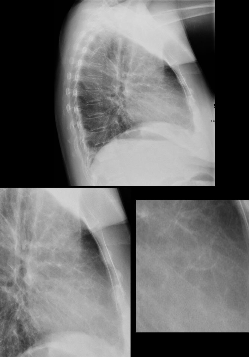
56 -year-old female with a history of amyloidosis (AL) presents for follow up following a pulmonary embolus. Lateral view of the chest shows diffuse reticular process best appreciated anteriorly suggesting thickening of the interlobular septa. (better appreciated in the magnified views in the lower panels). In the appropriate clinical setting these findings are compatible with the diagnosis of alveolar septal amyloidosis. Note the air fluid level in the stomach in this patient with known amyloidosis of the stomach with delayed gastric emptying.
Ashley Davidoff MD TheCommonVein.net 244 Lu 135741c07c
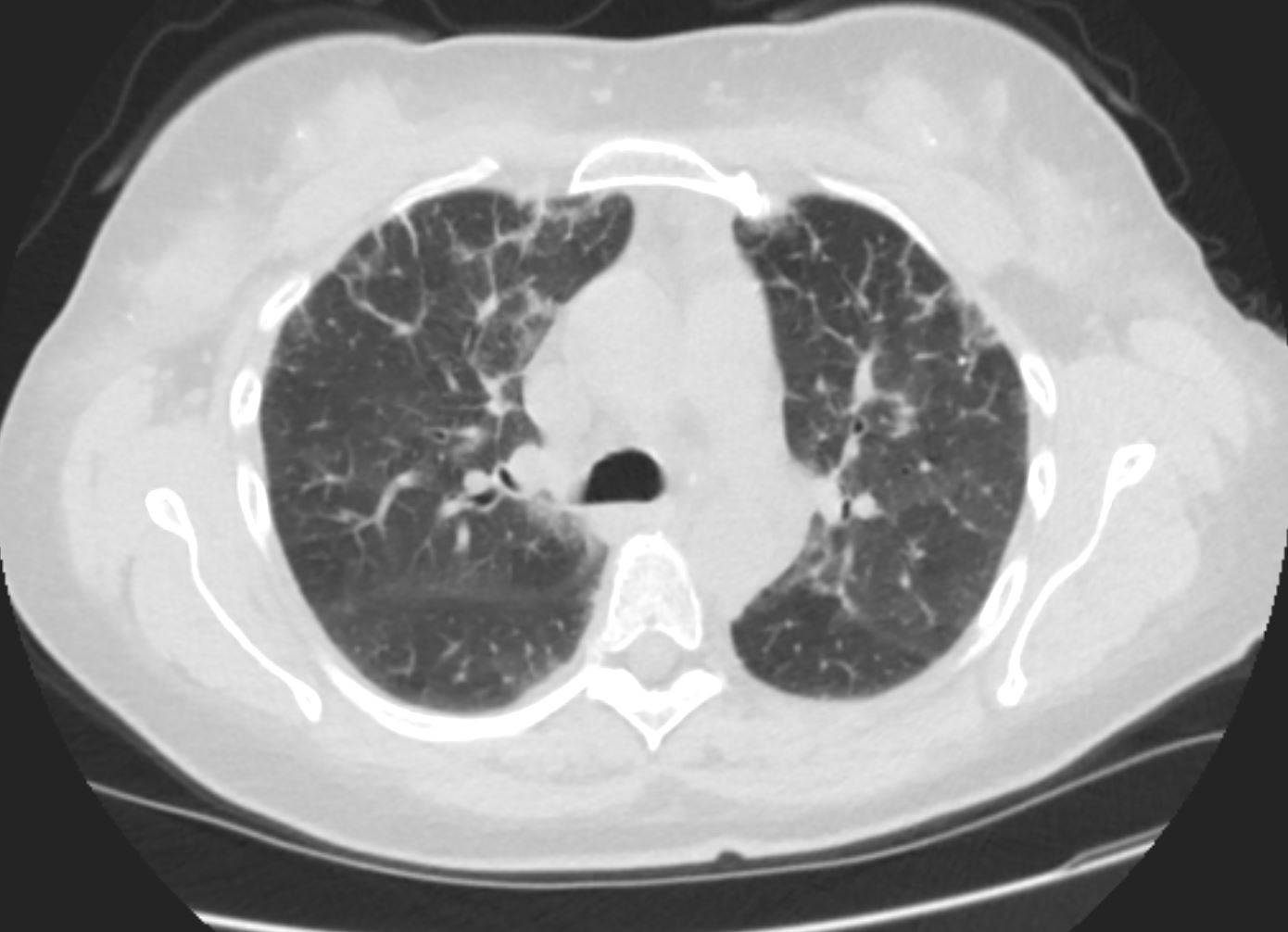
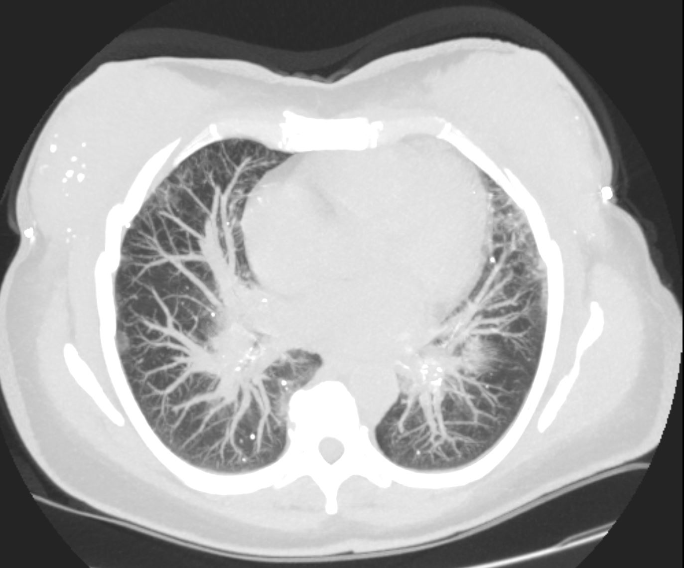
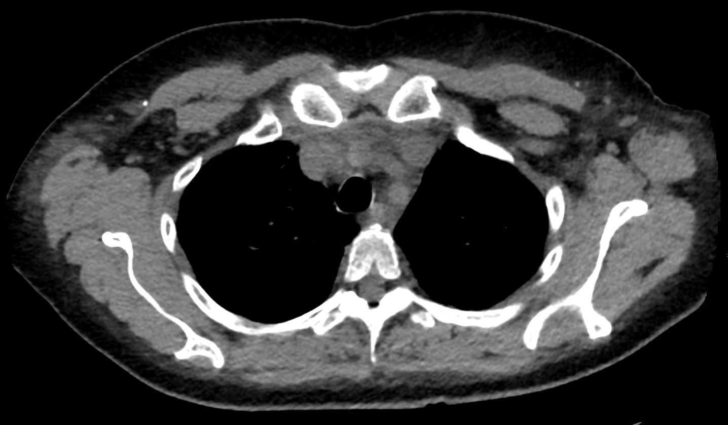
CT Alveolar Septal Amyloidosis and Dystrophic Calcifications
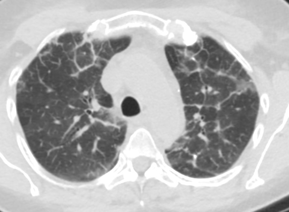
56 -year-old female with a history of amyloidosis (AL) presents for follow up. Axial CT of the chest shows diffuse reticular process best appreciated anteriorly with thickening of the interlobular septa. In the appropriate clinical setting these findings are compatible with the diagnosis of alveolar septal amyloidosis.
Ashley Davidoff MD TheCommonVein.net 244 Lu 135741d02
56 -year-old female with a history of amyloidosis (AL) presents for follow up. Axial CT of the chest shows diffuse reticular process best appreciated anteriorly with thickening of the interlobular septa. Punctate, dystrophic calcifications are seen in the interlobular septa (white arrowheads a, c and d), as well as in the pleura abutting the mediastinum (white arrowhead a). Image b shows similar calcifications, likely deposits of amyloid in the axilla with surrounding soft tissue changes
Ashley Davidoff MD TheCommonVein.net 244 Lu 135741d02cL
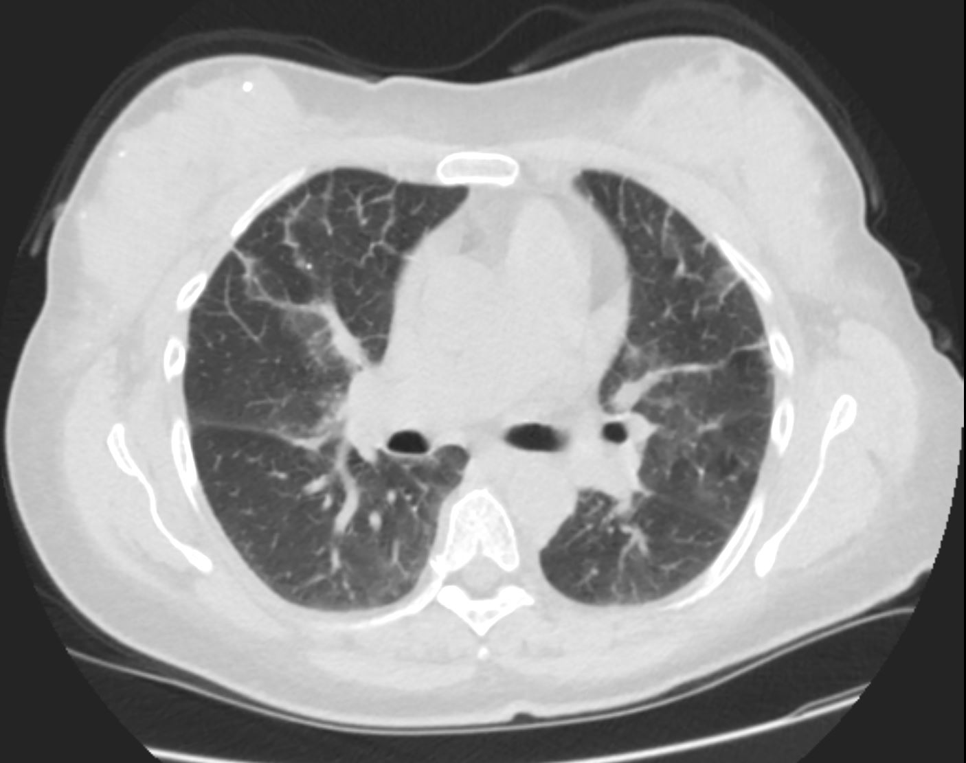
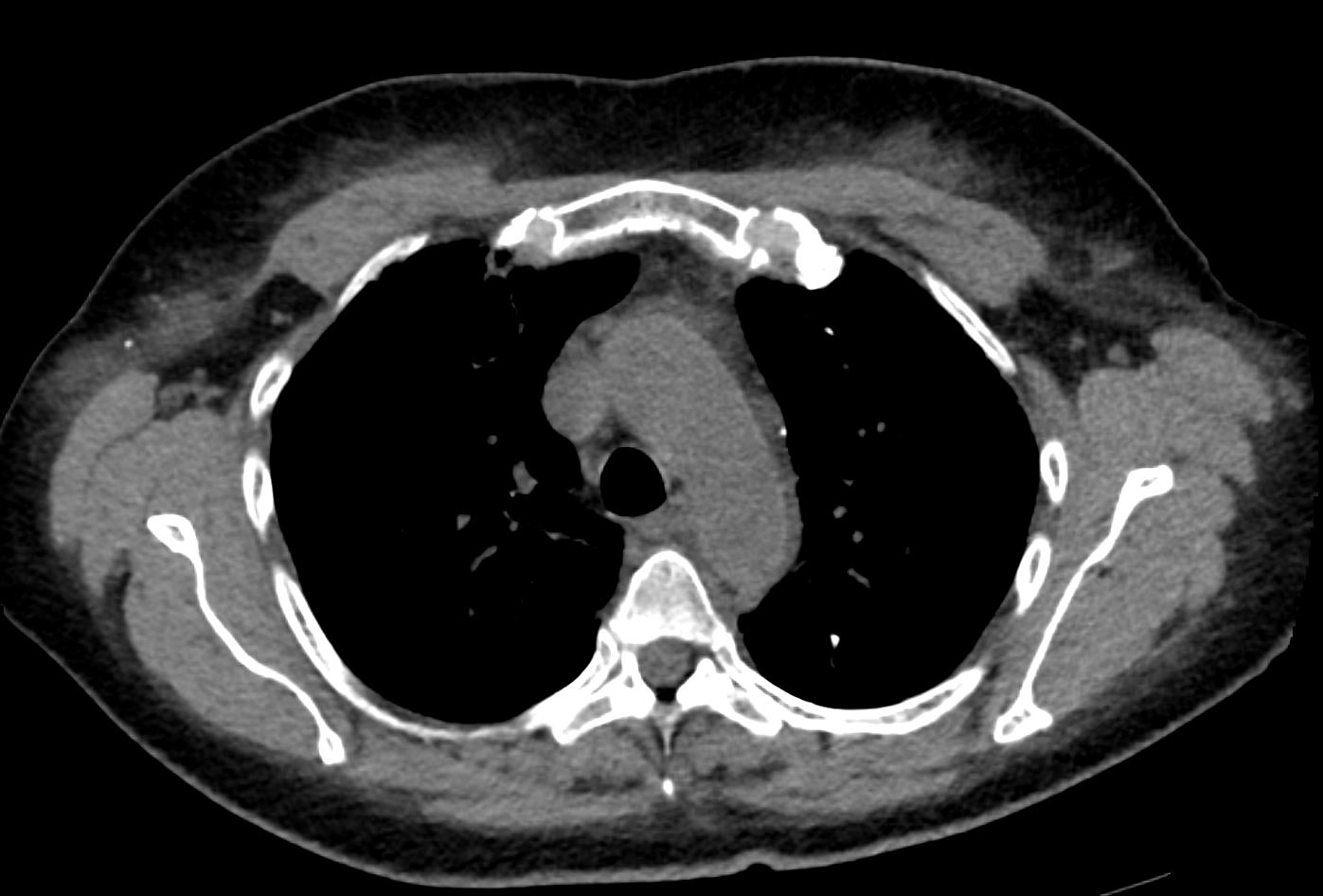
CT Alveolar Septal Amyloidosis
56 -year-old female with a history of amyloidosis (AL) presents for follow up. Axial CT of the chest shows diffuse reticular process best appreciated anteriorly with thickening of the interlobular septa. In the appropriate clinical setting these findings are compatible with the diagnosis of alveolar septal amyloidosis.
Ashley Davidoff MD TheCommonVein.net 244 Lu 135741d03
56 -year-old female with a history of amyloidosis (AL) presents for follow up. Axial CT of the chest shows diffuse reticular process best appreciated anteriorly with thickening of the interlobular septa. In the appropriate clinical setting these findings are compatible with the diagnosis of alveolar septal amyloidosis. There is a punctate dystrophic calcification in interlobular septum in the anterior segment of the right upper lobe as well as in the right breast
Ashley Davidoff MD TheCommonVein.net 244 Lu 135741d04
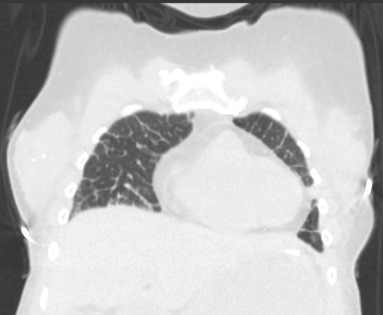
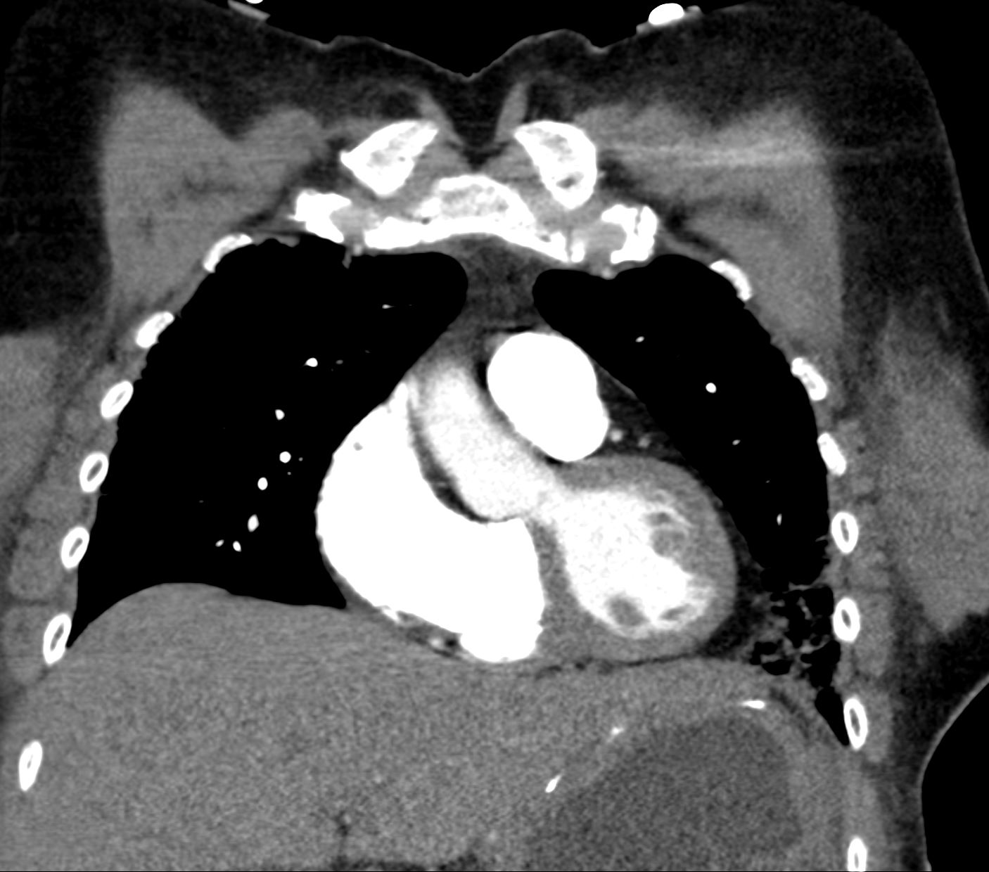
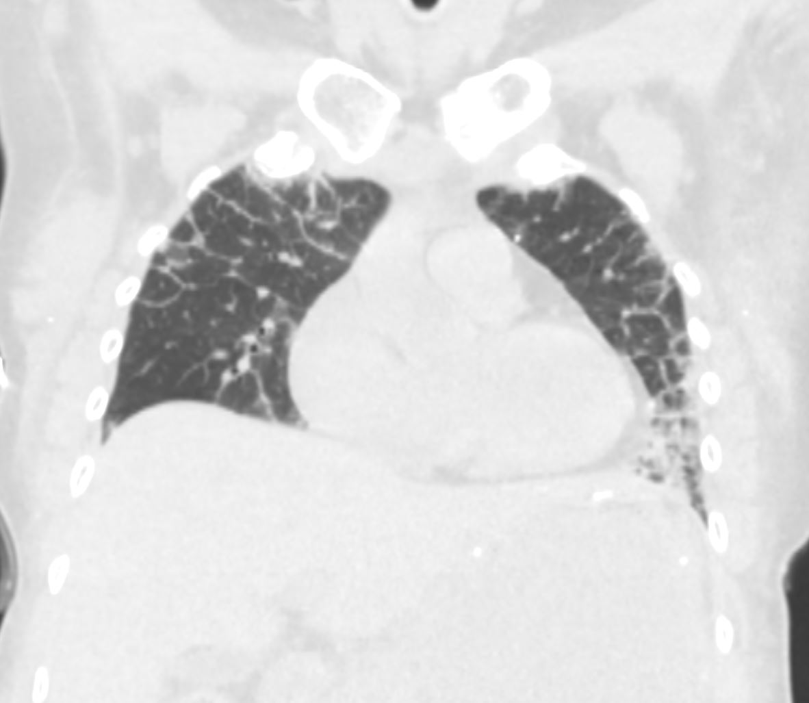
56 -year-old female with a history of amyloidosis (AL) presents for follow up. Coronal CT of the chest shows thickening of the interlobular septa most prominent in the periphery. In the appropriate clinical setting these findings are compatible with the diagnosis of alveolar septal amyloidosis.
Ashley Davidoff MD TheCommonVein.net 244 Lu 135741d14
56 -year-old female with a history of amyloidosis (AL) presents for follow up. Coronal CT of the chest shows thickening of the interlobular septa most prominent in the periphery. In the appropriate clinical setting these findings are compatible with the diagnosis of alveolar septal amyloidosis.
Ashley Davidoff MD TheCommonVein.net 244 Lu 135741d15
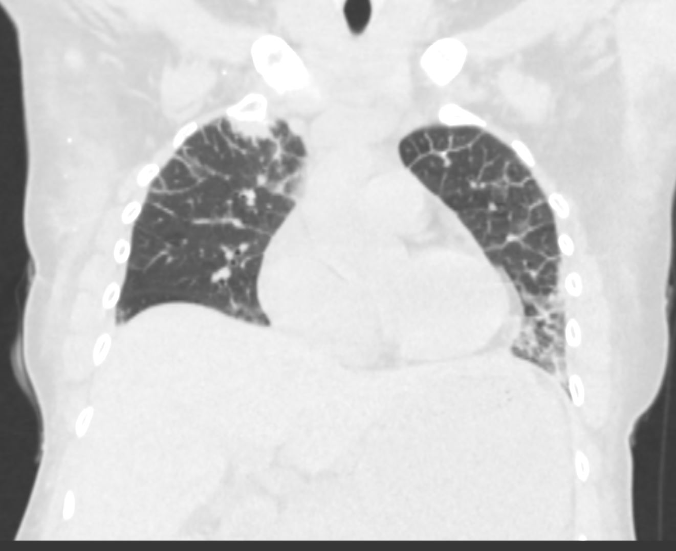
56 -year-old female with a history of amyloidosis (AL) presents for follow up. Coronal CT of the chest shows thickening of the interlobular septa most prominent in the periphery. In the appropriate clinical setting these findings are compatible with the diagnosis of alveolar septal amyloidosis.
Ashley Davidoff MD TheCommonVein.net 244 Lu 135741d16
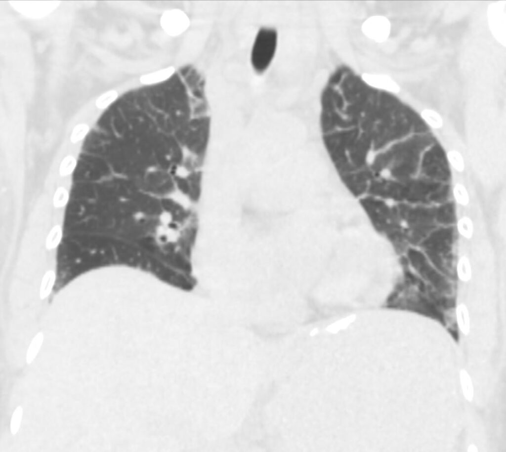
56 -year-old female with a history of amyloidosis (AL) presents for follow up. Coronal CT of the chest shows thickening of the interlobular septa most prominent in the periphery. In the appropriate clinical setting these findings are compatible with the diagnosis of alveolar septal amyloidosis.
Ashley Davidoff MD TheCommonVein.net 244 Lu 135741d17
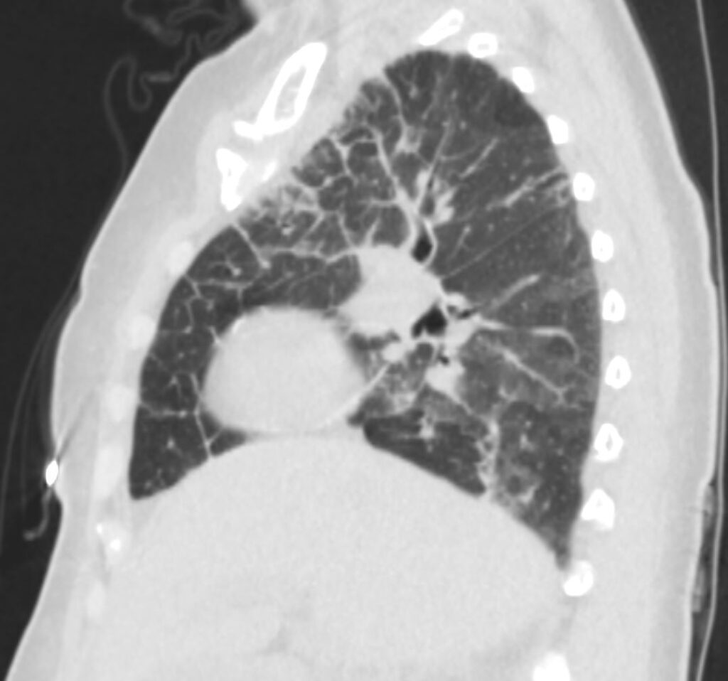
56 -year-old female with a history of amyloidosis (AL) presents for follow up. Sagittal CT of the chest shows thickening of the interlobular septa most prominent in the periphery and anteriorly. In the appropriate clinical setting these findings are compatible with the diagnosis of alveolar septal amyloidosis.
Ashley Davidoff MD TheCommonVein.net 244 Lu 135741d18
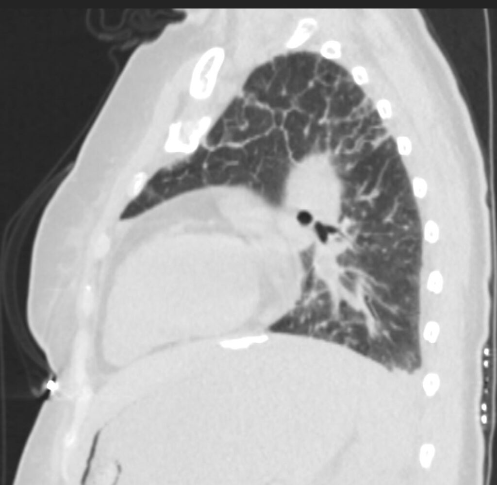
56 -year-old female with a history of amyloidosis (AL) presents for follow up. Sagittal CT of the chest shows thickening of the interlobular septa most prominent in the periphery and superiorly. In the appropriate clinical setting these findings are compatible with the diagnosis of alveolar septal amyloidosis.
Ashley Davidoff MD TheCommonVein.net 244 Lu 135741d19
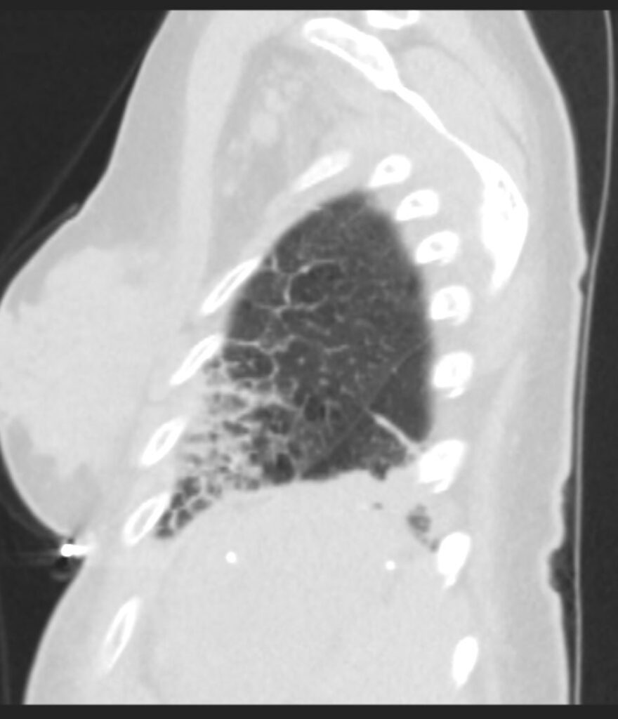
56 -year-old female with a history of amyloidosis (AL) presents for follow up. Sagittal CT of the chest shows thickening of the interlobular septa. Superiorly the septal thickening is smoother and finer and inferiorly the thickening is thicker and coarser. In the appropriate clinical setting these findings are compatible with the diagnosis of alveolar septal amyloidosis.
Ashley Davidoff MD TheCommonVein.net 244 Lu 135741d20
56 -year-old female with a history of amyloidosis (AL) presents for follow up. Axial CT with MIP shows extensive punctate calcifications throughout the lungs likely representing amyloid deposits. Calcifications also noted in the right breast
Ashley Davidoff MD TheCommonVein.net 244 Lu 135741d12
CT Alveolar Septal Amyloidosis Soft Tissue Changes and Dystrophic Calcifications
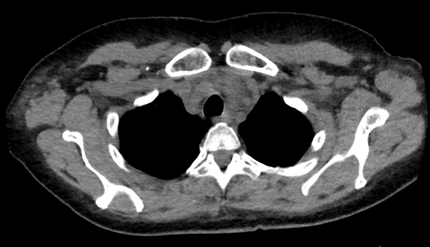
56 -year-old female with a history of amyloidosis (AL) presents for follow up. Axial CT of the chest shows diffuse soft tissue changes with thickening of the subcutaneous tissues in the right axilla and to lesser extent the left axilla surrounding the left lobe of the thyroid gland and in the subpectoral regions. There is a punctate dystrophic calcification in the right subpectoral region
Ashley Davidoff MD TheCommonVein.net 244 Lu 135741d05
56 -year-old female with a history of amyloidosis (AL) presents for follow up. Axial CT of the chest shows diffuse soft tissue changes with thickening of the subcutaneous tissues in the right axilla and to lesser extent the left axilla, and in the anterior superior mediastinum
Ashley Davidoff MD TheCommonVein.net 244 Lu 135741d06
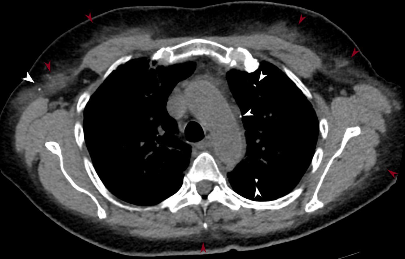
56 -year-old female with a history of amyloidosis (AL) presents for follow up. Axial CT of the chest shows punctate, dystrophic calcifications in the left upper lobe, pleura abutting the mediastinum and in the right axilla (white arrowheads). There is induration in the soft tissues suggesting amyloidosis (maroon arrowheads).
Ashley Davidoff MD TheCommonVein.net 244 Lu 135741d02m05L
56 -year-old female with a history of amyloidosis (AL) presents for follow up. Axial CT of the chest shows punctate, dystrophic calcifications in the superficial regions of the glandular tissue of the breasts, more prominent on the right. There is induration in the soft tissues in both axilla
Ashley Davidoff MD TheCommonVein.net 244 Lu 135741d07
56 -year-old female with a history of amyloidosis (AL) presents for follow up. Axial CT of the upper abdomen shows the fluid filled stomach with dystrophic amyloid calcifications in the gastric
Ashley Davidoff MD TheCommonVein.net 244 Lu 135741d11c
56-year-old female with a history of lambda AL amyloidosis presents for evaluation with most recent CT f6 months ago at which time occlusion of the proximal segmental distalmost left lower lobe artery was noted with hypoperfusion of the left lower lung parenchyma associated with multifocal consolidative opacities within the left lobe
CT without contrast shows mostly unchanged appearance of the lungs dominated by interlobular septal thickening in both the upper lobes and lower lobes and likely related to alveolar septal amyloidosis.
The parenchymal infiltrate in the inferior lingula and a band of infiltrate in the left lower lobe inferiorly have not changed since the last study. A wedge-shaped subsegmental defect in the lateral segment of the left lower lobe has improved and likely reflects resolving prior subsegmental infarction (Hampton’s hump)
Calcification in the region of the stomach wall and retroperitoneum likely reflect amyloid deposits in these regions. This finding correlates well with previous identification of a thickened gastric wall best appreciated on the study 2 years ago. The stomach also appears to be chronically distended and gastric stasis is included in the radiologic differential diagnosis