- Discoid
-

Linear Atelectasis
102 year old female with linear (discoid) atelectasis in the lingula.
Ashley Davidoff MD TheCommonVein.netLinear or Discoid Atelectasis
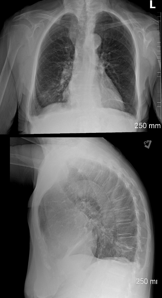
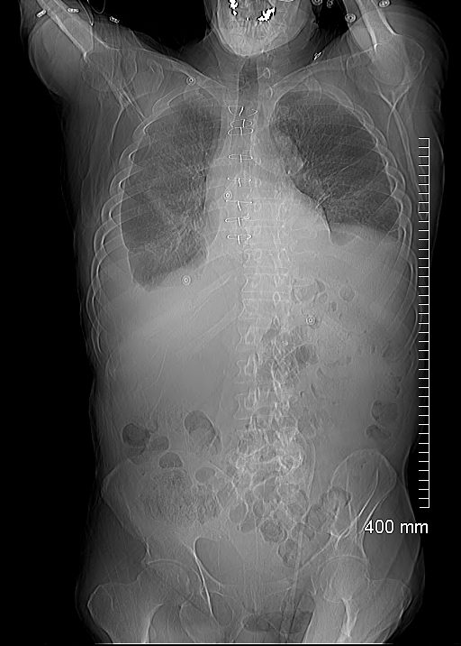
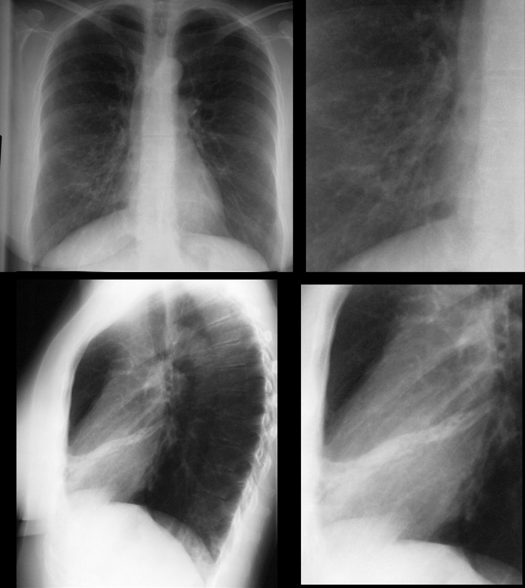
CXR PA and lateral 6 months prior (top row) shows no parenchymal abnormality. Loop recorder noted. 6 months later the 81 year male presents with a cough and the scout film shows discoid atelectasis in the left upper lung field with hyperinflation of the lower lung field (middle row)
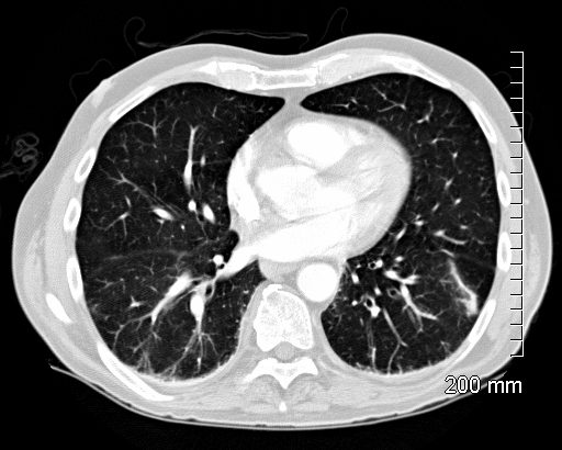
The sagittal CT reconstruction and axial image (lower row shows segmental atelectasis of the posterior segment of the left upper lobe. Hyperinflation of the lower lobe is noted.
Ashley Davidoff MD TheCommonVein.netLinear Atelectasis
60 year old male with linear (discoid) atelectasis in the middle lobe and the left upper lobe on CT. Note moderate sized bilateral pleural effusions
Ashley Davidoff MD TheCommonVein.netLinear Atelectasis
60 year old male with linear (discoid) atelectasis in the middle lobe and the left upper lobe on CT. Note moderate sized bilateral pleural effusion. Minor compressive atelectasis caused by the left effusion.
Ashley Davidoff MD TheCommonVein.netLinear Atelectasis
49-year-old male with linear (discoid) atelectasis in the middle lobe.
Ashley Davidoff MD TheCommonVein.netLinear Atelectasis
66 year old male with linear (discoid) atelectasis in the left lower lobe on CT
Ashley Davidoff MD TheCommonVein.net -
- Lobar
- Left Lung Collapse
-
White Out of the CXR with Passive Compressive Atelectasis of the Left Lung
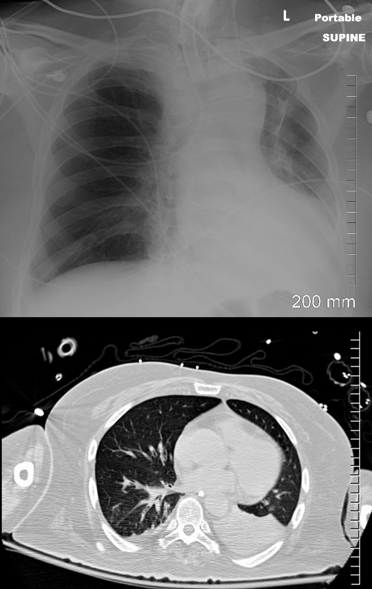
48 year-old male presents with a dyspnea. CXR shows a total white out of the left chest with pulmonary congestion. CT scan shows a large left pleural effusion with total atelectasis of the left lung. Incidental note is made of premature calcific coronary artery disease.
Ashley Davidoff MD TheCommonVein.netLeft Lung Collapse and Large Effusion s/p Stent
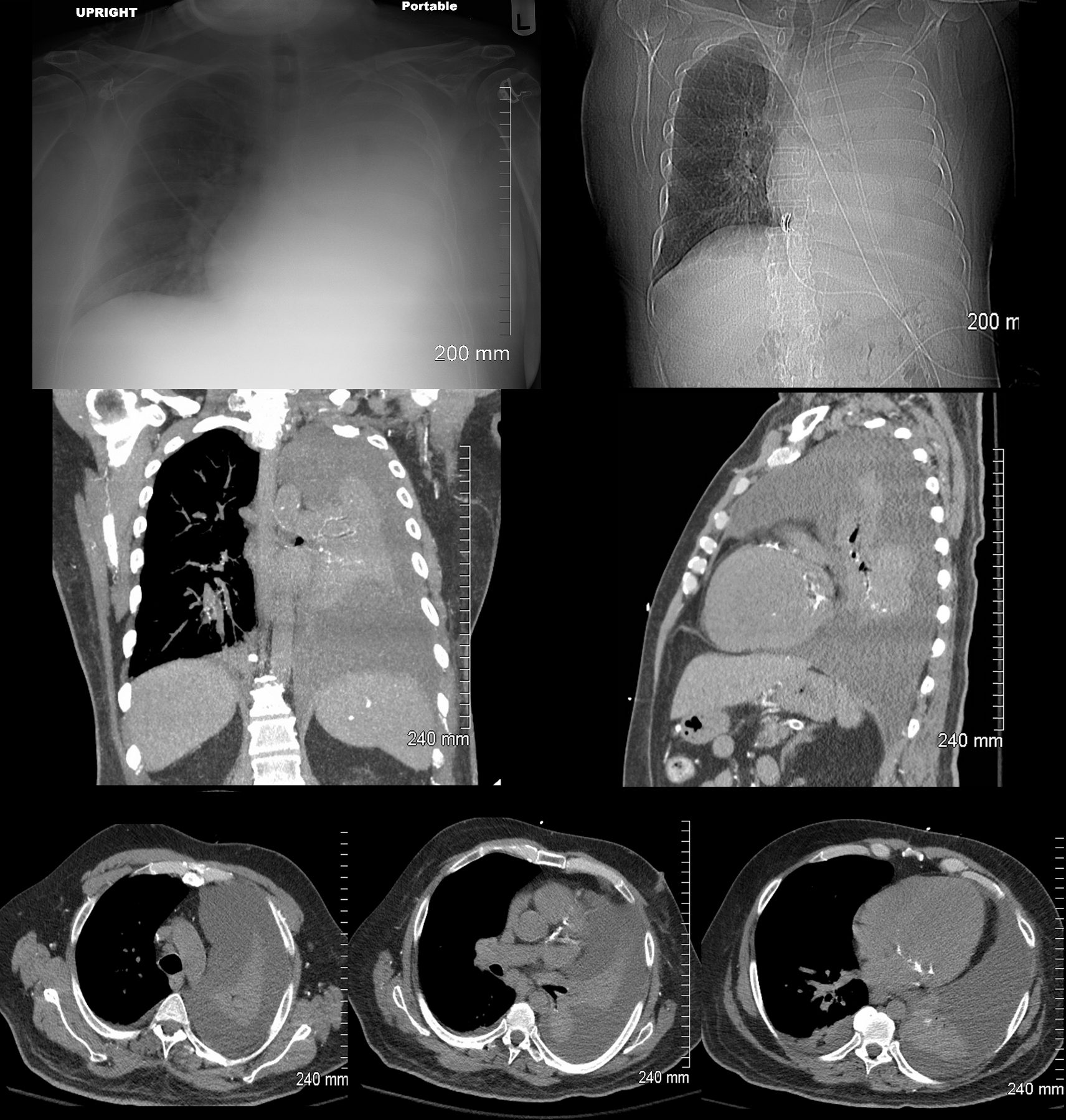
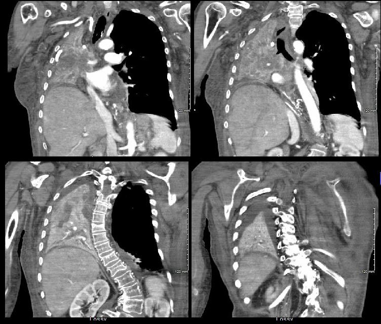
82-year-old female with dyspnea presents with an obstructing lesion of the left main stem bronchus and total atelectasis of the left lung and associated effusion. White out of the left lung with mediastinal shift are noted on the scout film (top left) and the axial images confirm complete collapse of the left lung with a large effusion. A proximal malignancy was found, and the left mainstem bronchus stented, unsuccessfully. Middle left image shows blocked stent.
Ashley Davidoff MD TheCommonVein.net
134720c - Left Upper Lobe
-
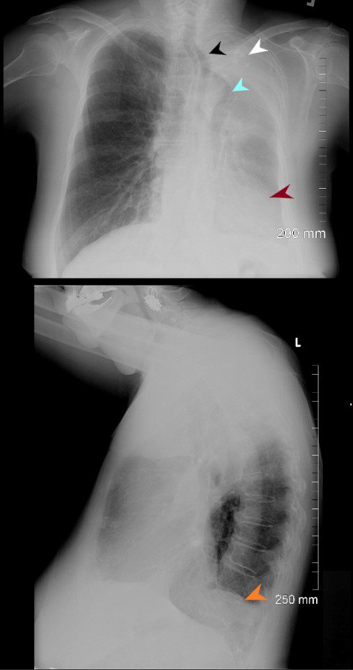
76-year old female presents with dyspnea. PA CXR shows airless consolidation of the left upper lobe (white arrowhead), volume loss of the left hemithorax, with leftward shift of the trachea (black arrowhead) and cardio mediastinal structures (maroon arrowhead). The right lung is hyperinflated with herniation across the midline (light blue arrowhead). The lateral examination shows an anterior airless consolidation, and elevation of the left hemidiaphragm orange arrowhead). Granulomatous calcifications are noted in the left upper lobe atelectatic segment
Ashley Davidoff MD TheCommonVein.net -
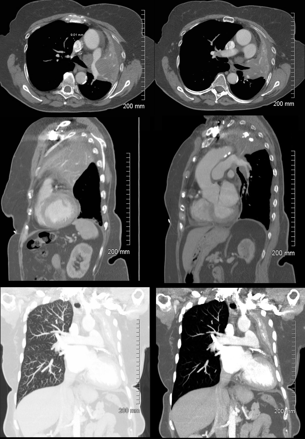
76-year-old female presents with dyspnea CT scan shows findings of left upper lobe atelectasis characterized by airless consolidation, with leftward shift of the cardio mediastinal structures. There is evidence of encasement of the pulmonary artery with a soft tissue mass surrounding it. There is occlusion of the left upper lobe bronchus. Granulomatous calcifications are noted in the left upper lobe atelectatic segment
Ashley Davidoff MD TheCommonVein.net - Central Squamous Cell Carcinoma
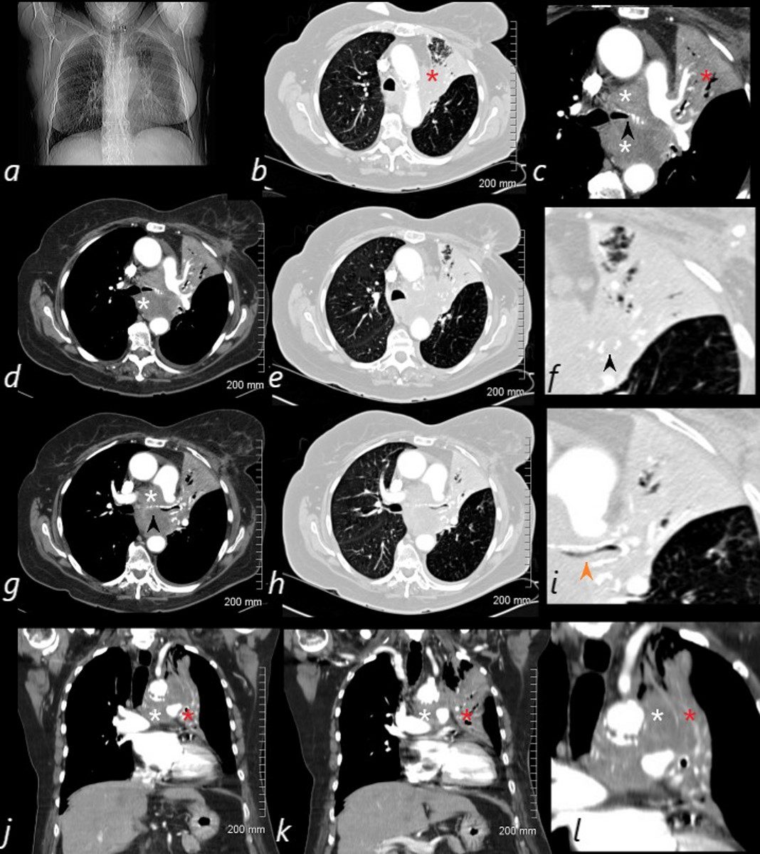
Left Upper Lobe Collapse by Central Squamous Cell Carcinoma
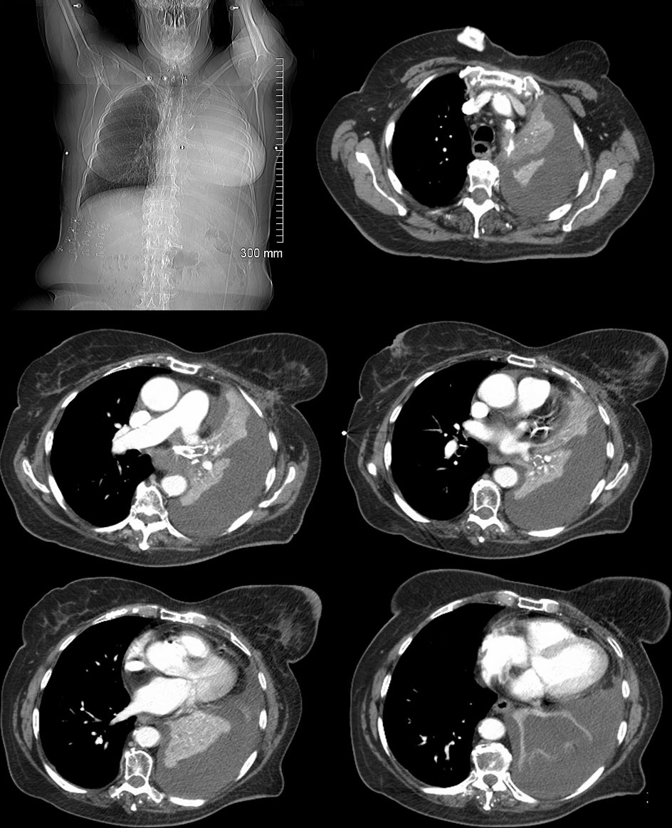
82-year-old female with dyspnea presents with an obstructing lesion of the left main stem bronchus and total atelectasis of the left lung caused by a central squamous cell carcinoma mass. The scout film (a) shows a vague area of increase density in the left upper lung field, with minimal elevation of the left mainstem bronchus. The central mass (white asterisk – is noted in c, d, g, j, k and l. Invasion and obstruction of the left main stem bronchus (black arrow – f, g) with invasion into the lumen without obstruction is notes in I – orange arrow. Post obstructive atelectasis of the left upper lobe is noted (red asterisk – b,c,j,k,l) with hyperinflated left lower lobe extending to the left apex.
Ashley Davidoff MD TheCommonVein.net - Posterior Segmental Discoid Atelectasis
-
CXR PA and lateral 6 months prior (top row) shows no parenchymal abnormality. Loop recorder noted. 6 months later the 81 year male presents with a cough and the scout film shows discoid atelectasis in the left upper lung field with hyperinflation of the lower lung field (middle row)
The sagittal CT reconstruction and axial image (lower row shows segmental atelectasis of the posterior segment of the left upper lobe. Hyperinflation of the lower lobe is noted.
Ashley Davidoff MD TheCommonVein.net - Lingula Atelectasis
56-year-old male presents with history of Central Non Small Lung Cancer with Lingula Atelectasis
Axial CT images show a central mass with lingula atelectasis. The scout film shows silhouetting of the left heart border. The CXR shows similar finding following stent placement in the lingula
Ashley Davidoff MD TheCommonVein.net - Left Lower Lobe
-
Left Lower Lobe Atelectasis
57-year old male presents with a cough. CXR shows silhouetting of the left hemidiaphragm and leftward mediastinal shift. CT scan shows an airless consolidation with leftward shift consistent with atelectasis.
Ashley Davidoff MD TheCommonVein.net -
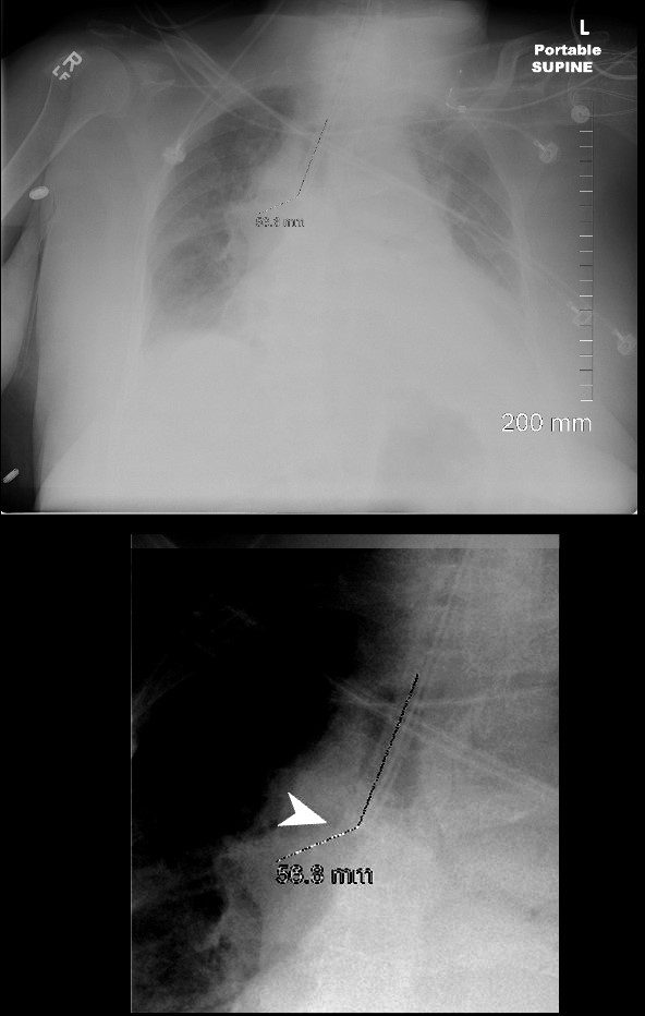
82-year-old female s/p intubation for endotracheal tube (ETT) check. CXR 2 hours prior showed “white out” of the left hemithorax with misplaced ETT which was in the right main and causing obstruction of the left main. This CXR performed 2 hour later, following repositioning of the tube, shows persistent malpositioning of the ETT which remains in the right mainstem bronchus (white arrowhead). However there is re-expansion of the left upper lung field but persistent opacification of the left lower lobe
Ashley Davidoff MD TheCommonVein.net - Right Lung
-
Complex Pleural Effusion and Atelectasis of the Right Lung
59F shows total collapse of right lung with an
occluded right main step bronchus(top right image)associated with a
right sided effusion. The occlusion is likely due to proximal cancer
Ashley Davidoff MD TheCommonVein.net - Right Upper Lobe
- Right Middle Lobe
-
Middle lobe Atelectasis thought to be due to Mucus Plug
On the PA of the CXR there is a ill defined loss of the right heart border. There is mild elevation of the right hemidiaphragm. The lateral exam is more convincing of a middle lobe consolidation
Ashley Davidoff MD TheCommonVein.net
30667c - Right Lower Lobe