
Bacterial
Granulomatous
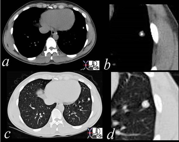
The nodule in the lingula has central calcification and is classical of granulomatous disease. No further work up for malignancy is required.
Ashley Davidoff, M.D. TheCommonVein.net Lung cancer P 034
TB Cavitating with Transbronchial Spread
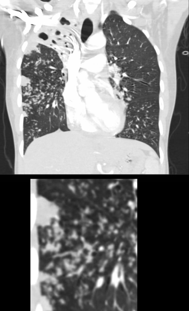
39-year-old immigrant Vietnamese male presents night sweats, fever, and cough. CT in the coronal plane of the chest shows a large cavitating lesion in the right upper lobe, with innumerable micronodules dominantly in the right midlung field, and to lesser extent in the right upper lung field. Some micronodules are probably present in the left lower lobe as well. Close to the largest subsegmental consolidation there is a bronchus which shows thickening of its wall.
Although it has the appearance has a “miliary” pattern, this term is usually referred to the hematogenous spread of the disease
Ashley Davidoff MD TheCommonvein.net 135786c
006Lu
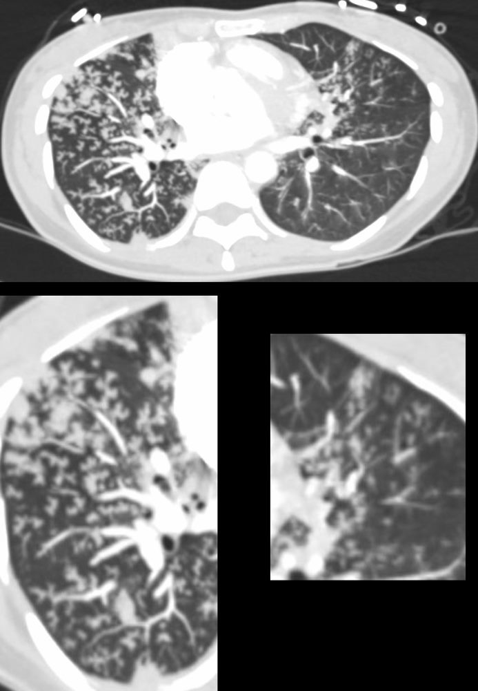
39-year-old immigrant Vietnamese male presents night sweats fever and cough. CT in the axial plane through the mid chest shows innumerable micronodules resulting from transbronchial spread with resultant tree in bud pattern scattered through the right lung (magnified in the right lower image). There are minimal similar changes in the lingula (magnified left lower image)..
Ashley Davidoff MD TheCommonvein.net 135789c 006Lu
Necrotizing Pneumonia
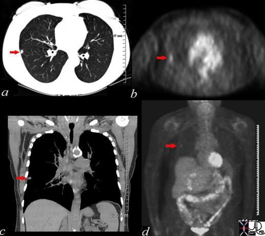
A small nodule (red arrow a, b,c) was identified on the CT in this 60-year-old female who is a smoker. The SUV at 1.3 was low (usually >2.5 in malignancy). At pathology, it was found to be a necrotizing pneumonia. Most necrotizing granulomas are related to infection.
Ashley Davidoff MD TheCommonVein.net Lung cancer P 035
Viral
Fungal
Acute Miliary Pulmonary Histoplasmosis
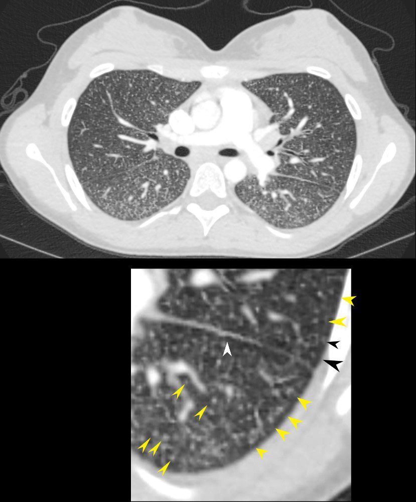
22-year-old female presents with flu like symptoms
CT of the chest at the level of the main pulmonary artery shows extensive diffuse miliary disease. Some of the nodules are centrilobular (yellow arrowheads). Some of the secondary lobules suggest mosaic attenuation (black arrowhead). The nodular and irregular appearance on the fissure (white arrowhead) indicates peri-lymphatic distribution.
Ashley Davidoff MD TheCommonvein.net 131708cL
Atypical